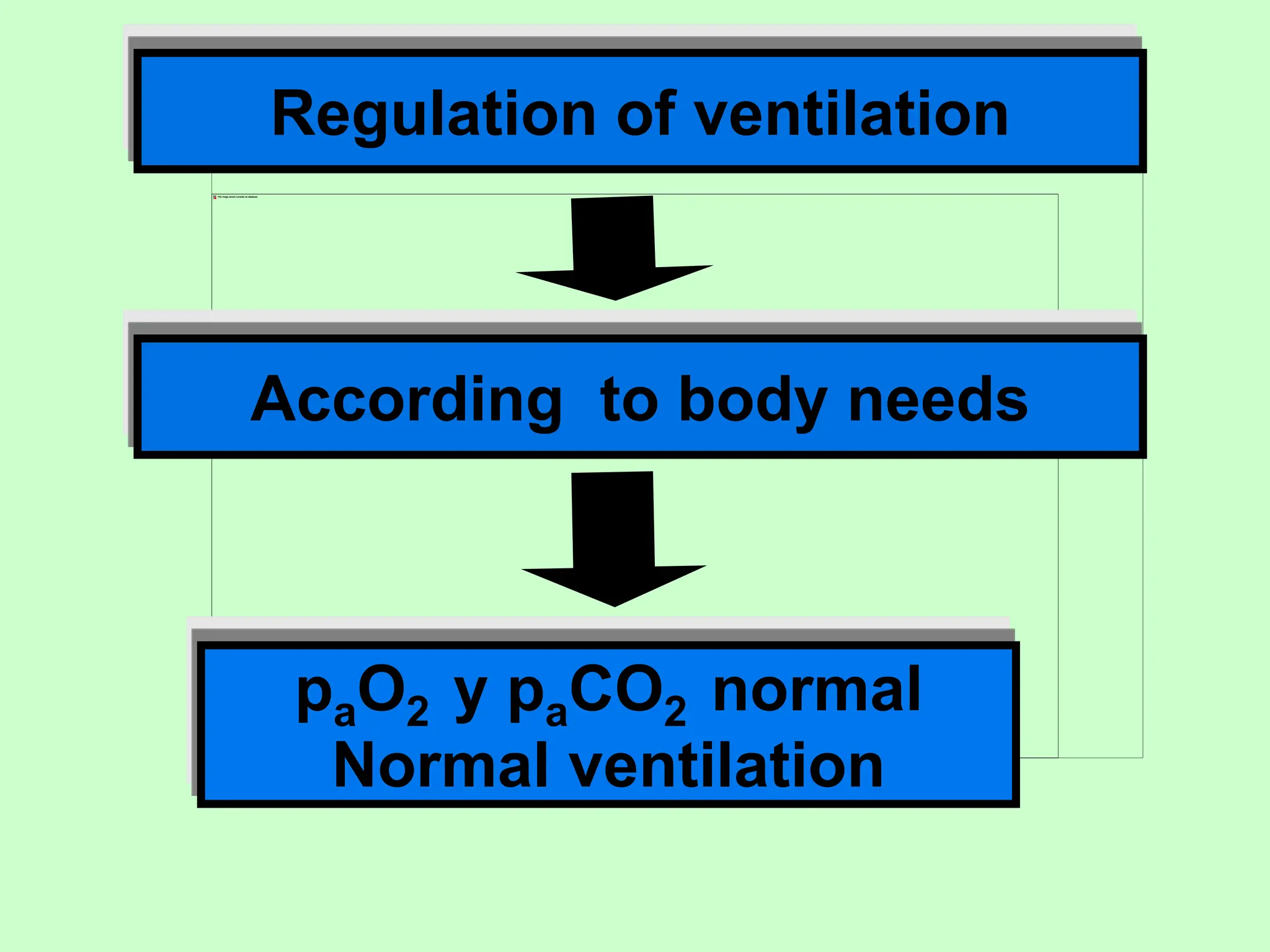
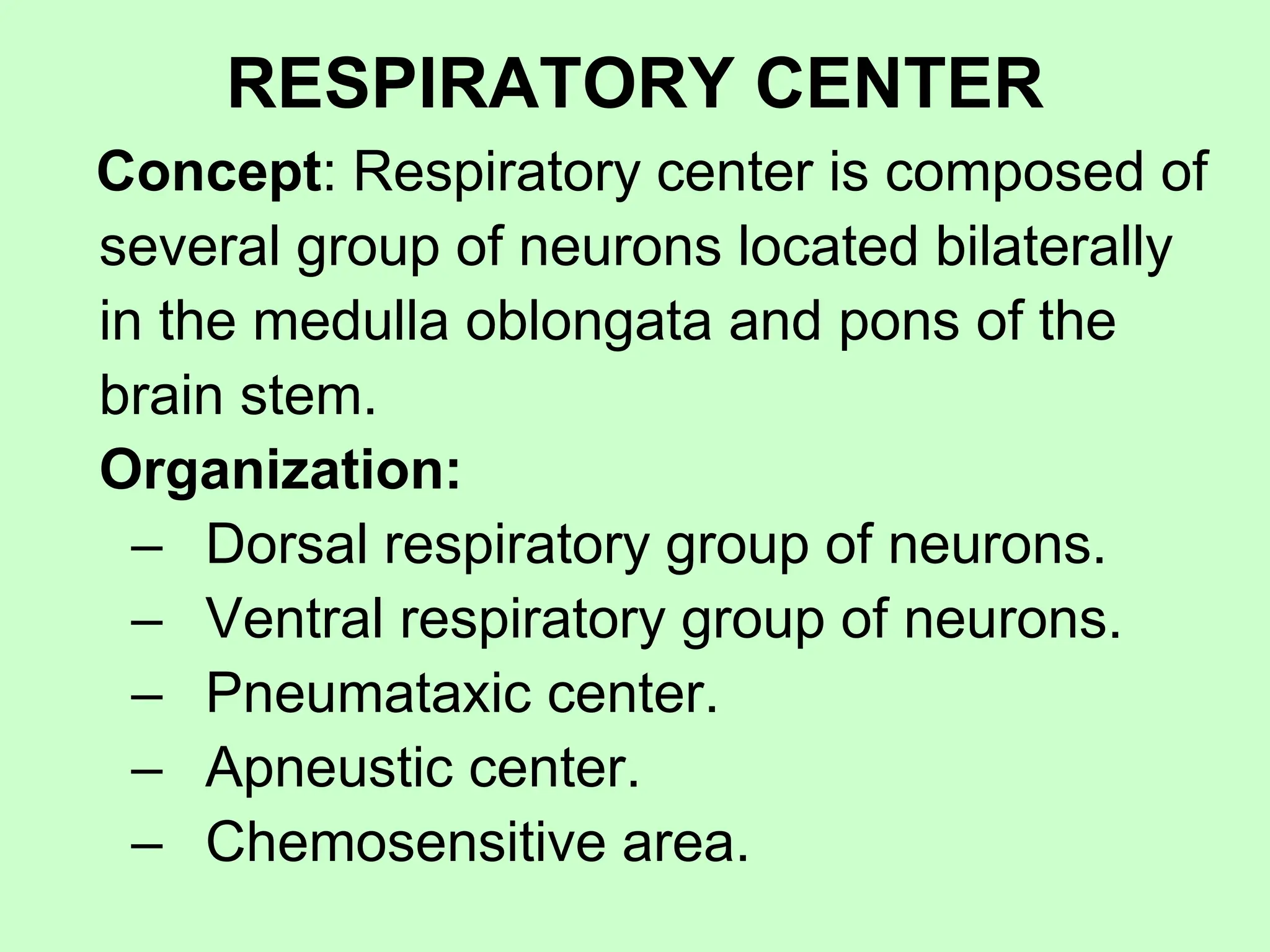
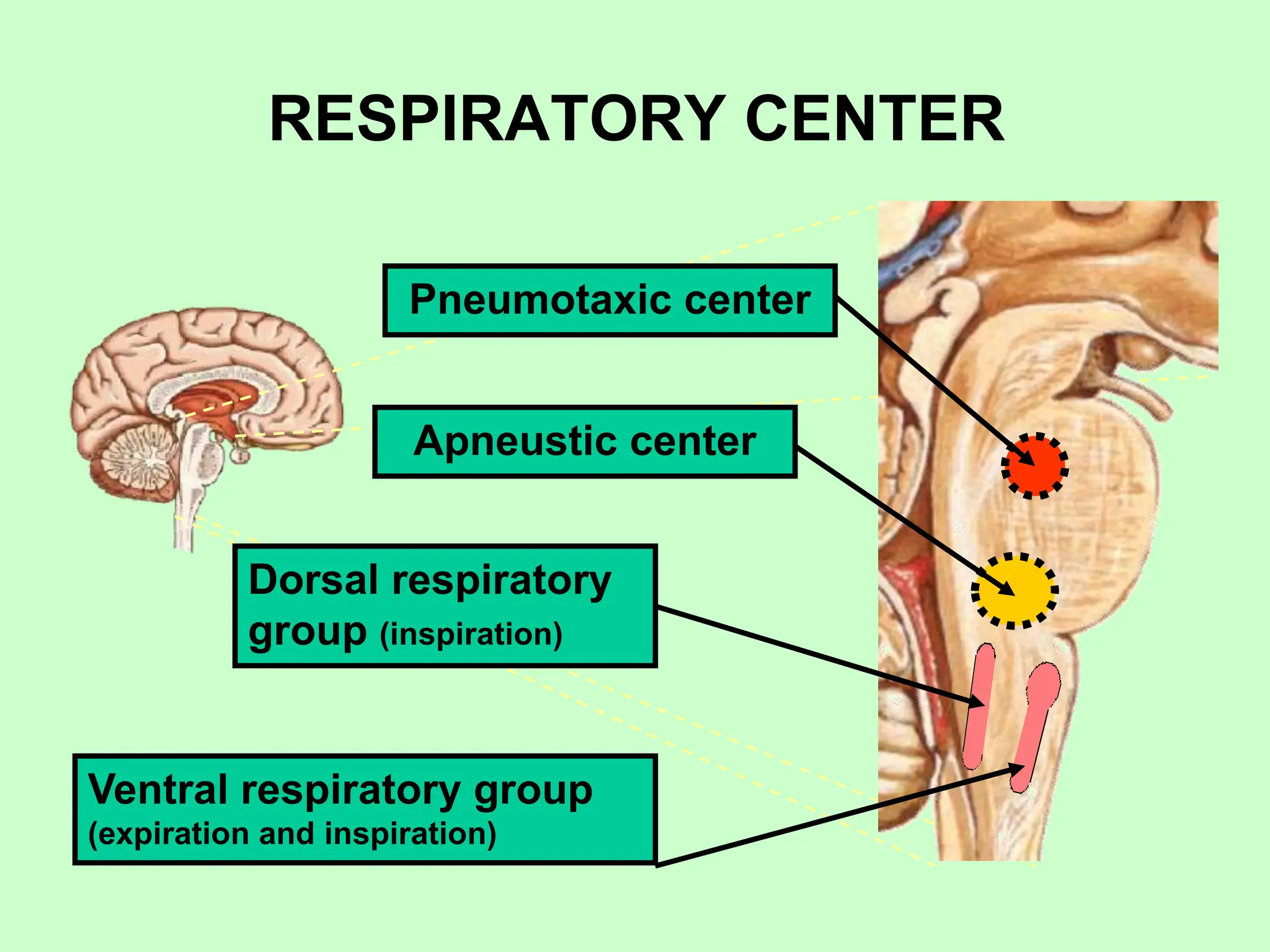
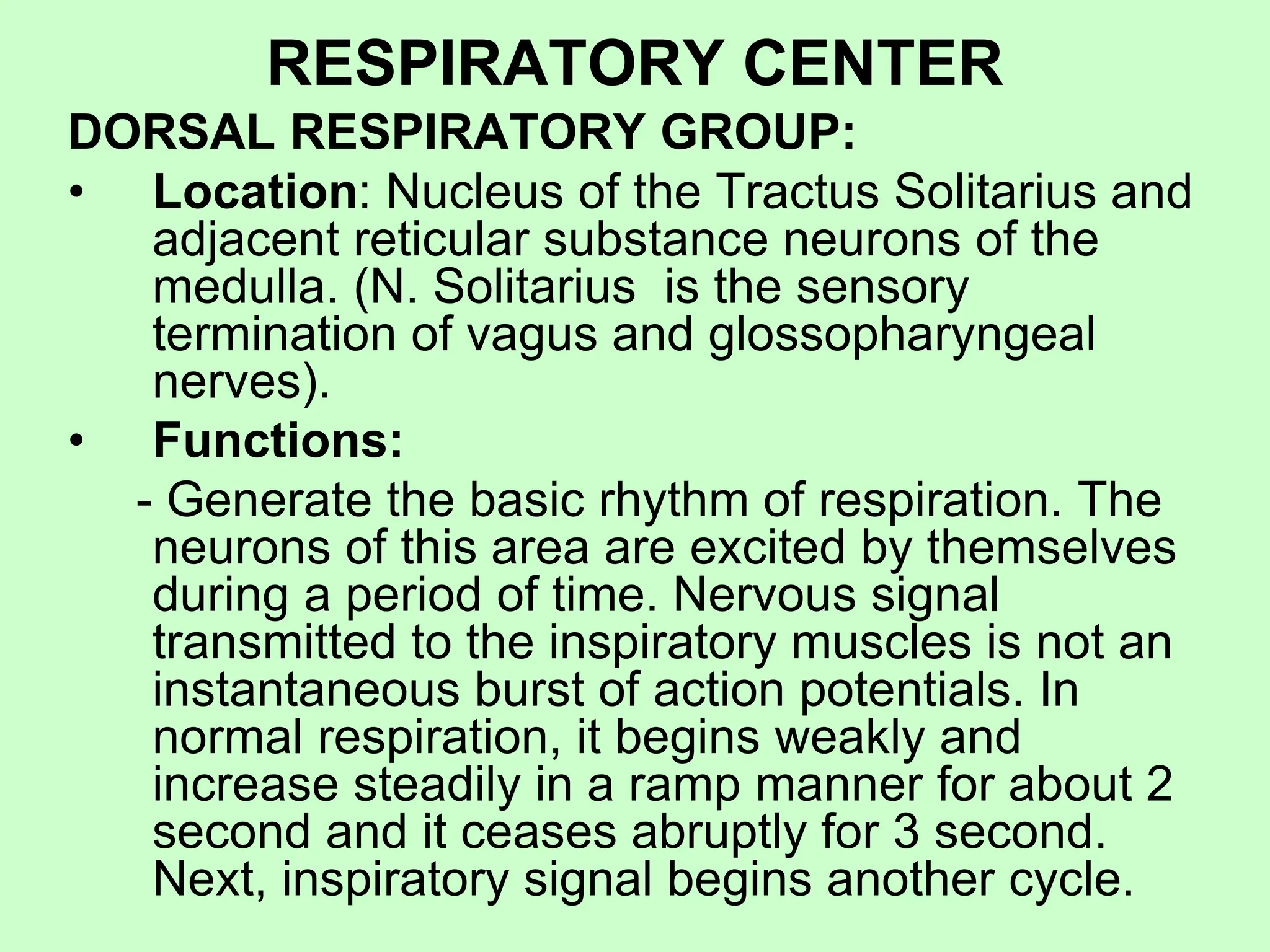
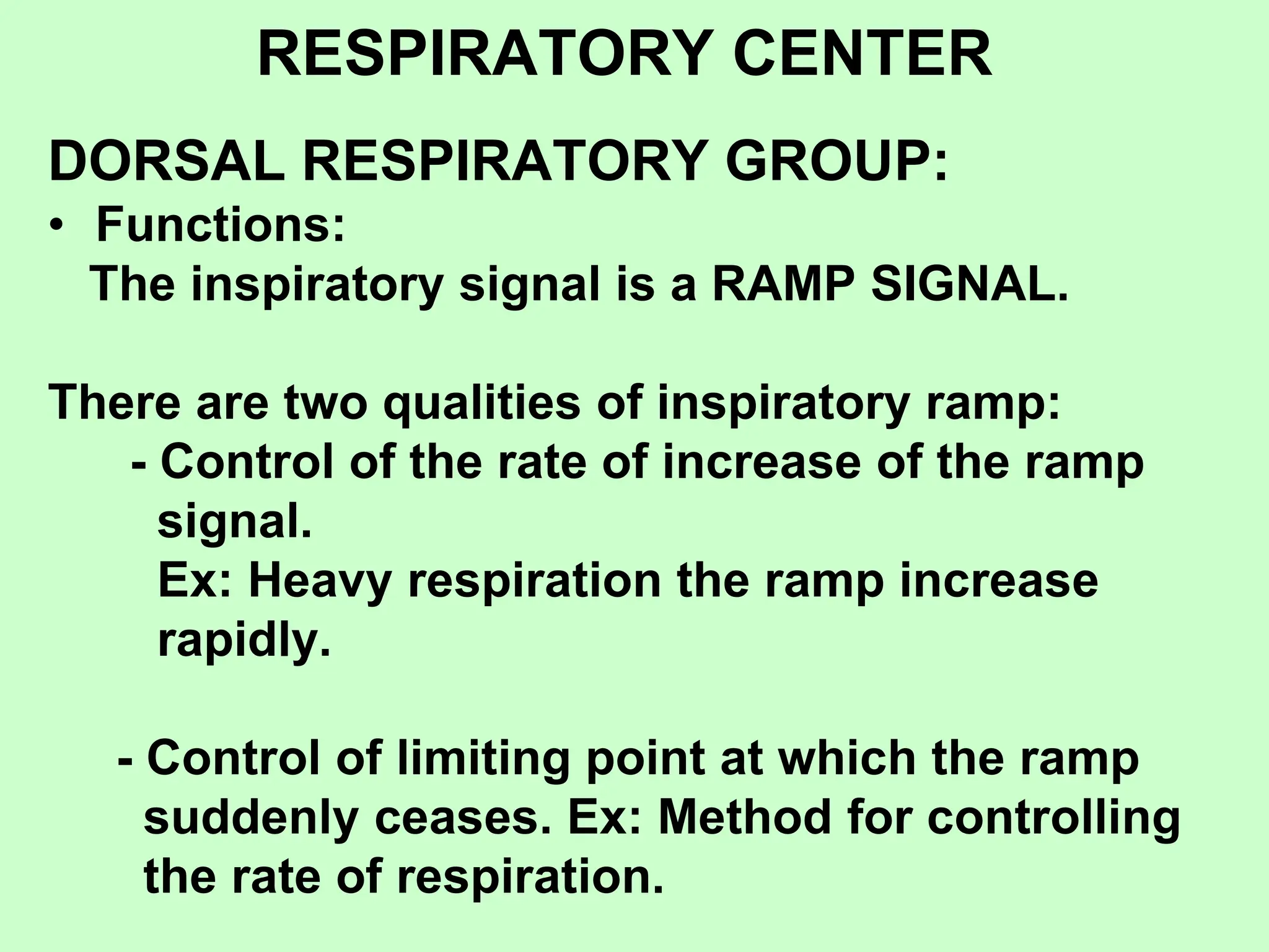
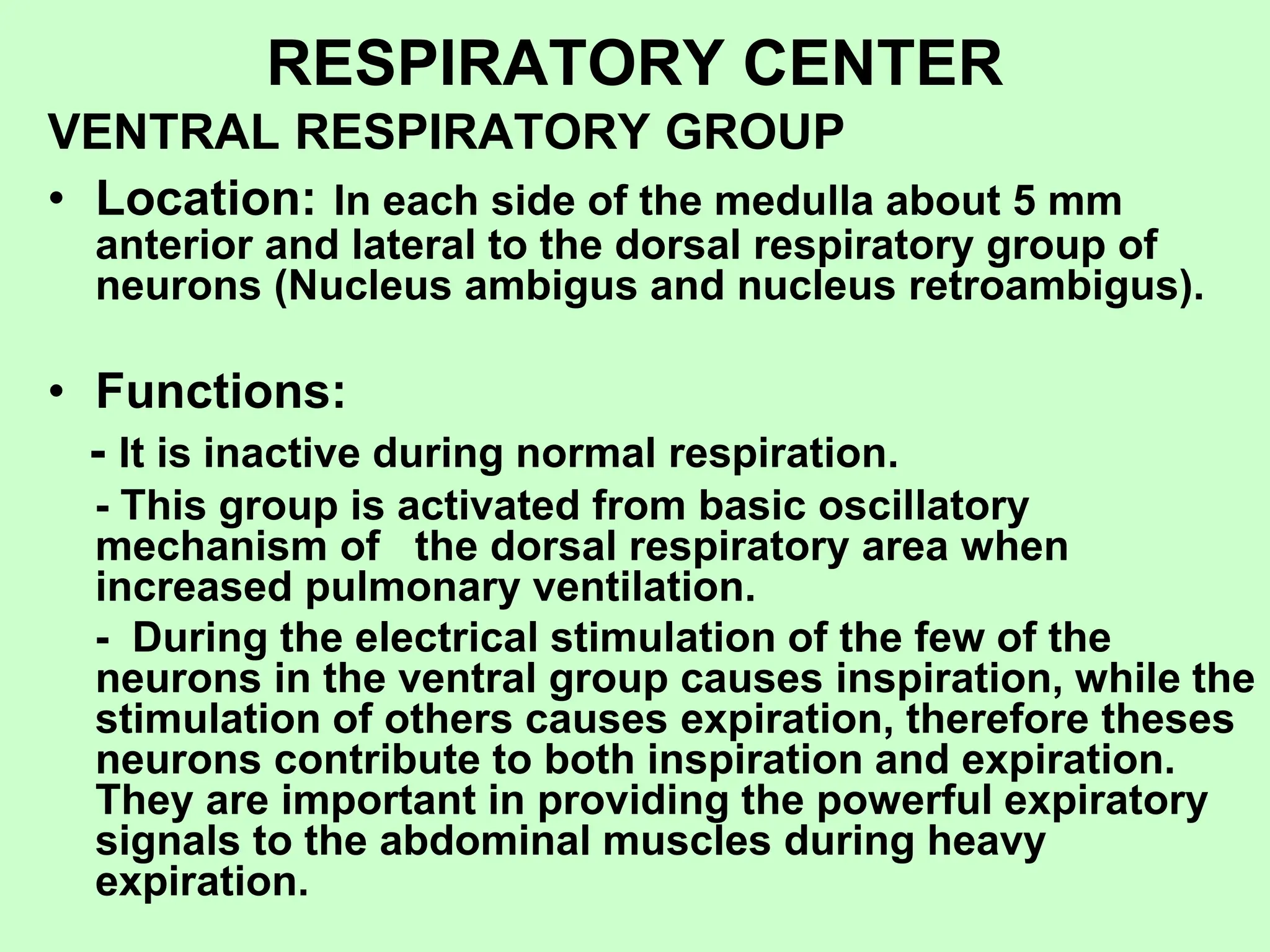
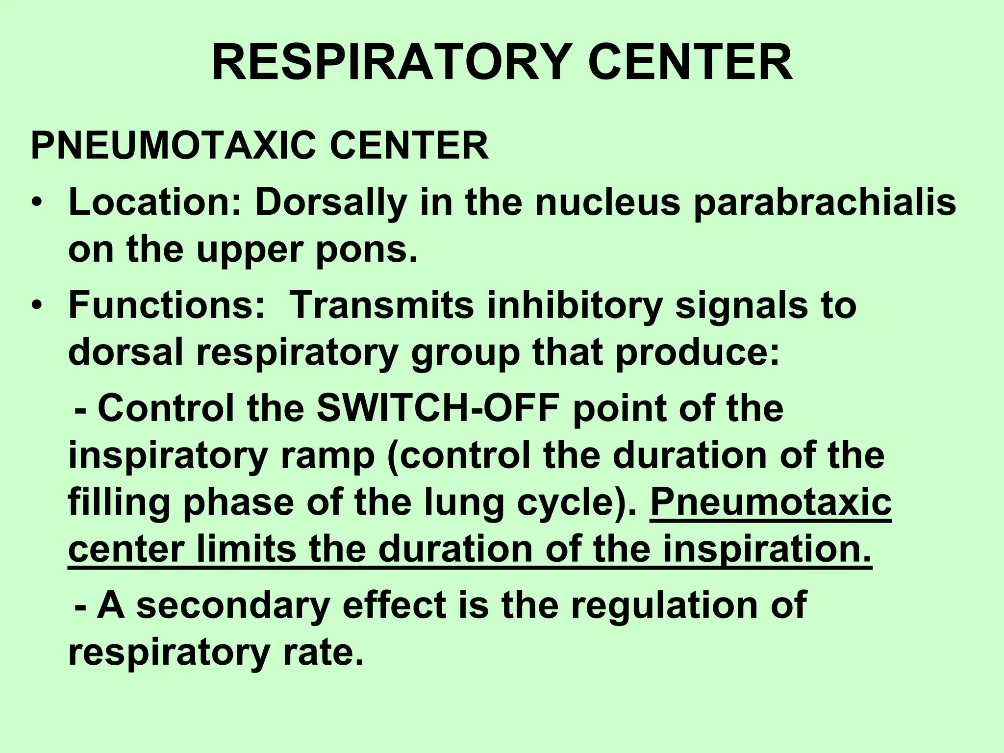
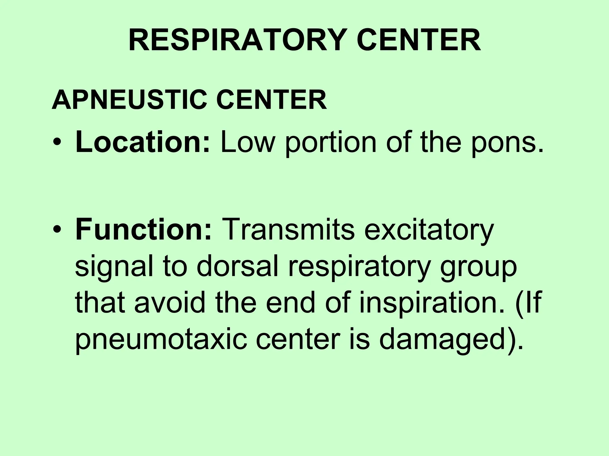
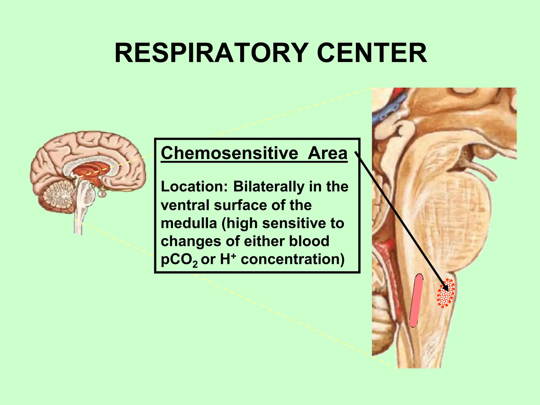
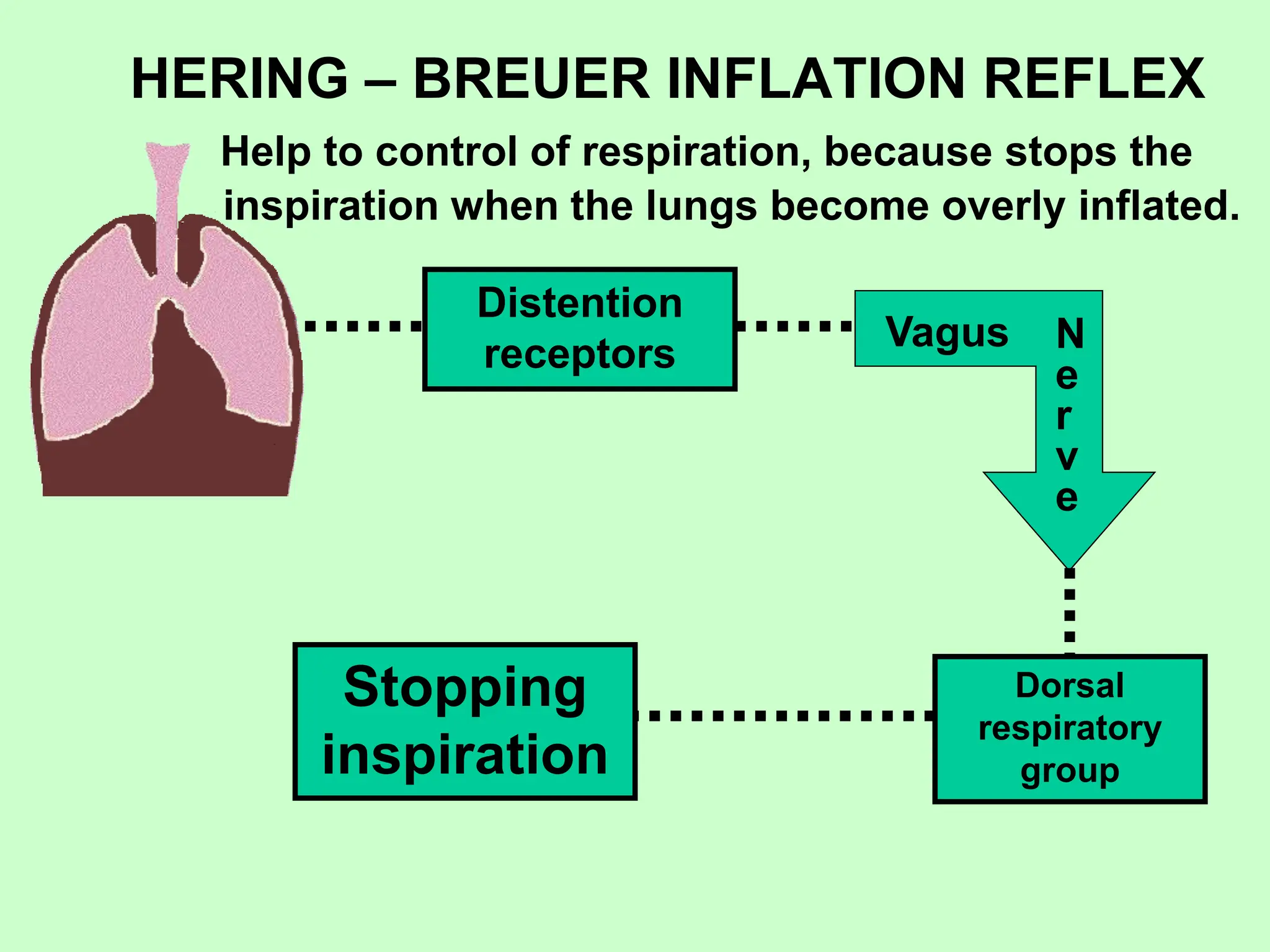
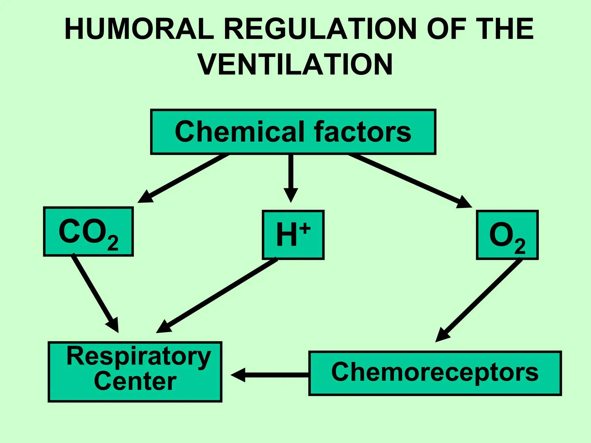
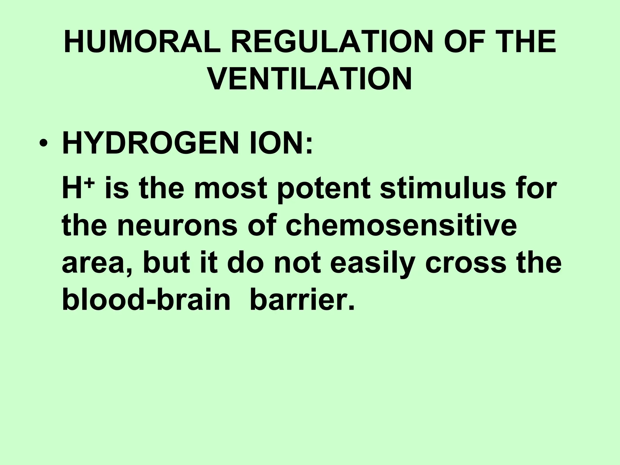
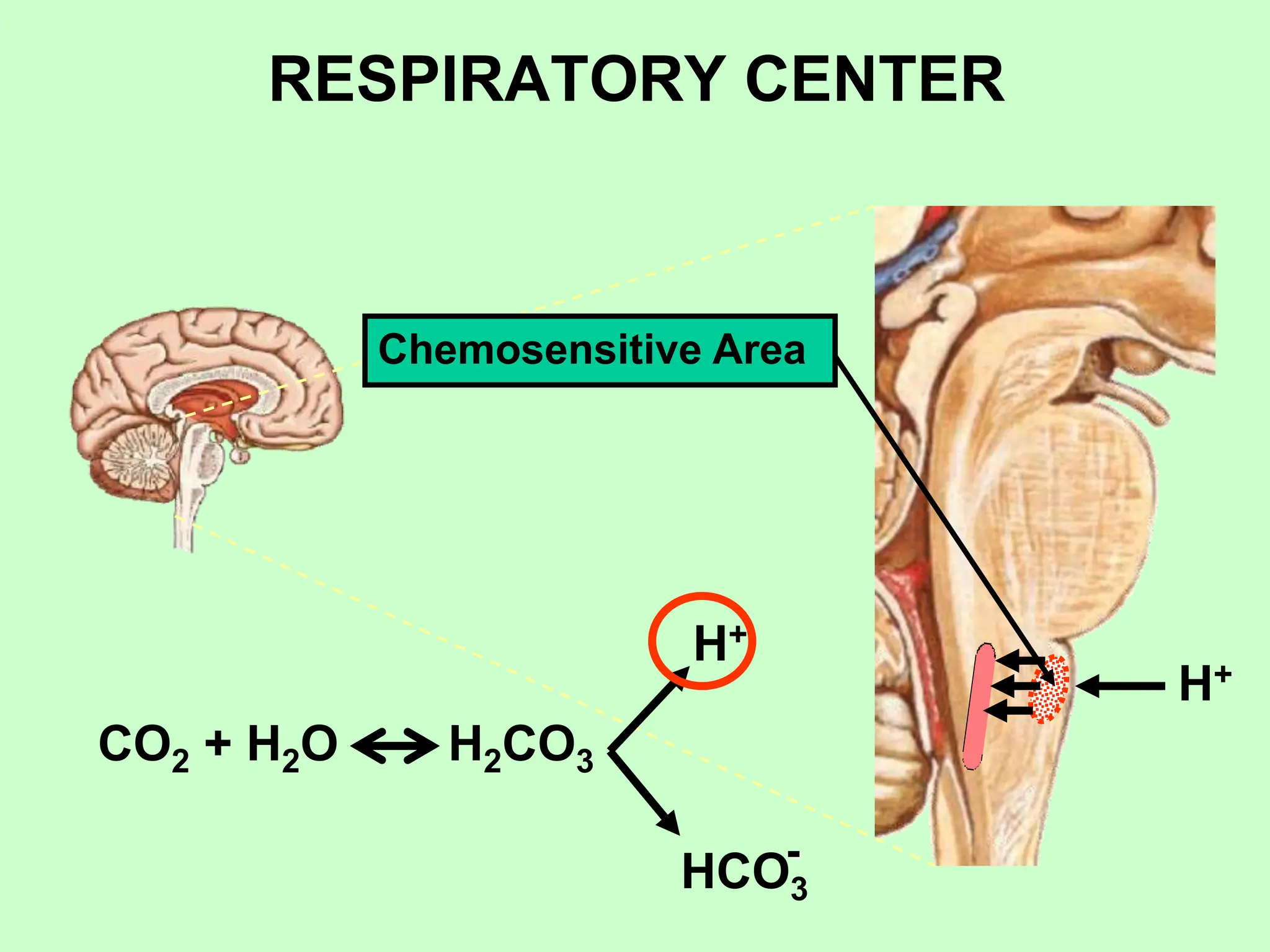
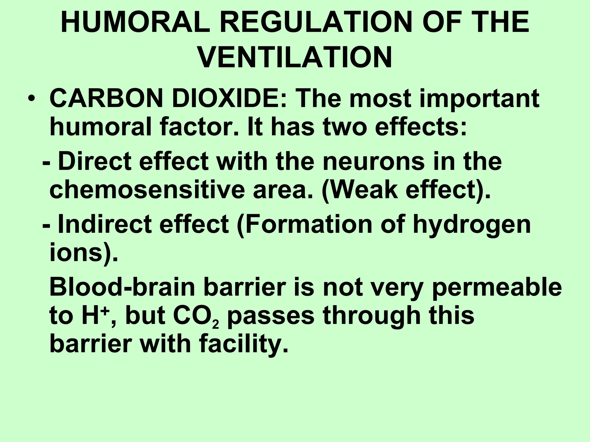
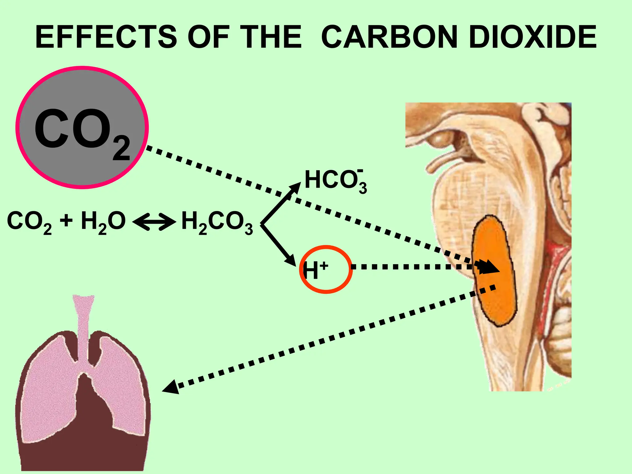
UNIVERSITY OF THE ZANZIBAR
SCHOOL OF HEALTH & MEDICAL
SCIENCES
ACADEMIC COURSE 2022 - 2023
PHYSIOLOGY
Pr.Diane Millo Martin.
Ferst Degree Specialist in
Physiology and Pathophysiology
Associate Professor
Visiting Lecturer
LECTURE:
REGULATION OF RESPIRATION
SUMMARY:
1. Nervous control of respiration.
Respiratory center.
2. Chemical control of respiration
a) Central chemoreceptor.
b) Peripheral chemoreceptor.
3. Regulation of respiration during
exercise.
4. Others factors.
BIBLIOGRAPHY
Textbook of Medical Physiology
Guyton & Hall.
13 edition.
Chapter 41
pages: 514 to 522.
Regulation of ventilation
According to body needs
paO2 y paCO2 normal
Normal ventilation
REGULATION OF RESPIRATION
VOLUNTARY INVOLUNTARY
RC
RESPIRATORY CENTER
Concept: Respiratory center is composed of
several group of neurons located bilaterally
in the medulla oblongata and pons of the
brain stem.
Organization:
– Dorsal respiratory group of neurons.
– Ventral respiratory group of neurons.
– Pneumataxic center.
– Apneustic center.
– Chemosensitive area.
RESPIRATORY CENTER
Dorsal respiratory
group (inspiration)
Ventral respiratory group
(expiration and inspiration)
Apneustic center
Pneumotaxic center
RESPIRATORY CENTER
DORSAL RESPIRATORY GROUP:
• Location: Nucleus of the Tractus Solitarius and
adjacent reticular substance neurons of the
medulla. (N. Solitarius is the sensory
termination of vagus and glossopharyngeal
nerves).
• Functions:
- Generate the basic rhythm of respiration. The
neurons of this area are excited by themselves
during a period of time. Nervous signal
transmitted to the inspiratory muscles is not an
instantaneous burst of action potentials. In
normal respiration, it begins weakly and
increase steadily in a ramp manner for about 2
second and it ceases abruptly for 3 second.
Next, inspiratory signal begins another cycle.
RESPIRATORY CENTER
DORSAL RESPIRATORY GROUP:
• Functions:
The inspiratory signal is a RAMP SIGNAL.
There are two qualities of inspiratory ramp:
- Control of the rate of increase of the ramp
signal.
Ex: Heavy respiration the ramp increase
rapidly.
- Control of limiting point at which the ramp
suddenly ceases. Ex: Method for controlling
the rate of respiration.
RESPIRATORY CENTER
VENTRAL RESPIRATORY GROUP
• Location: In each side of the medulla about 5 mm
anterior and lateral to the dorsal respiratory group of
neurons (Nucleus ambigus and nucleus retroambigus).
• Functions:
- It is inactive during normal respiration.
- This group is activated from basic oscillatory
mechanism of the dorsal respiratory area when
increased pulmonary ventilation.
- During the electrical stimulation of the few of the
neurons in the ventral group causes inspiration, while the
stimulation of others causes expiration, therefore theses
neurons contribute to both inspiration and expiration.
They are important in providing the powerful expiratory
signals to the abdominal muscles during heavy
expiration.
RESPIRATORY CENTER
PNEUMOTAXIC CENTER
• Location: Dorsally in the nucleus parabrachialis
on the upper pons.
• Functions: Transmits inhibitory signals to
dorsal respiratory group that produce:
- Control the SWITCH-OFF point of the
inspiratory ramp (control the duration of the
filling phase of the lung cycle). Pneumotaxic
center limits the duration of the inspiration.
- A secondary effect is the regulation of
respiratory rate.
RESPIRATORY CENTER
APNEUSTIC CENTER
• Location: Low portion of the pons.
• Function: Transmits excitatory
signal to dorsal respiratory group
that avoid the end of inspiration. (If
pneumotaxic center is damaged).
RESPIRATORY CENTER
Chemosensitive Area
Location: Bilaterally in the
ventral surface of the
medulla (high sensitive to
changes of either blood
pCO2 or H+ concentration)
HERING – BREUER INFLATION REFLEX
Help to control of respiration, because stops the
inspiration when the lungs become overly inflated.
Stopping
inspiration
Distention
receptors
Vagus N
e
r
v
e
Dorsal
respiratory
group
HUMORAL REGULATION OF THE
VENTILATION
H+
CO2 O2
Chemical factors
Respiratory
Center Chemoreceptors
HUMORAL REGULATION OF THE
VENTILATION
• HYDROGEN ION:
H+ is the most potent stimulus for
the neurons of chemosensitive
area, but it do not easily cross the
blood-brain barrier.
RESPIRATORY CENTER
Chemosensitive Area
H+
CO2 + H2O H2CO3
H+
HCO3
-
HUMORAL REGULATION OF THE
VENTILATION
• CARBON DIOXIDE: The most important
humoral factor. It has two effects:
- Direct effect with the neurons in the
chemosensitive area. (Weak effect).
- Indirect effect (Formation of hydrogen
ions).
Blood-brain barrier is not very permeable
to H+, but CO2 passes through this
barrier with facility.
EFFECTS OF THE CARBON DIOXIDE
CO2
CO2 + H2O H2CO3
H+
HCO3
-
HUMORAL REGULATION OF THE
VENTILATION
• OXYGEN: Changes O2 concentration don’t
have direct effect on the respiratory center. It
has an indirect effect acting through the
peripheral chemoreceptors.
• Location of chemoreceptors:
- Carotid bodies (Hering’s nerves to
glossopharyngeal).
- Aortic bodies (Vagus nerve).
• Stimuli: Decrease of O2, increase CO2 and H+
concentration.
REGULATION OF RESPIRATION
DURING EXERCISE
• In exercise oxygen consumption and
CO2 increases as 20-fold.
• Alveolar ventilation increases.
REGULATION OF RESPIRATION
DURING EXERCISE
WHAT CAUSES INTENSE VENTILATION
DURING EXERCISE?
The cerebral motor cortex transmits
motor impulses to the exercising
muscles and the same time transmits
collateral impulses into the brain stem to
excite the respiratory center that
increase the rate of ventilation.

Lecture IV Regulation of ventilation (DIANE)-1.ppt

  • 1.
    UNIVERSITY OF THEZANZIBAR SCHOOL OF HEALTH & MEDICAL SCIENCES ACADEMIC COURSE 2022 - 2023
  • 2.
  • 3.
    Pr.Diane Millo Martin. FerstDegree Specialist in Physiology and Pathophysiology Associate Professor Visiting Lecturer
  • 4.
  • 5.
    SUMMARY: 1. Nervous controlof respiration. Respiratory center. 2. Chemical control of respiration a) Central chemoreceptor. b) Peripheral chemoreceptor. 3. Regulation of respiration during exercise. 4. Others factors.
  • 6.
    BIBLIOGRAPHY Textbook of MedicalPhysiology Guyton & Hall. 13 edition. Chapter 41 pages: 514 to 522.
  • 7.
    Regulation of ventilation Accordingto body needs paO2 y paCO2 normal Normal ventilation
  • 8.
  • 9.
    RESPIRATORY CENTER Concept: Respiratorycenter is composed of several group of neurons located bilaterally in the medulla oblongata and pons of the brain stem. Organization: – Dorsal respiratory group of neurons. – Ventral respiratory group of neurons. – Pneumataxic center. – Apneustic center. – Chemosensitive area.
  • 10.
    RESPIRATORY CENTER Dorsal respiratory group(inspiration) Ventral respiratory group (expiration and inspiration) Apneustic center Pneumotaxic center
  • 11.
    RESPIRATORY CENTER DORSAL RESPIRATORYGROUP: • Location: Nucleus of the Tractus Solitarius and adjacent reticular substance neurons of the medulla. (N. Solitarius is the sensory termination of vagus and glossopharyngeal nerves). • Functions: - Generate the basic rhythm of respiration. The neurons of this area are excited by themselves during a period of time. Nervous signal transmitted to the inspiratory muscles is not an instantaneous burst of action potentials. In normal respiration, it begins weakly and increase steadily in a ramp manner for about 2 second and it ceases abruptly for 3 second. Next, inspiratory signal begins another cycle.
  • 12.
    RESPIRATORY CENTER DORSAL RESPIRATORYGROUP: • Functions: The inspiratory signal is a RAMP SIGNAL. There are two qualities of inspiratory ramp: - Control of the rate of increase of the ramp signal. Ex: Heavy respiration the ramp increase rapidly. - Control of limiting point at which the ramp suddenly ceases. Ex: Method for controlling the rate of respiration.
  • 13.
    RESPIRATORY CENTER VENTRAL RESPIRATORYGROUP • Location: In each side of the medulla about 5 mm anterior and lateral to the dorsal respiratory group of neurons (Nucleus ambigus and nucleus retroambigus). • Functions: - It is inactive during normal respiration. - This group is activated from basic oscillatory mechanism of the dorsal respiratory area when increased pulmonary ventilation. - During the electrical stimulation of the few of the neurons in the ventral group causes inspiration, while the stimulation of others causes expiration, therefore theses neurons contribute to both inspiration and expiration. They are important in providing the powerful expiratory signals to the abdominal muscles during heavy expiration.
  • 14.
    RESPIRATORY CENTER PNEUMOTAXIC CENTER •Location: Dorsally in the nucleus parabrachialis on the upper pons. • Functions: Transmits inhibitory signals to dorsal respiratory group that produce: - Control the SWITCH-OFF point of the inspiratory ramp (control the duration of the filling phase of the lung cycle). Pneumotaxic center limits the duration of the inspiration. - A secondary effect is the regulation of respiratory rate.
  • 15.
    RESPIRATORY CENTER APNEUSTIC CENTER •Location: Low portion of the pons. • Function: Transmits excitatory signal to dorsal respiratory group that avoid the end of inspiration. (If pneumotaxic center is damaged).
  • 16.
    RESPIRATORY CENTER Chemosensitive Area Location:Bilaterally in the ventral surface of the medulla (high sensitive to changes of either blood pCO2 or H+ concentration)
  • 17.
    HERING – BREUERINFLATION REFLEX Help to control of respiration, because stops the inspiration when the lungs become overly inflated. Stopping inspiration Distention receptors Vagus N e r v e Dorsal respiratory group
  • 18.
    HUMORAL REGULATION OFTHE VENTILATION H+ CO2 O2 Chemical factors Respiratory Center Chemoreceptors
  • 19.
    HUMORAL REGULATION OFTHE VENTILATION • HYDROGEN ION: H+ is the most potent stimulus for the neurons of chemosensitive area, but it do not easily cross the blood-brain barrier.
  • 20.
  • 21.
    HUMORAL REGULATION OFTHE VENTILATION • CARBON DIOXIDE: The most important humoral factor. It has two effects: - Direct effect with the neurons in the chemosensitive area. (Weak effect). - Indirect effect (Formation of hydrogen ions). Blood-brain barrier is not very permeable to H+, but CO2 passes through this barrier with facility.
  • 22.
    EFFECTS OF THECARBON DIOXIDE CO2 CO2 + H2O H2CO3 H+ HCO3 -
  • 23.
    HUMORAL REGULATION OFTHE VENTILATION • OXYGEN: Changes O2 concentration don’t have direct effect on the respiratory center. It has an indirect effect acting through the peripheral chemoreceptors. • Location of chemoreceptors: - Carotid bodies (Hering’s nerves to glossopharyngeal). - Aortic bodies (Vagus nerve). • Stimuli: Decrease of O2, increase CO2 and H+ concentration.
  • 24.
    REGULATION OF RESPIRATION DURINGEXERCISE • In exercise oxygen consumption and CO2 increases as 20-fold. • Alveolar ventilation increases.
  • 25.
    REGULATION OF RESPIRATION DURINGEXERCISE WHAT CAUSES INTENSE VENTILATION DURING EXERCISE? The cerebral motor cortex transmits motor impulses to the exercising muscles and the same time transmits collateral impulses into the brain stem to excite the respiratory center that increase the rate of ventilation.