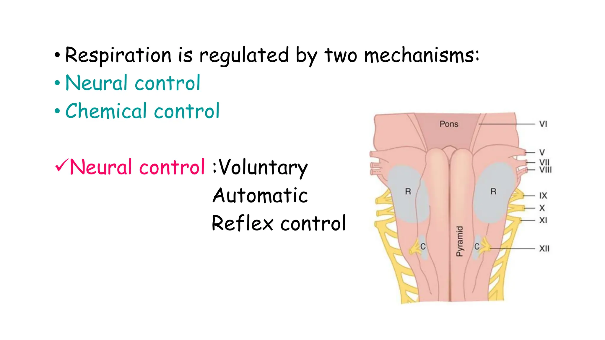
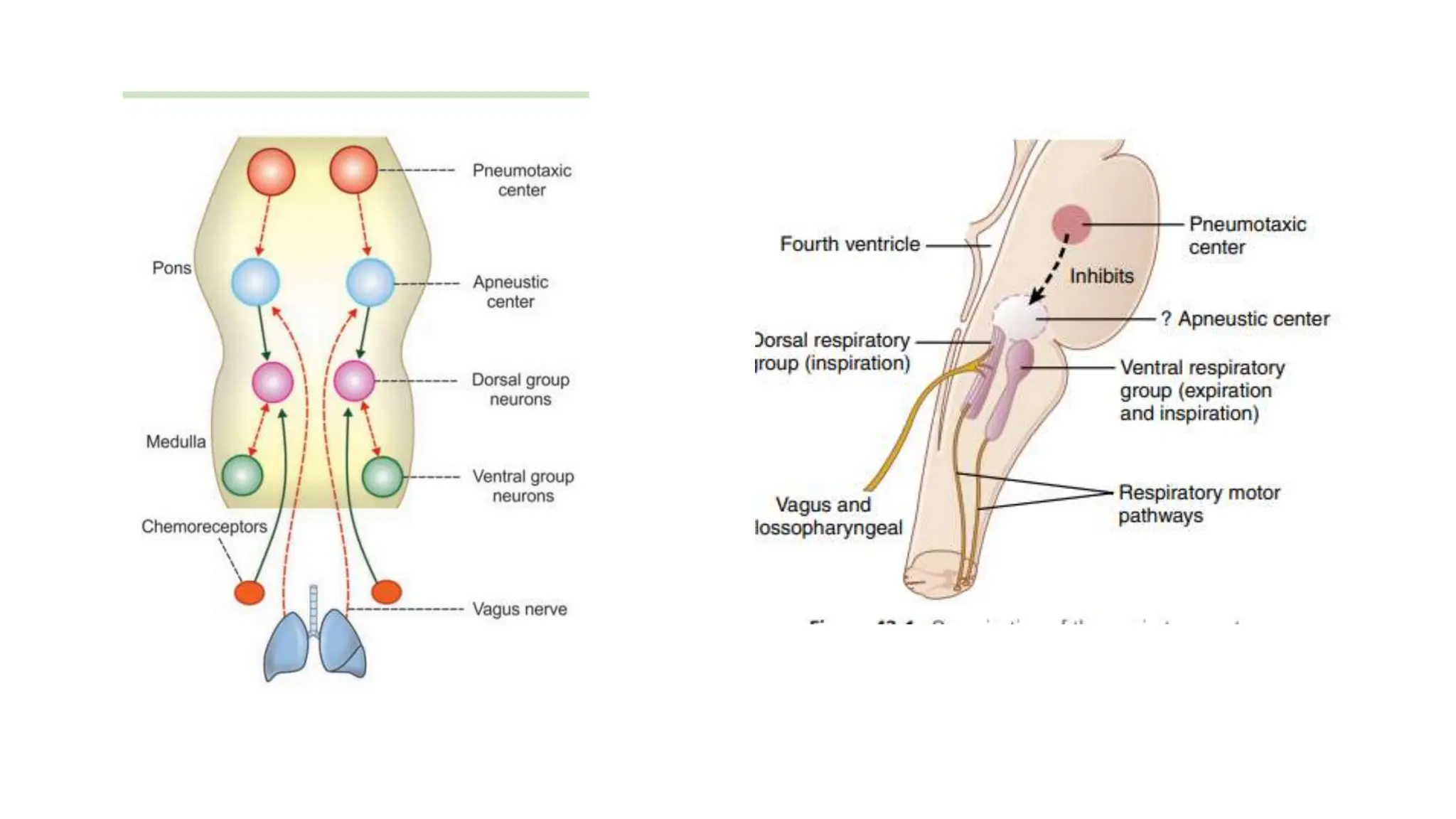
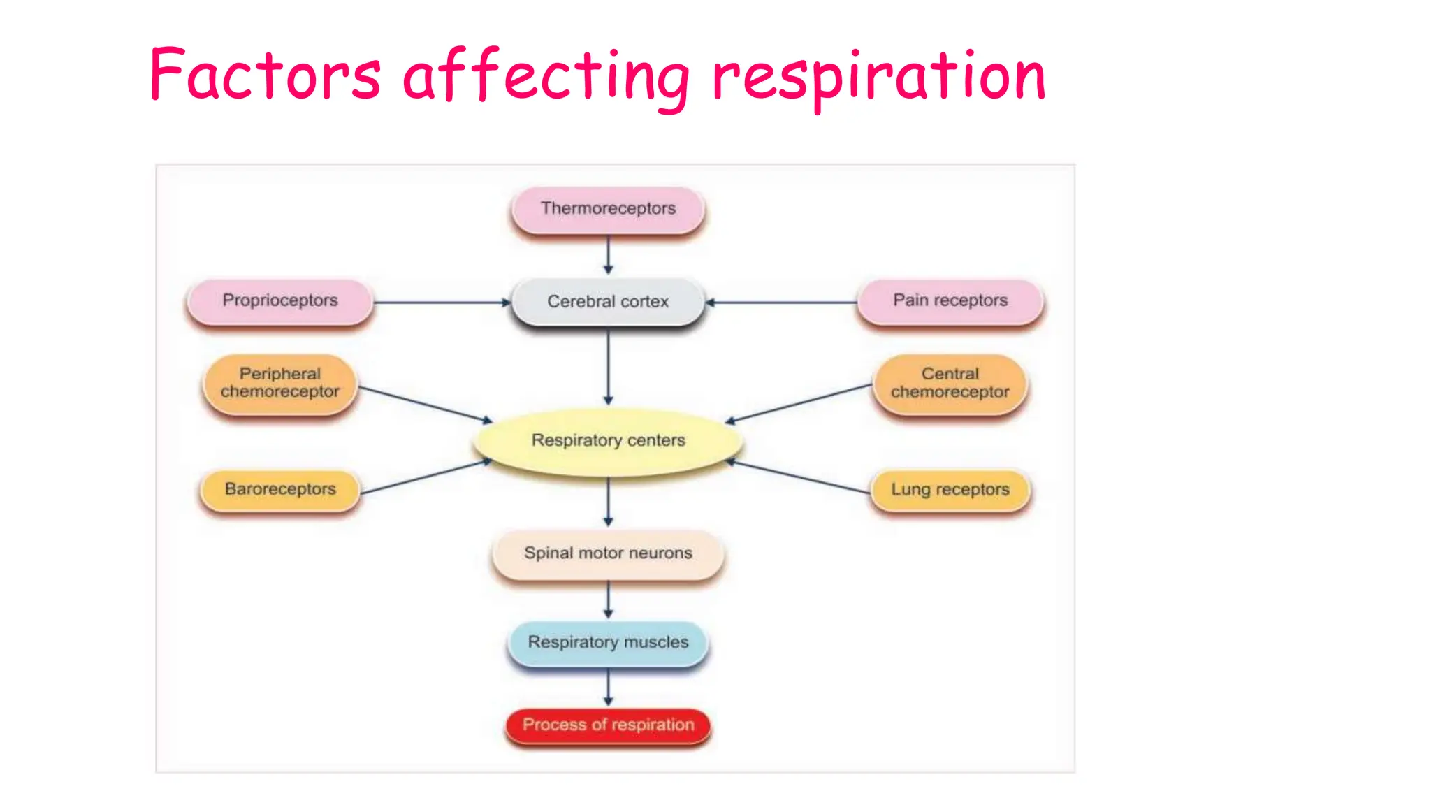
REGULATION OF RESPIRATION
• The main function of respiratory system is to
maintain Pco2 ,Po2 and pH of arterial blood constant
by adjusting ventilation.
• Spontaneous respiration is produced by rhythmic
discharges from the brain to the motor neurons
that innervate respiratory muscles.
• Respiration is regulated by two mechanisms:
• Neural control
• Chemical control
Neural control :Voluntary
Automatic
Reflex control
Voluntary control
• Respiration can be controlled voluntarily to some extent
because all the muscles concerned with respiration are
voluntary.
• The center for voluntary control is motor cortex which
send impulses through corticospinal tract to the
respiratory motor neurons.
• Voluntary hyperventilation and voluntary apnea are
possible
• The point at which breathing can no longer be
voluntarily inhibited is called breaking point.
• Normally it is 40 seconds
Reason : During apnea there is increase in
Pco2,increase in H+ and decrease in Po2.
Automatic control
• Automatic centre for control of respiration are
located in the pons and medulla.
• These centers are located bilaterally
• Nerve fibers mediating inspiration converge on
phrenic motor neurons(C3,4,5) and external
intercostal neurons in spinal cord.
• The fibres concerned with expiration converge on
spinal cord motor neuron that supply expiratory
muscles mainly internal intercostal muscles.
• The motor neurons to expiratory muscles are
inhibited when those supplying the inspiratory
muscles are activated through reciprocal innervation.
MEDULLARY CENTERS
• Rhythmic respiration is initiated by small group of
pacemaker cells in the pre-Bontziger complex on
both sides of medulla oblangata.
• These neurons can discharge spontaneously and are
responsible for the rhythmicity of respiration.
• Medulla also contain dorsal respiratory group and
ventral respiratory group.
DORSAL RESPIRATORY GROUP(DRG)
• DRG is located in and near the nucleus of tractus
solitarius(NTS).
• It is made up of inspiratory neurons which are
controlled by pacemaker cells in the Pre-Botziger
complex.
• They are active during inspiration.
• They receive impulses from lungs, chemoreceptors
and baro-receptors through vagus.
VENTRAL RESPIRATORY GROUP
• It is located in the ventrolateral part of medulla.
• It extends through nucleus ambiguus and nucleus
retro-ambiguus.
• It contains inspiratory and expiratory neurons.
• Expiratory neurons inhibit inspiratory neurons during
expiration.
Inspiratory ramp signal
• Pattern of action potential produced by the
respiratory center is called inspiratory ramp signal
• Initially there is a latent period then the intensity
of action potential increases and then decreases
followed by a latent period.
• The cycle is repeated and normally the duration of
one cycle is 4-5 seconds.
• The impulses pass to the inspiratory muscles and
produce inspiration
PONTINE CENTERS
• Rhythmic discharge of medullary neurons are
modified by neurons of pons and afferents coming
from the receptors in airways and lungs through
vagus.
• In the upper part of pons there is a pair of
respiratory centres called pneumotaxic centre.
• Pneumotaxic centers has inhibitory effect on I
neurons.
• When this area is stimulated ,I neurons are inhibited
and duration of IRS is reduced.
• Rate of respiration is increased and filling volume
will be decreased; respiration becomes shallow and
rapid.
• Lower part of pons contain another set of neurons
called apneustic center which is tonically active.
• It has excitatory effect on I neurons .
• Stimulation of these center increases duration of
IRS producing sustained contraction of inspiratory
muscles with expiratory gasps in between(apneusis).
Genesis of respiration
Genesis of inspiration:
• Apneustic center activates the inspiratory centre.
• Inspiratory center discharges over pathways in the
spinal cord to C3,4,5 and T1,2 and inspiration starts.
Genesis of expiration:
• Inspiratory neurons in the medulla send excitatory
impulses to pneumotaxic center which inturn inhibit
the apneustic center.
• Pulomonary stretch receptors in lungs get stimulated
during inspiration also send inhibitory impulses via
vagus nerve to the apneustic center.
• Pneumotaxic center stimulates expiratory neuron in
medulla which reciprocally inhibits the inspiratory
center.
• As a result inspiratory center stops discharging and
expiration follows.
• When the activity of inspiratory neurons are
increased the rate and depth of inspiration is also
increased.
Reflex control of respiration
Receptors are classified into :
• Receptors inside respiratory system
• Receptors outside respiratory system
Receptors inside respiratory centers:
• Stretch receptors
• Pulmonary irritant receptors
Stretch receptors
Hering –Breuer Reflexes:
Hering Breuer Inflation reflexes
• When there is increase in the tidal volume greater
than 1L,stretch receptors of lungs are stimulated .
• There is increased rate
of afferent impulses
passing through the
vagus to the inspiratory
centre and this leads to
inhibition of inspiratory
muscles
• This reflex operates
only when the tidal
volume exceeds 1-1.5 L
Hering Breuer Deflation reflex
• This reflex operates when there is excessive
deflation of lungs.
• Here there is stimulation of inspiration to bring back
the lung volume to normal.
Importance:
• It functions as an important feedback mechanism
and contribute to rhythmicity of respiration in NB
babies.
Irritant receptors.
• Pulmonary irritant receptors are found in airway
epithelial cells.
Stimulation of these receptors produce:
• Bronchoconstriction
• Hyperventilation
• Cough, sneezing etc
Cough reflex:
• This is a protective reflex .
• Stimulation of irritant receptors of larynx, trachea
and bigger bronchi produces cough.
Sneezing reflex:
• Stimulation of irritant receptors of nasal cavity and
upper airways produces sneezing.
• Sneezing is forced expiration against open glottis.
J reflex
• ‘J’ receptors are juxta-capillary receptors which are
present on the wall of the alveoli and have close
contact with the pulmonary capillaries.
• Stimulation of the ‘J’ receptors produces a reflex
response, which is characterized by apnea,
hyperventilation, bradycardia, hypotension and
weakness of skeletal muscles.
• Conditions when ‘J’ receptors are
stimulated :
• i. Pulmonary congestion
• ii. Pulmonary edema
• iii. Pneumonia
• iv. Over inflation of lungs
Receptors outside respiratory system
Those which stimulate respiration:
• Proprioceptors
• Nociceptors
• Thermoreceptors
• Emotions
Those which inhibit respiration:
• Baroreceptors
• Visceroceptors.
Proprioceptors
• These are receptors in muscles, tendons and joints.
• Stimulation of these receptors reflexly stimulates
I neurons which will increase ventilation at the
start of exercise .
•Nociceptors:
• These are pain receptors which when stimulated
stimulate the respiratory center.
• CHEMORECEPTORS:
• Acidosis and hypoxia stimulate chemoreceptors.
• THERMORECEPTORS:
• Thermoreceptors present in the hypothalamus are
stimulated when there is increase in body temperature
as in fever ,exercise.
• EMOTIONAL STIMULI
• Emotional stimuli produces impulses from hypothalamus
,limbic system which stimulate respiratory centre.
Those which inhibit Respiration:
Baroreceptors:
Increase in blood pressure stimulates baroreceptors
leading to inhibition of respiration.
Visceroceptors:
In visceral reflexes like vomiting, swallowing,
deglutition, there is reflex inhibition of respiration.
Factors affecting respiration
CHEMICAL REGULATION OF
RESPIRATION
• Changes in pO2,pCO2 and H+ concentration acts on
the respiratory centers in the medulla through a
set of receptors called as chemo receptors.
• Chemoreceptors are classified into:
 Central chemoreceptors
 Peripheral chemoreceptors.
Central chemoreceptors
• These are a set of neurons located on either side of
medulla near the exit of IX and X cranial nerves
• These can sense changes in H+ concentration in the
brain interstitial fluid.
• Central chemoreceptors are surrounded by brain
extracellular fluid.
• Blood brain barrier and blood CSF barrier are
easily crossed by carbondioxide.
• Whenever there is hypercapnia CO2 enters brain
tissue and CSF, where it is hydrated to form
H2CO3.
• This splits to form H+ and HCO3-.
• Increase in H+ concentration is only the direct
stimulant for central chemoreceptors.
• CO2 has only indirect action.
• Lack of oxygen does not have significant effect on
the central chemoreceptors
• Stimulation from central
chemoreceptors go to the
inspiratory center(DRG) and
stimulate respiration.
• Central chemoreceptors are
inhibited by anaesthesia,
cyanosis ,during sleep etc.
Peripheral chemoreceptors
• These are carotid body and aortic body.
• These are neurovascular structures
Location:
• Carotid body is located at the bifurcation of common
carotid artery.
• Aortic bodies are located in the arch of aorta.
Innervation of peripheral
chemoreceptors.
• Carotid body is supplied by a branch of ninth cranial
nerve(sinus nerve).
• Sensory impulses from carotid body are carried
through this nerve.
• Aortic body is supplied by a branch of vagus
nerve.(aortic nerve)
• Sinus nerve and aortic nerve together referred as
sino-aortic nerve.
• PCR contains two types of cells surrounded by
fenestrated sinusoidal capillaries.
• Type I cell/glomus cells
• Type II cells/Glial cells / supporting cells.
Type I cell or glomus cells.
• They contain dense granules with dopamine.
• They are in close association with afferent nerve
fibres of ninth cranial nerves.
Type II cells/glial cells or supporting cells.
• These cells surround 4-6 glomus cells .
• Function : Protection and support of glomus cells.
Mechanism of action of PCR
• Reduction in partial pressure of oxygen is the
most potent stimulus for PCR.
• If the arterial pO2 falls below 60 mm of Hg, the
peripheral chemoreceptors are strongly
stimulated which inturn will stimulate the
respiratory center .
• This leads to increase in rate and depth of
respiration which brings blood pO2 to normal
• In the absence of PCR, severe hypoxia depress
respiration by a direct inhibitory action on
respiratory centres.
Factors affecting PCR
Decrease in arterial pO2.
• Vascular stasis as in circulatory shock(stagnant
hypoxia).
• The PCR responds only to reduction in dissolved
oxygen in the blood.
• In anemic hypoxia and carbon-monoxide poisoning
there is no stimulation of respiration through PCR
Decrease in pCO2
• Peripheral chemoreceptors are also sensitive to
increased pCO2.
• Increased pCO2 stimulates peripheral chemo
receptors which inturn stimulates resp centre
• Increase in carbon dioxide washout until arterial
pCO2 becomes normal.
Effect of H+ concentration
Acidosis stimulates respiratory center and causes
hyperventilation
Alkalosis depress respiratory center and causes
hypoventilation.
Mediated by peripheral chemoreceptors.
NOTE : Action of CO2 on respiration is increased by
Hypoxia .
• Decreased by sleep, hypothermia and anaesthesia.
ABNORMALITIES
Respiratory center depression:
• Causes : Old age
• Anesthetics.
Periodic breathing : Consists of alternating waxing
and waning of respiration or alternate hyperpnea and
apnea.
• It may be normal or abnormal.
Types of periodic breathing
• 1.Voluntary hyperventilation
• Hyperventilation in normal subject is followed by a
period of apnea which in turn is followed by a few
shallow breaths and then another period of apnea
followed again by few breaths.
• This cycles lasts for some time before normal
breathing is resumed.
2.Cheyne-stokes respiration
• Seen in both physiological and pathological conditions
• Regular alternating period of hyperventilation and
apnea are seen.
Physiological causes:
• Deep sleep
• Infants
• High altitude
Pathological causes:
• Congestive cardiac failure
• Raised ICT, Uremia and morphine poisoning.
3.Biot’s breathing
• This type of breathing is always pathological.
• Irregular periods of apnea and hyperventilation.
• Changes are abrupt.
Causes : Meningitis
Medullary lesions.

Presentation of respiration and reflex associated with it

  • 2.
    REGULATION OF RESPIRATION •The main function of respiratory system is to maintain Pco2 ,Po2 and pH of arterial blood constant by adjusting ventilation. • Spontaneous respiration is produced by rhythmic discharges from the brain to the motor neurons that innervate respiratory muscles.
  • 3.
    • Respiration isregulated by two mechanisms: • Neural control • Chemical control Neural control :Voluntary Automatic Reflex control
  • 4.
    Voluntary control • Respirationcan be controlled voluntarily to some extent because all the muscles concerned with respiration are voluntary. • The center for voluntary control is motor cortex which send impulses through corticospinal tract to the respiratory motor neurons. • Voluntary hyperventilation and voluntary apnea are possible
  • 5.
    • The pointat which breathing can no longer be voluntarily inhibited is called breaking point. • Normally it is 40 seconds Reason : During apnea there is increase in Pco2,increase in H+ and decrease in Po2.
  • 7.
    Automatic control • Automaticcentre for control of respiration are located in the pons and medulla. • These centers are located bilaterally • Nerve fibers mediating inspiration converge on phrenic motor neurons(C3,4,5) and external intercostal neurons in spinal cord.
  • 8.
    • The fibresconcerned with expiration converge on spinal cord motor neuron that supply expiratory muscles mainly internal intercostal muscles. • The motor neurons to expiratory muscles are inhibited when those supplying the inspiratory muscles are activated through reciprocal innervation.
  • 9.
    MEDULLARY CENTERS • Rhythmicrespiration is initiated by small group of pacemaker cells in the pre-Bontziger complex on both sides of medulla oblangata. • These neurons can discharge spontaneously and are responsible for the rhythmicity of respiration. • Medulla also contain dorsal respiratory group and ventral respiratory group.
  • 10.
    DORSAL RESPIRATORY GROUP(DRG) •DRG is located in and near the nucleus of tractus solitarius(NTS). • It is made up of inspiratory neurons which are controlled by pacemaker cells in the Pre-Botziger complex. • They are active during inspiration. • They receive impulses from lungs, chemoreceptors and baro-receptors through vagus.
  • 11.
    VENTRAL RESPIRATORY GROUP •It is located in the ventrolateral part of medulla. • It extends through nucleus ambiguus and nucleus retro-ambiguus. • It contains inspiratory and expiratory neurons. • Expiratory neurons inhibit inspiratory neurons during expiration.
  • 12.
    Inspiratory ramp signal •Pattern of action potential produced by the respiratory center is called inspiratory ramp signal • Initially there is a latent period then the intensity of action potential increases and then decreases followed by a latent period.
  • 13.
    • The cycleis repeated and normally the duration of one cycle is 4-5 seconds. • The impulses pass to the inspiratory muscles and produce inspiration
  • 14.
    PONTINE CENTERS • Rhythmicdischarge of medullary neurons are modified by neurons of pons and afferents coming from the receptors in airways and lungs through vagus. • In the upper part of pons there is a pair of respiratory centres called pneumotaxic centre.
  • 15.
    • Pneumotaxic centershas inhibitory effect on I neurons. • When this area is stimulated ,I neurons are inhibited and duration of IRS is reduced. • Rate of respiration is increased and filling volume will be decreased; respiration becomes shallow and rapid.
  • 16.
    • Lower partof pons contain another set of neurons called apneustic center which is tonically active. • It has excitatory effect on I neurons . • Stimulation of these center increases duration of IRS producing sustained contraction of inspiratory muscles with expiratory gasps in between(apneusis).
  • 17.
    Genesis of respiration Genesisof inspiration: • Apneustic center activates the inspiratory centre. • Inspiratory center discharges over pathways in the spinal cord to C3,4,5 and T1,2 and inspiration starts.
  • 18.
    Genesis of expiration: •Inspiratory neurons in the medulla send excitatory impulses to pneumotaxic center which inturn inhibit the apneustic center. • Pulomonary stretch receptors in lungs get stimulated during inspiration also send inhibitory impulses via vagus nerve to the apneustic center.
  • 19.
    • Pneumotaxic centerstimulates expiratory neuron in medulla which reciprocally inhibits the inspiratory center. • As a result inspiratory center stops discharging and expiration follows. • When the activity of inspiratory neurons are increased the rate and depth of inspiration is also increased.
  • 22.
    Reflex control ofrespiration Receptors are classified into : • Receptors inside respiratory system • Receptors outside respiratory system Receptors inside respiratory centers: • Stretch receptors • Pulmonary irritant receptors
  • 23.
    Stretch receptors Hering –BreuerReflexes: Hering Breuer Inflation reflexes • When there is increase in the tidal volume greater than 1L,stretch receptors of lungs are stimulated .
  • 24.
    • There isincreased rate of afferent impulses passing through the vagus to the inspiratory centre and this leads to inhibition of inspiratory muscles • This reflex operates only when the tidal volume exceeds 1-1.5 L
  • 25.
    Hering Breuer Deflationreflex • This reflex operates when there is excessive deflation of lungs. • Here there is stimulation of inspiration to bring back the lung volume to normal. Importance: • It functions as an important feedback mechanism and contribute to rhythmicity of respiration in NB babies.
  • 26.
    Irritant receptors. • Pulmonaryirritant receptors are found in airway epithelial cells. Stimulation of these receptors produce: • Bronchoconstriction • Hyperventilation • Cough, sneezing etc
  • 27.
    Cough reflex: • Thisis a protective reflex . • Stimulation of irritant receptors of larynx, trachea and bigger bronchi produces cough. Sneezing reflex: • Stimulation of irritant receptors of nasal cavity and upper airways produces sneezing. • Sneezing is forced expiration against open glottis.
  • 28.
    J reflex • ‘J’receptors are juxta-capillary receptors which are present on the wall of the alveoli and have close contact with the pulmonary capillaries. • Stimulation of the ‘J’ receptors produces a reflex response, which is characterized by apnea, hyperventilation, bradycardia, hypotension and weakness of skeletal muscles.
  • 29.
    • Conditions when‘J’ receptors are stimulated : • i. Pulmonary congestion • ii. Pulmonary edema • iii. Pneumonia • iv. Over inflation of lungs
  • 30.
    Receptors outside respiratorysystem Those which stimulate respiration: • Proprioceptors • Nociceptors • Thermoreceptors • Emotions Those which inhibit respiration: • Baroreceptors • Visceroceptors.
  • 31.
    Proprioceptors • These arereceptors in muscles, tendons and joints. • Stimulation of these receptors reflexly stimulates I neurons which will increase ventilation at the start of exercise . •Nociceptors: • These are pain receptors which when stimulated stimulate the respiratory center.
  • 32.
    • CHEMORECEPTORS: • Acidosisand hypoxia stimulate chemoreceptors. • THERMORECEPTORS: • Thermoreceptors present in the hypothalamus are stimulated when there is increase in body temperature as in fever ,exercise. • EMOTIONAL STIMULI • Emotional stimuli produces impulses from hypothalamus ,limbic system which stimulate respiratory centre.
  • 33.
    Those which inhibitRespiration: Baroreceptors: Increase in blood pressure stimulates baroreceptors leading to inhibition of respiration. Visceroceptors: In visceral reflexes like vomiting, swallowing, deglutition, there is reflex inhibition of respiration.
  • 34.
  • 35.
    CHEMICAL REGULATION OF RESPIRATION •Changes in pO2,pCO2 and H+ concentration acts on the respiratory centers in the medulla through a set of receptors called as chemo receptors. • Chemoreceptors are classified into:  Central chemoreceptors  Peripheral chemoreceptors.
  • 36.
    Central chemoreceptors • Theseare a set of neurons located on either side of medulla near the exit of IX and X cranial nerves • These can sense changes in H+ concentration in the brain interstitial fluid. • Central chemoreceptors are surrounded by brain extracellular fluid.
  • 37.
    • Blood brainbarrier and blood CSF barrier are easily crossed by carbondioxide. • Whenever there is hypercapnia CO2 enters brain tissue and CSF, where it is hydrated to form H2CO3. • This splits to form H+ and HCO3-.
  • 38.
    • Increase inH+ concentration is only the direct stimulant for central chemoreceptors. • CO2 has only indirect action. • Lack of oxygen does not have significant effect on the central chemoreceptors
  • 39.
    • Stimulation fromcentral chemoreceptors go to the inspiratory center(DRG) and stimulate respiration. • Central chemoreceptors are inhibited by anaesthesia, cyanosis ,during sleep etc.
  • 41.
    Peripheral chemoreceptors • Theseare carotid body and aortic body. • These are neurovascular structures Location: • Carotid body is located at the bifurcation of common carotid artery. • Aortic bodies are located in the arch of aorta.
  • 43.
    Innervation of peripheral chemoreceptors. •Carotid body is supplied by a branch of ninth cranial nerve(sinus nerve). • Sensory impulses from carotid body are carried through this nerve. • Aortic body is supplied by a branch of vagus nerve.(aortic nerve)
  • 44.
    • Sinus nerveand aortic nerve together referred as sino-aortic nerve. • PCR contains two types of cells surrounded by fenestrated sinusoidal capillaries. • Type I cell/glomus cells • Type II cells/Glial cells / supporting cells.
  • 45.
    Type I cellor glomus cells. • They contain dense granules with dopamine. • They are in close association with afferent nerve fibres of ninth cranial nerves. Type II cells/glial cells or supporting cells. • These cells surround 4-6 glomus cells . • Function : Protection and support of glomus cells.
  • 46.
    Mechanism of actionof PCR • Reduction in partial pressure of oxygen is the most potent stimulus for PCR. • If the arterial pO2 falls below 60 mm of Hg, the peripheral chemoreceptors are strongly stimulated which inturn will stimulate the respiratory center .
  • 47.
    • This leadsto increase in rate and depth of respiration which brings blood pO2 to normal • In the absence of PCR, severe hypoxia depress respiration by a direct inhibitory action on respiratory centres.
  • 48.
    Factors affecting PCR Decreasein arterial pO2. • Vascular stasis as in circulatory shock(stagnant hypoxia). • The PCR responds only to reduction in dissolved oxygen in the blood. • In anemic hypoxia and carbon-monoxide poisoning there is no stimulation of respiration through PCR
  • 49.
    Decrease in pCO2 •Peripheral chemoreceptors are also sensitive to increased pCO2. • Increased pCO2 stimulates peripheral chemo receptors which inturn stimulates resp centre • Increase in carbon dioxide washout until arterial pCO2 becomes normal.
  • 50.
    Effect of H+concentration Acidosis stimulates respiratory center and causes hyperventilation Alkalosis depress respiratory center and causes hypoventilation. Mediated by peripheral chemoreceptors. NOTE : Action of CO2 on respiration is increased by Hypoxia . • Decreased by sleep, hypothermia and anaesthesia.
  • 52.
    ABNORMALITIES Respiratory center depression: •Causes : Old age • Anesthetics. Periodic breathing : Consists of alternating waxing and waning of respiration or alternate hyperpnea and apnea. • It may be normal or abnormal.
  • 53.
    Types of periodicbreathing • 1.Voluntary hyperventilation • Hyperventilation in normal subject is followed by a period of apnea which in turn is followed by a few shallow breaths and then another period of apnea followed again by few breaths. • This cycles lasts for some time before normal breathing is resumed.
  • 54.
    2.Cheyne-stokes respiration • Seenin both physiological and pathological conditions • Regular alternating period of hyperventilation and apnea are seen. Physiological causes: • Deep sleep • Infants • High altitude Pathological causes: • Congestive cardiac failure • Raised ICT, Uremia and morphine poisoning.
  • 55.
    3.Biot’s breathing • Thistype of breathing is always pathological. • Irregular periods of apnea and hyperventilation. • Changes are abrupt. Causes : Meningitis Medullary lesions.