Download as PPSX, PPTX

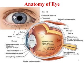
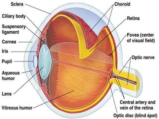
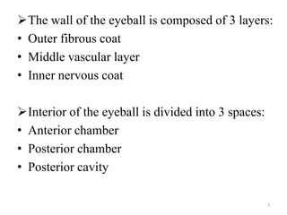
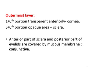
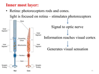
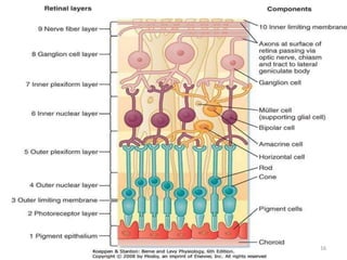
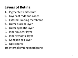
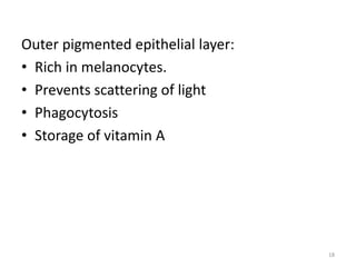
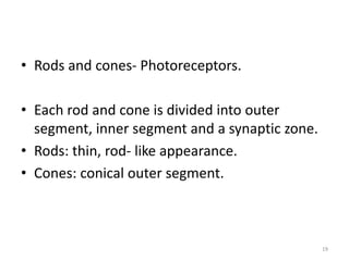
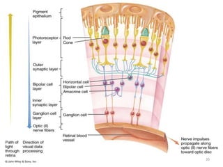
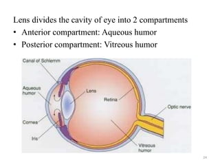
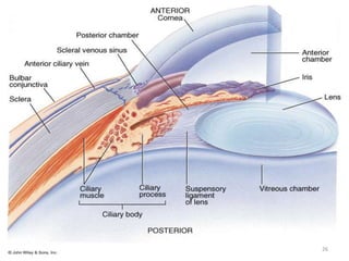
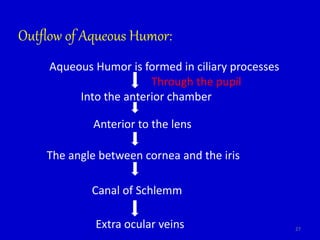
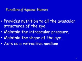
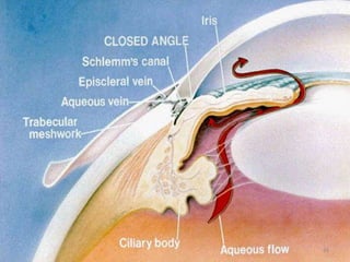
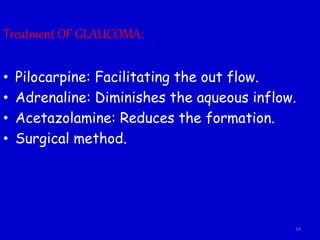


This document provides an overview of the anatomy and physiology of the eye. It begins with learning objectives focused on the functional anatomy of the eye. It then describes the two major parts of the eye as the optical system, which focuses light on the retina, and the neural system, which transmits the visual signal to the brain. The rest of the document details the layers of the eyes (sclera, cornea, retina), internal structures (choroid, ciliary body, iris), photoreceptors (rods and cones), aqueous humor, vitreous humor, and conditions like glaucoma. In summary, it provides a comprehensive review of the structures and functions that enable vision.


