Downloaded 1,598 times

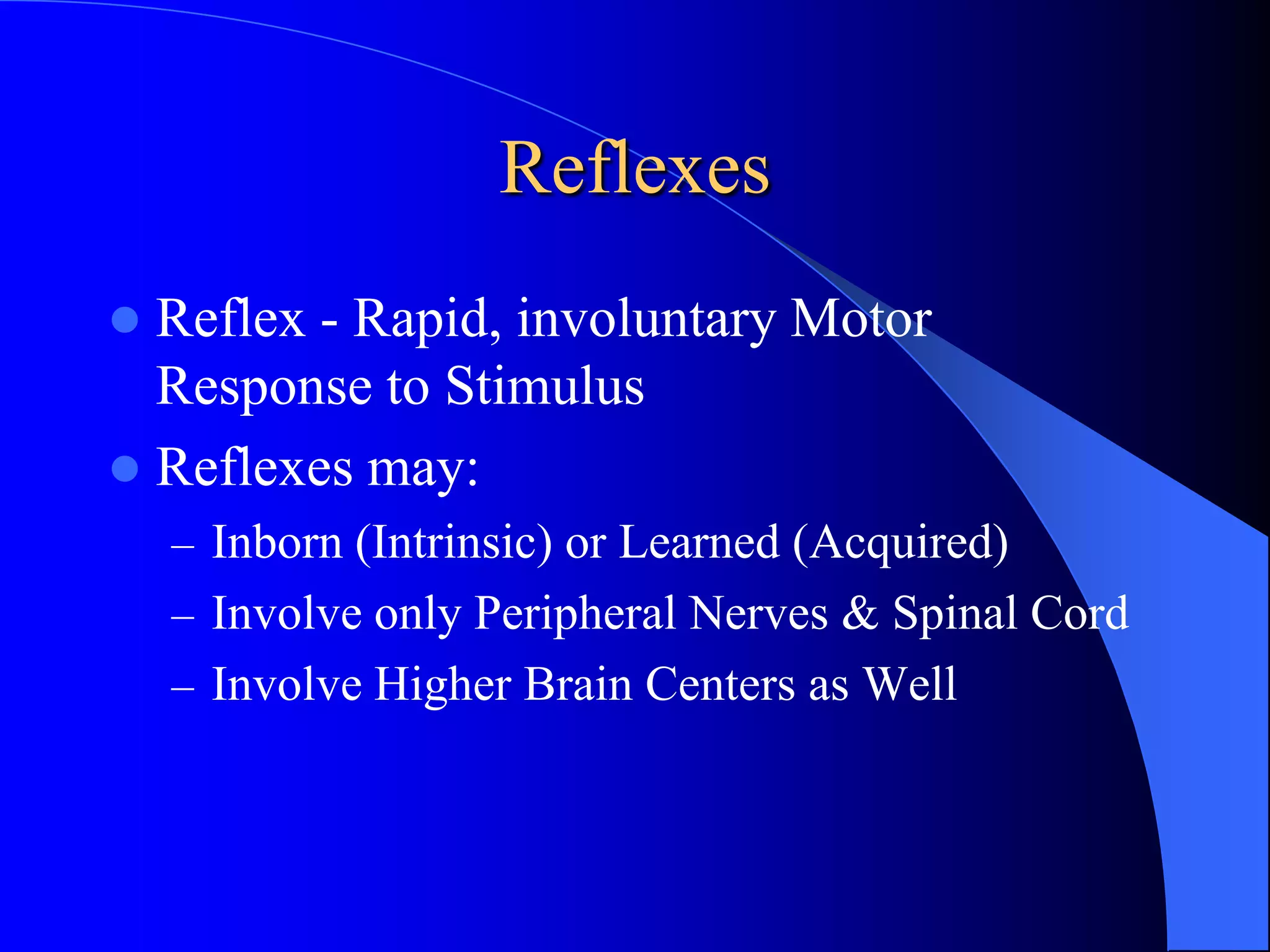
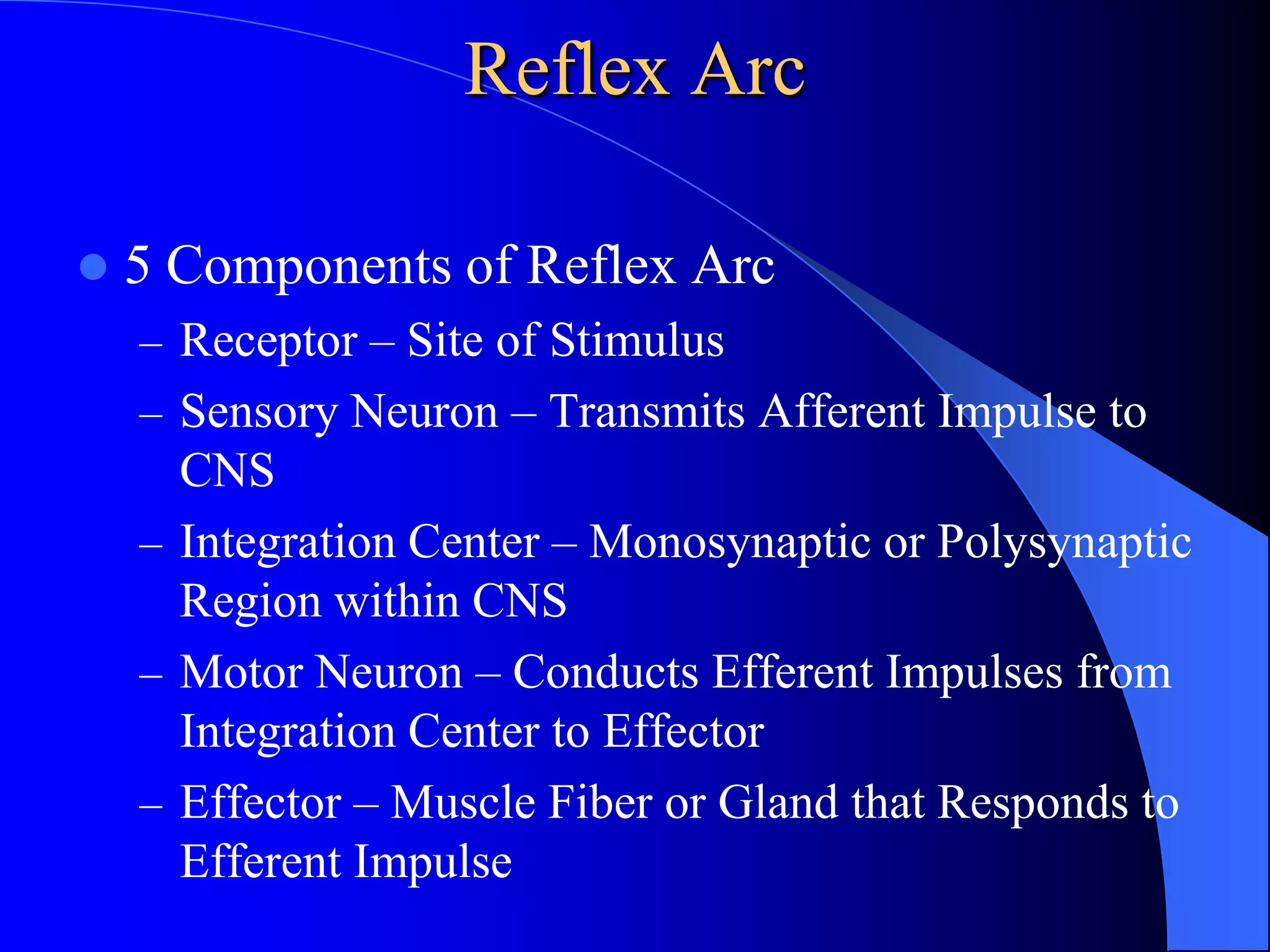


This document discusses reflexes, including: 1. Reflexes are rapid, involuntary motor responses to stimuli that may involve only peripheral nerves and spinal cord or higher brain centers as well. A reflex arc has 5 components: receptor, sensory neuron, integration center, motor neuron, and effector. 2. There are different types of reflexes including superficial, deep, segmental, intersegmental, suprasegmental, flexor, extensor, monosynaptic, and polysynaptic. Stretch reflexes and deep tendon reflexes help maintain healthy muscle tone through muscle spindles and golgi tendon organs. 3. Abnormal muscle tone includes hypertonia (too much tone) seen