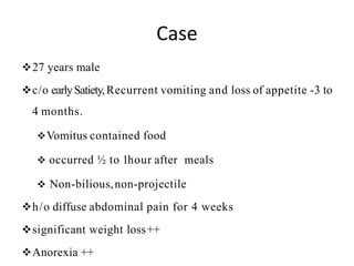
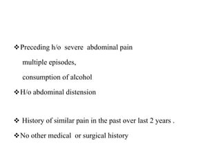
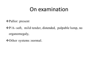
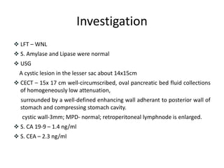
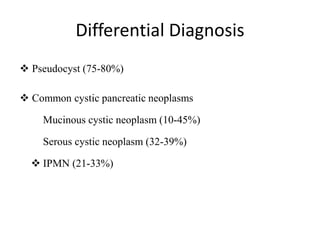
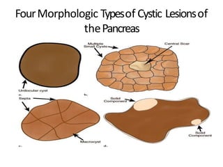
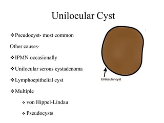
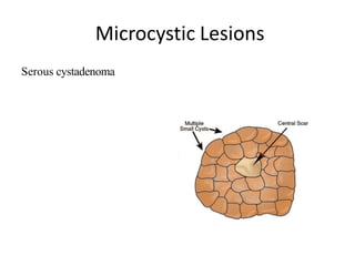
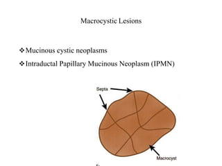
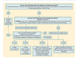
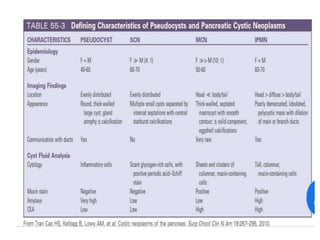
Approach to cystic lesion of
Pancreas
Dr. Kiran Pandey
MS Gen. Surgery Resident,
Bir Hospital, NAMS
Approach
 Is the lesion from pancreas?
 Lesion is solid or cystic?
 Neoplastic v/snon-neoplastic?
 SCAv/s MCN v/sIPMN?
 Management?
Case
27 years male
c/o earlySatiety,Recurrent vomiting and loss of appetite -3 to
4 months.
Vomitus contained food
 occurred ½ to 1hour after meals
 Non-bilious,non-projectile
h/o diffuse abdominal pain for 4 weeks
significant weight loss++
Anorexia ++
Preceding h/o severe abdominal pain
multiple episodes,
consumption of alcohol
H/o abdominal distension
 History of similar pain in the past over last 2 years .
No other medical or surgical history
On examination
Pallor: present
P/A- soft, mild tender, distended, palpable lump, no
organomegaly,
Other systems :normal.
Investigation
 LFT – WNL
 S. Amylase and Lipase were normal
 USG
A cystic lesion in the lesser sac about 14x15cm
 CECT – 15x 17 cm well-circumscribed, oval pancreatic bed fluid collections
of homogeneously low attenuation,
surrounded by a well-defined enhancing wall adherant to posterior wall of
stomach and compressing stomach cavity.
cystic wall-3mm; MPD- normal; retroperitoneal lymphnode is enlarged.
 S. CA 19-9 – 1.4 ng/ml
 S. CEA – 2.3 ng/ml
Must know
 Broad differential diagnosis
 Epidemiology of common lesions
 Clinical presentation
 Blood tests
 Imaging
 Histology
Differential Diagnosis
 Pseudocyst (75-80%)
 Common cystic pancreatic neoplasms
Mucinous cystic neoplasm (10-45%)
Serous cystic neoplasm (32-39%)
 IPMN (21-33%)
 Rare cystic pancreatic neoplasms
 Solid pseudopapillary tumor (<10%)
 Acinar cell cystadenocarcinoma (<1%)
 Lymphangioma
 Hemangioma
 Paraganglioma
 Solid pancreatic lesions with cystic degeneration
 Pancreatic adenocarcinoma (<1%)
 Neuroendocrine tumours
(insulinoma, glucagonoma, gastrinoma) (<10%)
 Metastasis
 Cystic teratoma
 Sarcoma
 Hydatid cyst
 Lymphatic cyst
 True epithelial cysts a/w-
von Hippel–Lindau disease
autosomal -dominant polycystic kidney disease
Imagingin Cystic lesions….
Imaging may be diagnostic, obviating further investigations
FourMorphologic Typesof Cystic Lesionsof
thePancreas
Unilocular Cyst
Pseudocyst- most common
Other causes-
IPMN occasionally
Unilocular serous cystadenoma
Lymphoepithelial cyst
Multiple
 von Hippel-Lindau
 Pseudocysts
Microcystic Lesions
Serous cystadenoma
Macrocystic Lesions
Mucinous cystic neoplasms
Intraductal Papillary Mucinous Neoplasm (IPMN)
Cysts with a solid component
Unilocular or multilocular
True cystic tumors or solid pancreatic neoplasms with cystic
component/degeneration
 Solid pseudopapillary tumor (SPEN)
 Mucinous cystic neoplasms
 IPMNs
 Islet cell tumor
 Adenocarcinoma
 Metastasis
Pancreatic Pseudocyst
Symptoms
Abdominal pain (80 – 90%)
 Lump in abdomen
 Nausea / vomiting ( due to gastric or duodenal compression)
 Early satiety
 Bloating, indigestion
 Jaundice ( due to compression of bile duct)
 Hemorrhage
Blood tests in suspected pseudocyst
Amylase/Lipase
Signs
 Tenderness
 Abdominal fullness
 Palpable mass
ImaginginPseudocyst…..
Ultrasonography
Most practical & Sensitivity (75 – 90%)
limited by patient habitus, operator experience and air in
stomach
CT scan
Gold standard for initial assessment and follow-up
Sensitivity 90- 100%
MRI
Better detail of content of cyst
MRCP
Establish the relationship of the pseudocyst to the pancreatic ducts
Endoscopic Ultrasonography (EUS +/- FNA)
Distinguishing pancreatic cystic lesions, helps in FNA
When a cysticlesions with…..
Sudden onset of pain consistent with pancreatitis pain
Imaging features of associatedpancreatitis
Unilocular cyst;and
Elevated amylase/lipase
Common Cystic neoplasm
MUCINOUS CYSTIC NEOPLASMS
Most common - 10% to 45% (MCA -67%, MCAC - 33%)
> 95% in women ( Mean ~ 50 yrs)
 Typically involve the body and tail of the pancreas
Never multifocal, occurring only in one location within
the pancreas.
Asymptomatic in 75% cases
 If symptoms, usually due to mass effect
 Adominal pain
 Palpable mass
Complex macrocystic mass with internal septations
 MRCP no communication between duct and the cyst
Contrast enhanced scans show enhancement of the cyst wall a
accentuate any septations and mural nodules
Presence of mural nodule and septal calcification s/o –
malignancy
 EUS can identify septations and cyst wall nodules In more
detail than MRI or CT
Allows cyst wall biopsy and cyst fluid aspiration for analysis
Cyst fluid analysis generally reveals
thick and mucoid material and low fluid amylase
elevated tumor markers (CEA)
mucinous epithelial cells by cytology
Mucinous Cystadenocarcinoma
Complex macrocystic lesion with internal septations
Peripheral and septal calcification indicative of malignancy
(arrowheads)
SEROUS CYSTADENOMAS
 Second MC Cystic tumor of the pancreas
formerly known as microcystic adenomas
 Occurring mostly in women (75%) with a mean 62 years
 Most (50% to 70%) occur in the body or tail of the pancreas
 An association with von Hippel-Lindau disease
 Mostly asymptomatic
 detected during evaluation for other unrelated conditions
Serous Cystadenoma
Lesion with numerous microcysts
“honey-comb” appearance
Lobulated outline Central stellate scar
Serous cystadenoma
 Pathognomonic image by CT scan:
 spongy mass with a central “sunburst” calcification -
10%
4 /5 CT and MRI features makes diagnosis
location in the pancreatic body and tail
 wall thickness < 2 mm
lobulated contour
lack of communication with the pancreatic duct
minimal wa ll enhancement
Intra-ductal PapillaryMucinousNeoplasm
Types
depend on involvement of duct
main pancreatic duct, isolated side branches, or a combination
of both
Benign (adenoma), borderline, or malignant
Malignant neoplasms account for 60% of IPMNs
 Equal frequency in men and women
 Median age at diagnosis - about 65 years
75% of patients are symptomatic
Abdominal pain and weight loss – MC complaints
Recurrent pancreatitis or Acute pancreatitis
Patients with malignant neoplasms are more likely to be
 older and more likely to present with jaundice or new-
onset diabetes
Diagnosis
Differentiation of IPMN from other cystic pancreatic masses may be
difficult at CT
Most reliable findings for the diagnosis
 Presence of a communication between the cystic lesion andthe main
pancreatic duct
 Presence of mural nodules projecting into the main pancreatic duct or
cystic lesions
Diffusely distended pancreatic duct with mucinous filling defects and grape-
like, cystic, space-occupying lesions
Sensitivity in diagnosing an IPMN
Highest for MRI with MRCP (88%),followed by ERCP (68%)and CT (42%)
Pathognomonic for IPMN in ERCP
A wide and gaping papilla with secretion of mucin and filling
defects in the dilated pancreatic duct –FISH MOUTH
AMPULLA
Features of Concern
Cysticlesionsof pancreas; will blood tests
help?
Amylase and/or Lipase??
CEA? Ca 19-9 ??
Not diagnostic of any of the cystic pancreatic tumors
Only provide corroborative evidence
 Serum amylaseor lipase levels
Increased - Pseudocyst, IPMN
 Serum CA19-9& CEA
Normal - benign cystic pancreatic tumors
Modestly elevated - MCNs and IPMNs, particularly
patients with malignancies
Markedlyelevated -retention cyst secondary to obstruction
of the main pancreatic duct by anadenocarcinoma.
Viscosity Amylase Cytology
Pseudocyst Low High Inflamm.
SCA Low Low 5% +
MCA High Low 40% +
MCAC High Low 67% +
CEA CA 15-3 CA 72-4
Pseudocyst Low Low Low
SCA Low Low Low
MCA High High Low
MCAC High High High
1 Lewandrowski KB, et al. Ann Surg 1993,217:41-7.
2 Brugge WR, et al. N Engl J Med 2004,351:1218-26.
Cyst Fluid Analysis
Comingbackto ourcase…
What could be our
diagnosis???
Pancreatic Pseudocyst
Thank You

Approach to the cystic lesion of pancrease

  • 1.
    Approach to cysticlesion of Pancreas Dr. Kiran Pandey MS Gen. Surgery Resident, Bir Hospital, NAMS
  • 2.
    Approach  Is thelesion from pancreas?  Lesion is solid or cystic?  Neoplastic v/snon-neoplastic?  SCAv/s MCN v/sIPMN?  Management?
  • 3.
    Case 27 years male c/oearlySatiety,Recurrent vomiting and loss of appetite -3 to 4 months. Vomitus contained food  occurred ½ to 1hour after meals  Non-bilious,non-projectile h/o diffuse abdominal pain for 4 weeks significant weight loss++ Anorexia ++
  • 4.
    Preceding h/o severeabdominal pain multiple episodes, consumption of alcohol H/o abdominal distension  History of similar pain in the past over last 2 years . No other medical or surgical history
  • 5.
    On examination Pallor: present P/A-soft, mild tender, distended, palpable lump, no organomegaly, Other systems :normal.
  • 6.
    Investigation  LFT –WNL  S. Amylase and Lipase were normal  USG A cystic lesion in the lesser sac about 14x15cm  CECT – 15x 17 cm well-circumscribed, oval pancreatic bed fluid collections of homogeneously low attenuation, surrounded by a well-defined enhancing wall adherant to posterior wall of stomach and compressing stomach cavity. cystic wall-3mm; MPD- normal; retroperitoneal lymphnode is enlarged.  S. CA 19-9 – 1.4 ng/ml  S. CEA – 2.3 ng/ml
  • 8.
    Must know  Broaddifferential diagnosis  Epidemiology of common lesions  Clinical presentation  Blood tests  Imaging  Histology
  • 9.
    Differential Diagnosis  Pseudocyst(75-80%)  Common cystic pancreatic neoplasms Mucinous cystic neoplasm (10-45%) Serous cystic neoplasm (32-39%)  IPMN (21-33%)
  • 10.
     Rare cysticpancreatic neoplasms  Solid pseudopapillary tumor (<10%)  Acinar cell cystadenocarcinoma (<1%)  Lymphangioma  Hemangioma  Paraganglioma
  • 11.
     Solid pancreaticlesions with cystic degeneration  Pancreatic adenocarcinoma (<1%)  Neuroendocrine tumours (insulinoma, glucagonoma, gastrinoma) (<10%)  Metastasis  Cystic teratoma  Sarcoma
  • 12.
     Hydatid cyst Lymphatic cyst  True epithelial cysts a/w- von Hippel–Lindau disease autosomal -dominant polycystic kidney disease
  • 13.
    Imagingin Cystic lesions…. Imagingmay be diagnostic, obviating further investigations
  • 14.
    FourMorphologic Typesof CysticLesionsof thePancreas
  • 15.
    Unilocular Cyst Pseudocyst- mostcommon Other causes- IPMN occasionally Unilocular serous cystadenoma Lymphoepithelial cyst Multiple  von Hippel-Lindau  Pseudocysts
  • 16.
  • 17.
    Macrocystic Lesions Mucinous cysticneoplasms Intraductal Papillary Mucinous Neoplasm (IPMN)
  • 18.
    Cysts with asolid component Unilocular or multilocular True cystic tumors or solid pancreatic neoplasms with cystic component/degeneration  Solid pseudopapillary tumor (SPEN)  Mucinous cystic neoplasms  IPMNs  Islet cell tumor  Adenocarcinoma  Metastasis
  • 19.
    Pancreatic Pseudocyst Symptoms Abdominal pain(80 – 90%)  Lump in abdomen  Nausea / vomiting ( due to gastric or duodenal compression)  Early satiety  Bloating, indigestion  Jaundice ( due to compression of bile duct)  Hemorrhage
  • 20.
    Blood tests insuspected pseudocyst Amylase/Lipase Signs  Tenderness  Abdominal fullness  Palpable mass
  • 21.
    ImaginginPseudocyst….. Ultrasonography Most practical &Sensitivity (75 – 90%) limited by patient habitus, operator experience and air in stomach CT scan Gold standard for initial assessment and follow-up Sensitivity 90- 100%
  • 22.
    MRI Better detail ofcontent of cyst MRCP Establish the relationship of the pseudocyst to the pancreatic ducts Endoscopic Ultrasonography (EUS +/- FNA) Distinguishing pancreatic cystic lesions, helps in FNA
  • 23.
    When a cysticlesionswith….. Sudden onset of pain consistent with pancreatitis pain Imaging features of associatedpancreatitis Unilocular cyst;and Elevated amylase/lipase
  • 24.
  • 25.
    MUCINOUS CYSTIC NEOPLASMS Mostcommon - 10% to 45% (MCA -67%, MCAC - 33%) > 95% in women ( Mean ~ 50 yrs)  Typically involve the body and tail of the pancreas Never multifocal, occurring only in one location within the pancreas.
  • 26.
    Asymptomatic in 75%cases  If symptoms, usually due to mass effect  Adominal pain  Palpable mass
  • 27.
    Complex macrocystic masswith internal septations  MRCP no communication between duct and the cyst Contrast enhanced scans show enhancement of the cyst wall a accentuate any septations and mural nodules Presence of mural nodule and septal calcification s/o – malignancy
  • 28.
     EUS canidentify septations and cyst wall nodules In more detail than MRI or CT Allows cyst wall biopsy and cyst fluid aspiration for analysis Cyst fluid analysis generally reveals thick and mucoid material and low fluid amylase elevated tumor markers (CEA) mucinous epithelial cells by cytology
  • 29.
    Mucinous Cystadenocarcinoma Complex macrocysticlesion with internal septations Peripheral and septal calcification indicative of malignancy (arrowheads)
  • 30.
    SEROUS CYSTADENOMAS  SecondMC Cystic tumor of the pancreas formerly known as microcystic adenomas  Occurring mostly in women (75%) with a mean 62 years  Most (50% to 70%) occur in the body or tail of the pancreas  An association with von Hippel-Lindau disease  Mostly asymptomatic  detected during evaluation for other unrelated conditions
  • 31.
    Serous Cystadenoma Lesion withnumerous microcysts “honey-comb” appearance Lobulated outline Central stellate scar
  • 32.
    Serous cystadenoma  Pathognomonicimage by CT scan:  spongy mass with a central “sunburst” calcification - 10% 4 /5 CT and MRI features makes diagnosis location in the pancreatic body and tail  wall thickness < 2 mm lobulated contour lack of communication with the pancreatic duct minimal wa ll enhancement
  • 33.
    Intra-ductal PapillaryMucinousNeoplasm Types depend oninvolvement of duct main pancreatic duct, isolated side branches, or a combination of both Benign (adenoma), borderline, or malignant Malignant neoplasms account for 60% of IPMNs
  • 34.
     Equal frequencyin men and women  Median age at diagnosis - about 65 years 75% of patients are symptomatic Abdominal pain and weight loss – MC complaints Recurrent pancreatitis or Acute pancreatitis Patients with malignant neoplasms are more likely to be  older and more likely to present with jaundice or new- onset diabetes
  • 35.
    Diagnosis Differentiation of IPMNfrom other cystic pancreatic masses may be difficult at CT Most reliable findings for the diagnosis  Presence of a communication between the cystic lesion andthe main pancreatic duct  Presence of mural nodules projecting into the main pancreatic duct or cystic lesions Diffusely distended pancreatic duct with mucinous filling defects and grape- like, cystic, space-occupying lesions Sensitivity in diagnosing an IPMN Highest for MRI with MRCP (88%),followed by ERCP (68%)and CT (42%)
  • 36.
    Pathognomonic for IPMNin ERCP A wide and gaping papilla with secretion of mucin and filling defects in the dilated pancreatic duct –FISH MOUTH AMPULLA
  • 37.
  • 40.
    Cysticlesionsof pancreas; willblood tests help? Amylase and/or Lipase?? CEA? Ca 19-9 ?? Not diagnostic of any of the cystic pancreatic tumors Only provide corroborative evidence
  • 41.
     Serum amylaseorlipase levels Increased - Pseudocyst, IPMN  Serum CA19-9& CEA Normal - benign cystic pancreatic tumors Modestly elevated - MCNs and IPMNs, particularly patients with malignancies Markedlyelevated -retention cyst secondary to obstruction of the main pancreatic duct by anadenocarcinoma.
  • 42.
    Viscosity Amylase Cytology PseudocystLow High Inflamm. SCA Low Low 5% + MCA High Low 40% + MCAC High Low 67% + CEA CA 15-3 CA 72-4 Pseudocyst Low Low Low SCA Low Low Low MCA High High Low MCAC High High High 1 Lewandrowski KB, et al. Ann Surg 1993,217:41-7. 2 Brugge WR, et al. N Engl J Med 2004,351:1218-26. Cyst Fluid Analysis
  • 44.
  • 45.
  • 46.