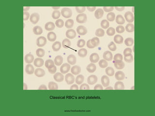
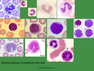
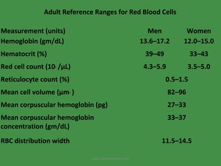
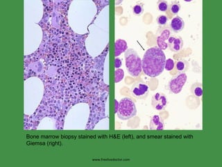
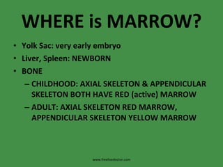
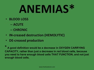
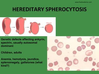
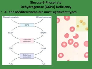
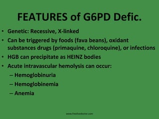
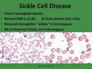
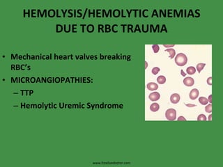
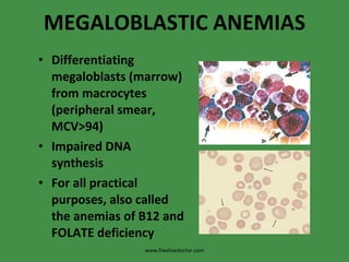
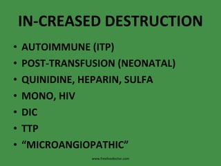
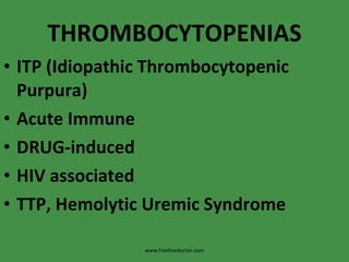
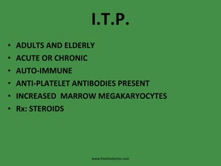
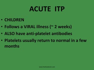
RBC and BLEEDING DISORDERS www.freelivedoctor.com
RBC and Bleeding Disorders NORMAL Anatomy, histology Development Physiology ANEMIAS Blood loss: acute, chronic Hemolytic Diminished erythropoesis POLYCYTHEMIA BLEEDING DISORDERS www.freelivedoctor.com
www.freelivedoctor.com Classical RBC’s and platelets,
www.freelivedoctor.com Classical features of peripheral white cells
www.freelivedoctor.com Adult Reference Ranges for Red Blood Cells     Measurement (units) Men Women Hemoglobin (gm/dL) 13.6–17.2 12.0–15.0 Hematocrit (%) 39–49 33–43 Red cell count (10 6  /µL) 4.3–5.9 3.5–5.0 Reticulocyte count (%) 0.5–1.5 Mean cell volume (µm 3  ) 82–96 Mean corpuscular hemoglobin (pg) 27–33 Mean corpuscular hemoglobin concentration (gm/dL) 33–37 RBC distribution width 11.5–14.5
www.freelivedoctor.com Bone marrow biopsy stained with H&E (left), and smear stained with Giemsa (right).
WHERE is MARROW? Yolk Sac: very early embryo Liver, Spleen: NEWBORN BONE CHILDHOOD: AXIAL SKELETON & APPENDICULAR SKELETON BOTH HAVE RED (active) MARROW ADULT: AXIAL SKELETON RED MARROW, APPENDICULAR SKELETON YELLOW MARROW www.freelivedoctor.com
MARROW FEATURES CELLULARITY MEGAKARYOCYTES M:E RATIO MYELOID MATURATION ERYTHROID MATURATION LYMPHS, PLASMA CELLS STORAGE IRON, i.e., HEMOSIDERIN “ FOREIGN CELLS” www.freelivedoctor.com
MARROW “ DIFFERENTIATION” www.freelivedoctor.com
www.freelivedoctor.com
ANEMIAS* BLOOD LOSS ACUTE CHRONIC IN-creased destruction (HEMOLYTIC) DE-creased production *  A good definition would be a decrease in OXYGEN CARRYING CAPACITY, rather than just a decrease in red blood cells, because you need to have enough blood cells THAT FUNCTION, and not just enough blood cells. www.freelivedoctor.com
Features   of ALL anemias Pallor, where? Tiredness Weakness Dyspnea, why? Palpitations Heart Failure (high output), why? www.freelivedoctor.com
www.freelivedoctor.com Blood Loss Acute: trauma Chronic: lesions of gastrointestinal tract, gynecologic disturbances. The features of chronic blood loss anemia are the same as iron deficiency anemia, and is defined as a situation in which the production cannot keep up with the loss
HEMOLYTIC HEREDITARY MEMBRANE disorders: e.g., spherocytosis ENZYME disorders: e.g., G6PD deficciency HGB disorders (hemoglobinopathies) ACQUIRED MEMBRANE disorders (PNH) ANTIBODY MEDIATED, transfusion or autoantibodies MECHANICAL TRAUMA INFECTIONS DRUGS, TOXINS HYPERSPLENISM www.freelivedoctor.com
IMPAIRED PRODUCTION Disturbance of proliferation and differentiation of stem cells: aplastic anemias, pure RBC aplasia, renal failure Disturbance of proliferation and maturation of erythroblasts Defective DNA synthesis:  (Megaloblastic) Defective heme synthesis:  (Fe) Deficient globin synthesis: (Thalassemias) www.freelivedoctor.com
MODIFIERS MCV, microcytosis, macrocytosis MCH  MCHC, hypochromic RDW, anisocytosis www.freelivedoctor.com
HEMOLYTIC ANEMIAS Life span LESS than 120 days Marrow hyperplasia (M:E), EPO+ Increased catabolic products, e.g., bilirubin, serum HGB, hemosiderin www.freelivedoctor.com
HEMOLYSIS INTRA-vascular (vessels) EXTRA-vascular (spleen) www.freelivedoctor.com
HEREDITARY SPHEROCYTOSIS Genetic defects affecting ankyrin, spectrin, usually autosomal dominant Children, adults Anemia, hemolysis, jaundice, splenomegaly, gallstones (what kind?) www.freelivedoctor.com
Glucose-6-Phosphate  Dehydrogenase (G6PD) Deficiency A -   and Mediterranean are most significant types www.freelivedoctor.com
FEATURES of G6PD Defic. Genetic: Recessive, X-linked Can be triggered by foods (fava beans), oxidant substances drugs (primaquine, chloroquine), or infections HGB can precipitate as HEINZ bodies Acute intravascular hemolysis can occur: Hemoglobinuria Hemoglobinemia Anemia www.freelivedoctor.com
Sickle Cell Disease Classic hemoglobinopathy Normal HGB is  α 2  β 2:  β -chain defects (Val->Glu) Reduced hemoglobin “sickles” in homozygous 8% of American blacks are heterozygous www.freelivedoctor.com
Clinical features of HGB-S disease Severe anemia Jaundice PAIN (pain CRISIS) Vaso-occlusive disease: EVEREWHERE, but clinically significant bone, spleen (autosplenectomy) Infections: Pneumococcus, Hem. Influ . www.freelivedoctor.com
www.freelivedoctor.com
THALASSEMIAS A WIDE VARIETY of diseases involving GLOBIN synthesis, COMPLEX genetics Alpha  or  beta  chains deficient synthesis involved Often termed MAJOR or MINOR, depending on severity,  silent carriers and “traits” are seen HEMOLYSIS is uniformly a feature, a microcytic anemia A “crew cut” skull x-ray appearance may be   seen www.freelivedoctor.com
Hemoglobin H Disease Deletion of THREE alpha chain genes HGB-H is primarilly Asian HGB-H has a HIGH affinity for oxygen HGB-H is unstable and therefore has classical hemolytic behavior www.freelivedoctor.com
HYDROPS FETALIS FOUR alpha chain genes are deleted, so this is the MOST SEVERE form of thalassemia Many/most never make it to term Children born will have a SEVERE hemolytic anemia as in the erythroblastosis fetalis of Rh disease: Pallor (as in all anemias) Edema (hence the name “hydrops”) Massive hepatosplenomegaly (hemolysis) www.freelivedoctor.com
Paroxysmal Nocturnal Hemoglobinuria  (PNH) ACQUIRED, NOT INHERITED like all the previous hemolytic anemias were ACQUIRED mutations in phosphatidylinositol glycan A (PIGA) It is “P” and “N” only 25% of the time G lycosylphos P hatidyl I nositol  www.freelivedoctor.com
Immunohemolytic Anemia All of these have the presence of antibodies and/or compliment present on RBC surfaces NOT all are AUTOimmune, some are caused by drugs Antibodies can be WARM  (IgG) COLD  AGGLUTININ (IgM) COLD HEMOLYSIN (paroxysmal) (IgG) www.freelivedoctor.com
IMMUNOHEMOLYTIC ANEMIAS WARM  (IgG), will NOT hemolyze at room temp Primary Idiopathic (most common) Secondary (Tumors, especially leuk/lymph, drugs) COLD AGGLUTININS:  (IgM), WILL hemolyze at room temp Mycoplasma pneumoniae, HIV, mononucleosis COLD HEMOLYSINS: (IgG) Cold Paroxysmal Hemoglobinuria, hemo-LYSIS in body, ALSO often follows mycoplasma pneumoniae www.freelivedoctor.com
COOMBS TEST DIRECT: Patient’s CELLS are tested for surface Ab’s INDIRECT: Patient’s SERUM is tested for Ab’s. www.freelivedoctor.com
HEMOLYSIS/HEMOLYTIC ANEMIAS DUE TO RBC TRAUMA Mechanical heart valves breaking RBC’s MICROANGIOPATHIES: TTP Hemolytic Uremic Syndrome www.freelivedoctor.com
NON-Hemolytic Anemias: i.e., DE-creased Production “ Megaloblastic” Anemias B12 Deficiency (Pernicious Anemia) Folate Deficiency Iron Deficiency Anemia of Chronic Disease Aplastic Anemia “ Pure” Red Cell Aplasia OTHER forms of Marrow Failure www.freelivedoctor.com
MEGALOBLASTIC ANEMIAS Differentiating megaloblasts (marrow) from macrocytes (peripheral smear, MCV>94) Impaired DNA synthesis For all practical purposes, also called the anemias of B12 and FOLATE deficiency www.freelivedoctor.com
www.freelivedoctor.com Decreased intake Inadequate diet, vegetarianism Impaired absorption Intrinsic factor deficiency    Pernicious anemia      Gastrectomy      Malabsorption states    Diffuse intestinal disease, e.g.,   lymphoma, systemic sclerosis Ileal resection, ileitis    Competitive parasitic uptake    Fish tapeworm infestation      Bacterial overgrowth in blind loops and   diverticula of bowel Increased requirement Pregnancy, hyperthyroidism,   disseminated cancer
Vit-B12 Physiology Oral ingestion Combines with INTRINSIC FACTOR in the gastric mucosa Absorbed in the terminal ileum DEFECTS at ANY of these sites can produce a MEGALOBLASTIC anemia www.freelivedoctor.com
Please remember that ALL megaloblastic anemias are also MACROCYTIC (MCV>94  or  MCV~100), and that not only are the RBC’s BIG, but so are the neutrophils, and neutrophilic precursors in the bone marrow too, and even more so, HYPERSEGMENTED!!! www.freelivedoctor.com
PERNICIOUS ANEMIA MEGALOBLASTIC anemia LEUKOPENIA and HYPERSEGS JAUNDICE NEUROLOGIC posterolateral spinal tracts ACHLORHYDRIA Can’t absorb B12 LOW serum B12 Flunk Schilling test, i.e., can’t absorb B12, using a radioactive tracer www.freelivedoctor.com
FOLATE DEFICIENCY MEGALOBLASTIC AMEMIAS Decreased Intake: diet, etoh-ism, infancy Impaired Absorption: intestinal disease DRUGS: anticonvulsants, BCPs, CHEMO Increased Loss: Hemodialysis Increased Requirement: Pregnancy, infancy Impaired Usage  www.freelivedoctor.com
Fe Deficiency Anemia Due to increased loss or decreased ingestion, almost always, in USA, nowadays, increased loss is the reason Macrocytic (low MCV), Hypochromic (low MCHC) THE ONLY WAY WE CAN LOSE IRON IS BY LOSING BLOOD www.freelivedoctor.com
Fe Transferrin Ferritin (GREAT test) Hemosiderin www.freelivedoctor.com
Clinical Fe-Defic-Anemia Adult men: GI Blood Loss PRE menopausal women: menorrhagia POST menopausal women: GI Blood Loss www.freelivedoctor.com
www.freelivedoctor.com
2 BEST lab tests: Serum Ferritin Prussian blue hemosiderin stain of marrow (also called an “iron” stain) www.freelivedoctor.com
Anemia of Chronic Disease CHRONIC INFECTIONS CHRONIC IMMUNE DISORDERS NEOPLASMS LIVER, KIDNEY failure www.freelivedoctor.com
APLASTIC ANEMIAS ALMOST ALWAYS involve platelet and WBC suppression as well Some are idiopathic, but MOST are related to drugs, radiation FANCONI’s ANEMIA is the only one that is inherited, and NOT acquired Act at STEM CELL level, except for “pure” red cell aplasia www.freelivedoctor.com
APLASTIC ANEMIAS www.freelivedoctor.com
APLASTIC ANEMIAS CHLORAMPHENICOL OTHER ANTIBIOTICS CHEMO INSECTICIDES VIRUSES EBV HEPATITIS VZ www.freelivedoctor.com
MYELOPHTHISIC  ANEMIAS Are anemias caused by metastatic tumor cells replacing the bone marrow extensively www.freelivedoctor.com
POLYCYTHEMIA Relative  (e.g., hemoconcentration) Absolute POLYCYTHEMIA  VERA  (Primary)  ( LOW  EPO) POLYCYTHEMIA  (Secondary) (HIGH EPO) HIGH ALTITUDE EPO TUMORS EPO “Doping” CVAC, the trendy California bubble pods www.freelivedoctor.com
P. VERA A “myeloproliferative” disease ALL cell lines are increased, not just RBCs www.freelivedoctor.com
BLEEDING DISORDERS (aka, Hemorrhagic “DIATHESES”) Blood vessel wall abnormalities √ Reduced platelets √ Decreased platelet function √ Abnormal clotting factors √ DIC (Disseminated INTRA-vascular Coagulation) www.freelivedoctor.com
VESSEL WALL ABNORMALITIES (NON-thrombotic cytopenic purpuras ) Infections, especially, meningococcemia, and rickettsia Drug reactions causing a leukocytoclastic vasculitis Scurvy, Ehlers-Danlos, Cushing syndrome Henoch-Schönlein purpura (mesangial deposits too) Hereditary hemorrhagic telangiectasia Amyloid www.freelivedoctor.com
THROMBOCYTOPENIAS Like RBCs: DE-creased production IN-creased destruction Sequestration (Hypersplenism) Dilutional Normal value 150K-300K www.freelivedoctor.com
DE-CREASED PRODUCTION APLASTIC ANEMIA ACUTE LEUKEMIAS ALCOHOL, THIAZIDES, CHEMO MEASLES, HIV MEGALOBLASTIC ANEMIAS MYELODYSPLASTIC SYNDROMES www.freelivedoctor.com
IN-CREASED DESTRUCTION AUTOIMMUNE (ITP) POST-TRANSFUSION (NEONATAL) QUINIDINE, HEPARIN, SULFA MONO, HIV DIC TTP “ MICROANGIOPATHIC” www.freelivedoctor.com
THROMBOCYTOPENIAS ITP (Idiopathic Thrombocytopenic Purpura) Acute Immune DRUG-induced HIV associated TTP, Hemolytic Uremic Syndrome www.freelivedoctor.com
I.T.P. ADULTS AND ELDERLY ACUTE OR CHRONIC AUTO-IMMUNE ANTI-PLATELET ANTIBODIES PRESENT INCREASED  MARROW MEGAKARYOCYTES Rx: STEROIDS www.freelivedoctor.com
ACUTE  ITP CHILDREN Follows a VIRAL illness (~ 2 weeks) ALSO have anti-platelet antibodies Platelets usually return to normal in a few months www.freelivedoctor.com
DRUGS Quinine Quinidine Sulfonamide antibiotics HEPARIN www.freelivedoctor.com
HIV BOTH DE-creased production AND IN-creased destruction factors are present www.freelivedoctor.com
Thrombotic  Microangiopathies BOTH are very SERIOUS CONDITIONS with a HIGH mortality: TTP (THROMBOTIC THROMBOCYTOPENIC PURPURA) H.U.S. (HEMOLYTIC UREMIC SYNDROME) These can also be called “consumptive” coagulopathies, just like a DIC www.freelivedoctor.com
“ QUALITATIVE” platelet disorders Mostly congenital (genetic): Bernard-Soulier syndrome (Glycoprotein-1-b deficiency) Glanzmann’s thrombasthenia (Glyc.-IIB/IIIA deficiency) Storage pool disorders, i.e., platelets mis-function AFTER they degranulate ACQUIRED:  ASPIRIN, ASPIRIN, ASPIRIN www.freelivedoctor.com
BLEEDING DISORDERS due to CLOTTING FACTOR DEFICIENCIES NOT spontaneous, but following surgery or trauma ALL factor deficiencies are possible Factor VIII and IX both are the classic X-linked recessive hemophilias, A and B, respectively ACQUIRED disorders often due to Vitamin-K deficiencies von Willebrand disease the most common,  1% www.freelivedoctor.com
von Willebrand Disease 1% prevalence, most common bleeding disorder Spontaneous and wound bleeding Usually autosomal dominant Gazillions of variants, genetics even more complex Prolonged BLEEDING TIME, NL platelet count vWF is von Willebrand Factor, which complexes with Factor VIII, it is the von Willebrand Factor which is defective in von Willebrand disease Usually BOTH platelet and FactorVIII-vWF disorders are present www.freelivedoctor.com
HEMOPHILIA A The “classic” HEMOPHILIA Factor VIII decreased Co-factor of Factor IX to activate Factor X Sex-linked recessive Hemorrhage usually NOT spontaneous Wide variety of severities Prolonged PTT (intrinsic) only Rx: Recombinant Factor VIII www.freelivedoctor.com
HEMOPHILIA B The “Christmas” HEMOPHILIA Factor IX decreased Sex-linked recessive Hemorrhage usually NOT spontaneous Wide variety of severities Prolonged PTT (intrinsic) only Rx: Recombinant Factor IX www.freelivedoctor.com
DIC, Disseminated INTRA-vascular, Coagulation ENDOTHELIAL INJURY WIDESPREAD FIBRIN DEPOSITION HIGH MORTALITY ALL MAJOR ORGANS COMMONLY INVOLVED www.freelivedoctor.com
DIC, Disseminated INTRA-vascular, Coagulation Extremely SERIOUS condition NOT a disease in itself but secondary to many conditions Obstetric: MAJOR OB complications, toxemia, sepsis, abruption Infections: Gm-, meningococcemia, RMSF, fungi, Malaria Many neoplasms, acute promyelocytic leukemia Massive tissue injury: trauma, burns, surgery “ Consumptive” coagulopathy www.freelivedoctor.com
Common Coagulation TESTS PTT (intrinsic) PT   INR (extrinsic) Platelet count, aggregation Bleeding Time, so EASY to do Fibrinogen Factor Assays www.freelivedoctor.com

Rbc

  • 1.
    RBC and BLEEDINGDISORDERS www.freelivedoctor.com
  • 2.
    RBC and BleedingDisorders NORMAL Anatomy, histology Development Physiology ANEMIAS Blood loss: acute, chronic Hemolytic Diminished erythropoesis POLYCYTHEMIA BLEEDING DISORDERS www.freelivedoctor.com
  • 3.
  • 4.
  • 5.
    www.freelivedoctor.com Adult ReferenceRanges for Red Blood Cells Measurement (units) Men Women Hemoglobin (gm/dL) 13.6–17.2 12.0–15.0 Hematocrit (%) 39–49 33–43 Red cell count (10 6 /µL) 4.3–5.9 3.5–5.0 Reticulocyte count (%) 0.5–1.5 Mean cell volume (µm 3 ) 82–96 Mean corpuscular hemoglobin (pg) 27–33 Mean corpuscular hemoglobin concentration (gm/dL) 33–37 RBC distribution width 11.5–14.5
  • 6.
    www.freelivedoctor.com Bone marrowbiopsy stained with H&E (left), and smear stained with Giemsa (right).
  • 7.
    WHERE is MARROW?Yolk Sac: very early embryo Liver, Spleen: NEWBORN BONE CHILDHOOD: AXIAL SKELETON & APPENDICULAR SKELETON BOTH HAVE RED (active) MARROW ADULT: AXIAL SKELETON RED MARROW, APPENDICULAR SKELETON YELLOW MARROW www.freelivedoctor.com
  • 8.
    MARROW FEATURES CELLULARITYMEGAKARYOCYTES M:E RATIO MYELOID MATURATION ERYTHROID MATURATION LYMPHS, PLASMA CELLS STORAGE IRON, i.e., HEMOSIDERIN “ FOREIGN CELLS” www.freelivedoctor.com
  • 9.
    MARROW “ DIFFERENTIATION”www.freelivedoctor.com
  • 10.
  • 11.
    ANEMIAS* BLOOD LOSSACUTE CHRONIC IN-creased destruction (HEMOLYTIC) DE-creased production * A good definition would be a decrease in OXYGEN CARRYING CAPACITY, rather than just a decrease in red blood cells, because you need to have enough blood cells THAT FUNCTION, and not just enough blood cells. www.freelivedoctor.com
  • 12.
    Features of ALL anemias Pallor, where? Tiredness Weakness Dyspnea, why? Palpitations Heart Failure (high output), why? www.freelivedoctor.com
  • 13.
    www.freelivedoctor.com Blood LossAcute: trauma Chronic: lesions of gastrointestinal tract, gynecologic disturbances. The features of chronic blood loss anemia are the same as iron deficiency anemia, and is defined as a situation in which the production cannot keep up with the loss
  • 14.
    HEMOLYTIC HEREDITARY MEMBRANEdisorders: e.g., spherocytosis ENZYME disorders: e.g., G6PD deficciency HGB disorders (hemoglobinopathies) ACQUIRED MEMBRANE disorders (PNH) ANTIBODY MEDIATED, transfusion or autoantibodies MECHANICAL TRAUMA INFECTIONS DRUGS, TOXINS HYPERSPLENISM www.freelivedoctor.com
  • 15.
    IMPAIRED PRODUCTION Disturbanceof proliferation and differentiation of stem cells: aplastic anemias, pure RBC aplasia, renal failure Disturbance of proliferation and maturation of erythroblasts Defective DNA synthesis: (Megaloblastic) Defective heme synthesis: (Fe) Deficient globin synthesis: (Thalassemias) www.freelivedoctor.com
  • 16.
    MODIFIERS MCV, microcytosis,macrocytosis MCH MCHC, hypochromic RDW, anisocytosis www.freelivedoctor.com
  • 17.
    HEMOLYTIC ANEMIAS Lifespan LESS than 120 days Marrow hyperplasia (M:E), EPO+ Increased catabolic products, e.g., bilirubin, serum HGB, hemosiderin www.freelivedoctor.com
  • 18.
    HEMOLYSIS INTRA-vascular (vessels)EXTRA-vascular (spleen) www.freelivedoctor.com
  • 19.
    HEREDITARY SPHEROCYTOSIS Geneticdefects affecting ankyrin, spectrin, usually autosomal dominant Children, adults Anemia, hemolysis, jaundice, splenomegaly, gallstones (what kind?) www.freelivedoctor.com
  • 20.
    Glucose-6-Phosphate Dehydrogenase(G6PD) Deficiency A - and Mediterranean are most significant types www.freelivedoctor.com
  • 21.
    FEATURES of G6PDDefic. Genetic: Recessive, X-linked Can be triggered by foods (fava beans), oxidant substances drugs (primaquine, chloroquine), or infections HGB can precipitate as HEINZ bodies Acute intravascular hemolysis can occur: Hemoglobinuria Hemoglobinemia Anemia www.freelivedoctor.com
  • 22.
    Sickle Cell DiseaseClassic hemoglobinopathy Normal HGB is α 2 β 2: β -chain defects (Val->Glu) Reduced hemoglobin “sickles” in homozygous 8% of American blacks are heterozygous www.freelivedoctor.com
  • 23.
    Clinical features ofHGB-S disease Severe anemia Jaundice PAIN (pain CRISIS) Vaso-occlusive disease: EVEREWHERE, but clinically significant bone, spleen (autosplenectomy) Infections: Pneumococcus, Hem. Influ . www.freelivedoctor.com
  • 24.
  • 25.
    THALASSEMIAS A WIDEVARIETY of diseases involving GLOBIN synthesis, COMPLEX genetics Alpha or beta chains deficient synthesis involved Often termed MAJOR or MINOR, depending on severity, silent carriers and “traits” are seen HEMOLYSIS is uniformly a feature, a microcytic anemia A “crew cut” skull x-ray appearance may be seen www.freelivedoctor.com
  • 26.
    Hemoglobin H DiseaseDeletion of THREE alpha chain genes HGB-H is primarilly Asian HGB-H has a HIGH affinity for oxygen HGB-H is unstable and therefore has classical hemolytic behavior www.freelivedoctor.com
  • 27.
    HYDROPS FETALIS FOURalpha chain genes are deleted, so this is the MOST SEVERE form of thalassemia Many/most never make it to term Children born will have a SEVERE hemolytic anemia as in the erythroblastosis fetalis of Rh disease: Pallor (as in all anemias) Edema (hence the name “hydrops”) Massive hepatosplenomegaly (hemolysis) www.freelivedoctor.com
  • 28.
    Paroxysmal Nocturnal Hemoglobinuria (PNH) ACQUIRED, NOT INHERITED like all the previous hemolytic anemias were ACQUIRED mutations in phosphatidylinositol glycan A (PIGA) It is “P” and “N” only 25% of the time G lycosylphos P hatidyl I nositol www.freelivedoctor.com
  • 29.
    Immunohemolytic Anemia Allof these have the presence of antibodies and/or compliment present on RBC surfaces NOT all are AUTOimmune, some are caused by drugs Antibodies can be WARM (IgG) COLD AGGLUTININ (IgM) COLD HEMOLYSIN (paroxysmal) (IgG) www.freelivedoctor.com
  • 30.
    IMMUNOHEMOLYTIC ANEMIAS WARM (IgG), will NOT hemolyze at room temp Primary Idiopathic (most common) Secondary (Tumors, especially leuk/lymph, drugs) COLD AGGLUTININS: (IgM), WILL hemolyze at room temp Mycoplasma pneumoniae, HIV, mononucleosis COLD HEMOLYSINS: (IgG) Cold Paroxysmal Hemoglobinuria, hemo-LYSIS in body, ALSO often follows mycoplasma pneumoniae www.freelivedoctor.com
  • 31.
    COOMBS TEST DIRECT:Patient’s CELLS are tested for surface Ab’s INDIRECT: Patient’s SERUM is tested for Ab’s. www.freelivedoctor.com
  • 32.
    HEMOLYSIS/HEMOLYTIC ANEMIAS DUETO RBC TRAUMA Mechanical heart valves breaking RBC’s MICROANGIOPATHIES: TTP Hemolytic Uremic Syndrome www.freelivedoctor.com
  • 33.
    NON-Hemolytic Anemias: i.e.,DE-creased Production “ Megaloblastic” Anemias B12 Deficiency (Pernicious Anemia) Folate Deficiency Iron Deficiency Anemia of Chronic Disease Aplastic Anemia “ Pure” Red Cell Aplasia OTHER forms of Marrow Failure www.freelivedoctor.com
  • 34.
    MEGALOBLASTIC ANEMIAS Differentiatingmegaloblasts (marrow) from macrocytes (peripheral smear, MCV>94) Impaired DNA synthesis For all practical purposes, also called the anemias of B12 and FOLATE deficiency www.freelivedoctor.com
  • 35.
    www.freelivedoctor.com Decreased intakeInadequate diet, vegetarianism Impaired absorption Intrinsic factor deficiency    Pernicious anemia      Gastrectomy      Malabsorption states    Diffuse intestinal disease, e.g., lymphoma, systemic sclerosis Ileal resection, ileitis    Competitive parasitic uptake    Fish tapeworm infestation      Bacterial overgrowth in blind loops and diverticula of bowel Increased requirement Pregnancy, hyperthyroidism, disseminated cancer
  • 36.
    Vit-B12 Physiology Oralingestion Combines with INTRINSIC FACTOR in the gastric mucosa Absorbed in the terminal ileum DEFECTS at ANY of these sites can produce a MEGALOBLASTIC anemia www.freelivedoctor.com
  • 37.
    Please remember thatALL megaloblastic anemias are also MACROCYTIC (MCV>94 or MCV~100), and that not only are the RBC’s BIG, but so are the neutrophils, and neutrophilic precursors in the bone marrow too, and even more so, HYPERSEGMENTED!!! www.freelivedoctor.com
  • 38.
    PERNICIOUS ANEMIA MEGALOBLASTICanemia LEUKOPENIA and HYPERSEGS JAUNDICE NEUROLOGIC posterolateral spinal tracts ACHLORHYDRIA Can’t absorb B12 LOW serum B12 Flunk Schilling test, i.e., can’t absorb B12, using a radioactive tracer www.freelivedoctor.com
  • 39.
    FOLATE DEFICIENCY MEGALOBLASTICAMEMIAS Decreased Intake: diet, etoh-ism, infancy Impaired Absorption: intestinal disease DRUGS: anticonvulsants, BCPs, CHEMO Increased Loss: Hemodialysis Increased Requirement: Pregnancy, infancy Impaired Usage  www.freelivedoctor.com
  • 40.
    Fe Deficiency AnemiaDue to increased loss or decreased ingestion, almost always, in USA, nowadays, increased loss is the reason Macrocytic (low MCV), Hypochromic (low MCHC) THE ONLY WAY WE CAN LOSE IRON IS BY LOSING BLOOD www.freelivedoctor.com
  • 41.
    Fe Transferrin Ferritin(GREAT test) Hemosiderin www.freelivedoctor.com
  • 42.
    Clinical Fe-Defic-Anemia Adultmen: GI Blood Loss PRE menopausal women: menorrhagia POST menopausal women: GI Blood Loss www.freelivedoctor.com
  • 43.
  • 44.
    2 BEST labtests: Serum Ferritin Prussian blue hemosiderin stain of marrow (also called an “iron” stain) www.freelivedoctor.com
  • 45.
    Anemia of ChronicDisease CHRONIC INFECTIONS CHRONIC IMMUNE DISORDERS NEOPLASMS LIVER, KIDNEY failure www.freelivedoctor.com
  • 46.
    APLASTIC ANEMIAS ALMOSTALWAYS involve platelet and WBC suppression as well Some are idiopathic, but MOST are related to drugs, radiation FANCONI’s ANEMIA is the only one that is inherited, and NOT acquired Act at STEM CELL level, except for “pure” red cell aplasia www.freelivedoctor.com
  • 47.
  • 48.
    APLASTIC ANEMIAS CHLORAMPHENICOLOTHER ANTIBIOTICS CHEMO INSECTICIDES VIRUSES EBV HEPATITIS VZ www.freelivedoctor.com
  • 49.
    MYELOPHTHISIC ANEMIASAre anemias caused by metastatic tumor cells replacing the bone marrow extensively www.freelivedoctor.com
  • 50.
    POLYCYTHEMIA Relative (e.g., hemoconcentration) Absolute POLYCYTHEMIA VERA (Primary) ( LOW EPO) POLYCYTHEMIA (Secondary) (HIGH EPO) HIGH ALTITUDE EPO TUMORS EPO “Doping” CVAC, the trendy California bubble pods www.freelivedoctor.com
  • 51.
    P. VERA A“myeloproliferative” disease ALL cell lines are increased, not just RBCs www.freelivedoctor.com
  • 52.
    BLEEDING DISORDERS (aka,Hemorrhagic “DIATHESES”) Blood vessel wall abnormalities √ Reduced platelets √ Decreased platelet function √ Abnormal clotting factors √ DIC (Disseminated INTRA-vascular Coagulation) www.freelivedoctor.com
  • 53.
    VESSEL WALL ABNORMALITIES(NON-thrombotic cytopenic purpuras ) Infections, especially, meningococcemia, and rickettsia Drug reactions causing a leukocytoclastic vasculitis Scurvy, Ehlers-Danlos, Cushing syndrome Henoch-Schönlein purpura (mesangial deposits too) Hereditary hemorrhagic telangiectasia Amyloid www.freelivedoctor.com
  • 54.
    THROMBOCYTOPENIAS Like RBCs:DE-creased production IN-creased destruction Sequestration (Hypersplenism) Dilutional Normal value 150K-300K www.freelivedoctor.com
  • 55.
    DE-CREASED PRODUCTION APLASTICANEMIA ACUTE LEUKEMIAS ALCOHOL, THIAZIDES, CHEMO MEASLES, HIV MEGALOBLASTIC ANEMIAS MYELODYSPLASTIC SYNDROMES www.freelivedoctor.com
  • 56.
    IN-CREASED DESTRUCTION AUTOIMMUNE(ITP) POST-TRANSFUSION (NEONATAL) QUINIDINE, HEPARIN, SULFA MONO, HIV DIC TTP “ MICROANGIOPATHIC” www.freelivedoctor.com
  • 57.
    THROMBOCYTOPENIAS ITP (IdiopathicThrombocytopenic Purpura) Acute Immune DRUG-induced HIV associated TTP, Hemolytic Uremic Syndrome www.freelivedoctor.com
  • 58.
    I.T.P. ADULTS ANDELDERLY ACUTE OR CHRONIC AUTO-IMMUNE ANTI-PLATELET ANTIBODIES PRESENT INCREASED MARROW MEGAKARYOCYTES Rx: STEROIDS www.freelivedoctor.com
  • 59.
    ACUTE ITPCHILDREN Follows a VIRAL illness (~ 2 weeks) ALSO have anti-platelet antibodies Platelets usually return to normal in a few months www.freelivedoctor.com
  • 60.
    DRUGS Quinine QuinidineSulfonamide antibiotics HEPARIN www.freelivedoctor.com
  • 61.
    HIV BOTH DE-creasedproduction AND IN-creased destruction factors are present www.freelivedoctor.com
  • 62.
    Thrombotic MicroangiopathiesBOTH are very SERIOUS CONDITIONS with a HIGH mortality: TTP (THROMBOTIC THROMBOCYTOPENIC PURPURA) H.U.S. (HEMOLYTIC UREMIC SYNDROME) These can also be called “consumptive” coagulopathies, just like a DIC www.freelivedoctor.com
  • 63.
    “ QUALITATIVE” plateletdisorders Mostly congenital (genetic): Bernard-Soulier syndrome (Glycoprotein-1-b deficiency) Glanzmann’s thrombasthenia (Glyc.-IIB/IIIA deficiency) Storage pool disorders, i.e., platelets mis-function AFTER they degranulate ACQUIRED: ASPIRIN, ASPIRIN, ASPIRIN www.freelivedoctor.com
  • 64.
    BLEEDING DISORDERS dueto CLOTTING FACTOR DEFICIENCIES NOT spontaneous, but following surgery or trauma ALL factor deficiencies are possible Factor VIII and IX both are the classic X-linked recessive hemophilias, A and B, respectively ACQUIRED disorders often due to Vitamin-K deficiencies von Willebrand disease the most common, 1% www.freelivedoctor.com
  • 65.
    von Willebrand Disease1% prevalence, most common bleeding disorder Spontaneous and wound bleeding Usually autosomal dominant Gazillions of variants, genetics even more complex Prolonged BLEEDING TIME, NL platelet count vWF is von Willebrand Factor, which complexes with Factor VIII, it is the von Willebrand Factor which is defective in von Willebrand disease Usually BOTH platelet and FactorVIII-vWF disorders are present www.freelivedoctor.com
  • 66.
    HEMOPHILIA A The“classic” HEMOPHILIA Factor VIII decreased Co-factor of Factor IX to activate Factor X Sex-linked recessive Hemorrhage usually NOT spontaneous Wide variety of severities Prolonged PTT (intrinsic) only Rx: Recombinant Factor VIII www.freelivedoctor.com
  • 67.
    HEMOPHILIA B The“Christmas” HEMOPHILIA Factor IX decreased Sex-linked recessive Hemorrhage usually NOT spontaneous Wide variety of severities Prolonged PTT (intrinsic) only Rx: Recombinant Factor IX www.freelivedoctor.com
  • 68.
    DIC, Disseminated INTRA-vascular,Coagulation ENDOTHELIAL INJURY WIDESPREAD FIBRIN DEPOSITION HIGH MORTALITY ALL MAJOR ORGANS COMMONLY INVOLVED www.freelivedoctor.com
  • 69.
    DIC, Disseminated INTRA-vascular,Coagulation Extremely SERIOUS condition NOT a disease in itself but secondary to many conditions Obstetric: MAJOR OB complications, toxemia, sepsis, abruption Infections: Gm-, meningococcemia, RMSF, fungi, Malaria Many neoplasms, acute promyelocytic leukemia Massive tissue injury: trauma, burns, surgery “ Consumptive” coagulopathy www.freelivedoctor.com
  • 70.
    Common Coagulation TESTSPTT (intrinsic) PT  INR (extrinsic) Platelet count, aggregation Bleeding Time, so EASY to do Fibrinogen Factor Assays www.freelivedoctor.com

Editor's Notes

  • #2 RBC 7.4 microns in diameter
  • #3 Topics
  • #4 Classical RBC’s and platelets, as in in the lab or your office with WRIGHT’s stain.
  • #5 Classical features of peripheral white cells
  • #7 Bone marrow biopsy stained with H&E (left), and smear stained with Giemsa (right). BOTH have special advantages!
  • #9 This is a very intense slide, you may have to write a lot of stuff down and listen well!
  • #11 This is also a very INTENSE slide!
  • #12 Platelet analogy.
  • #20 Note lack of a central pallor and a microcytosis, i.e., low MCV
  • #21 G6PD converts glucose-6-phosphate into 6-phosphoglucono-δ-lactone and is the rate-limiting enzyme of the pentose phosphate pathway .
  • #22 Heinz bodies, precipitated hemoglobin are seen quite well on a methylene blue stain.
  • #25 At first the spleen may be enlarged (left) because of HYPERPLENISM due to hemolysis, later it may infarct itself due to small vessel occlussive disease and be quite small (right), perhaps only 1/10 its normal size.
  • #27 H= High affinity
  • #29 PIGA makes GPI, defective PIGA makes defective or inadequate GPI. Why does the term hemoglobinuria imply hemolysis? What does “paroxysmal” mean” Ans: Sudden, UN-controllable
  • #31 What is the difference between an “agglutinin” and a “hemolysin”? Ans: “-lysis” implies complement fixation
  • #32 The Coombs is a routine test used in the workup of just about ALL kinds of hemolytic anemias
  • #34 Anemias of diminished erythropoesis. Doesn’t this just really boil down to three items?
  • #35 Megaloblasts on top, macrocytes on bottom. What is the difference between a megaloblast and a macrocyte? What is the difference between a megaloblast and an erythroblast?
  • #36 This is the HARD way to remember megaloblastic anemias
  • #37 This is the EASY way
  • #42 A great diagram of the iron cycle. Know what heme, transferrin, ferritin, and hemosiderin are in the iron cycle
  • #44 Relate hypochromia, microcytosis, anisocytosis to the Wintrobe indices: Ans: MCHC, MCV, RDW, respectively
  • #46 Most are hypochromic (low MCHC), and microcytic (low MCV) like Fe deficiency anemias but have NORMAL iron stores (i.e., hemosiderin).
  • #47 Fanconi syndrome and Fanconi anemia are two completely different disorders, but named after the same guy, even though the “syndrome was NOT described by him. Fanconi’s Anemia is characterized by  short stature , skeletal anomalies, increased incidence of solid tumors and leukemias, bone marrow failure ( aplastic anemia ), and cellular sensitivity to DNA damaging agents such as  mitomycin C . If you understand the cell differentiation concept, why would an aplastic anemia be less likely to involve lymphocytes?
  • #48 The NORMAL adult RED bone marrow in the axial skeleton should be about 50% cells and 50% fat. What is this? Perhaps around 90:10?
  • #49 Does this sound like the usual suspects again?
  • #52 What do you think the most serious consequence might be for a person with increased RBCs and platelets?
  • #53 Doesn’t this really boil down to TWO things? 1) Reduced platelet function, and 2) everything else?
  • #54 Normal platelets, but DAMAGED vessel walls
  • #55 At what platelet count level does SPONTANEOUS bleeding generally occur? Ans: 20K Platelets normally 150K-300K
  • #57 Note the last three items are ALL in the same category
  • #58 At what platelet count level does SPONTANEOUS bleeding generally occur? Ans: 20K Platelets normally 150K-300K
  • #59 Any thrombocytopenia of increased destruction should have INCREASED megakaryocytes in the marrow! JUST LIKE a hemolytic anemia has an erythroid HYPER-plasia, same principle!
  • #68 For all practical purposes, the same as Hemophilia A. How to differentiate? Factor assays! Note the AMAZING similarities between Hemophilia A and B
  • #69 What is a “consumptive” coagulopathy? Ans: the platelets and many clotting factors are “consumed”, i.e., used up!