Downloaded 38 times
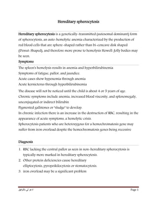

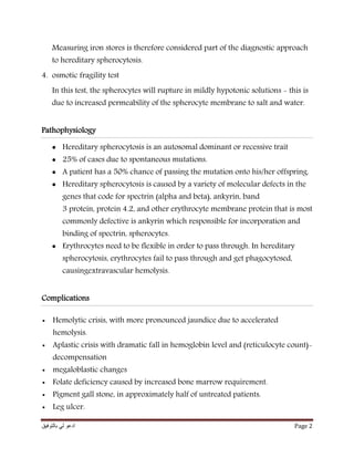
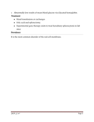

Hereditary spherocytosis is a genetically transmitted form of anemia where red blood cells are sphere-shaped rather than disk-shaped, making them prone to hemolysis. Symptoms include fatigue, pallor, jaundice, and in acute cases hypoxemia and hyperbilirubinemia. Diagnosis involves observing the lack of central pallor in red blood cells and increased fragility in hypotonic solutions. The condition is caused by defects in genes coding for membrane proteins, preventing red blood cells from maintaining their disk shape and surviving passage through the spleen. Treatment options include blood transfusions, folic acid supplementation, and splenectomy.