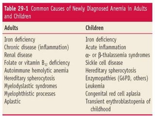
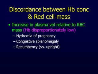
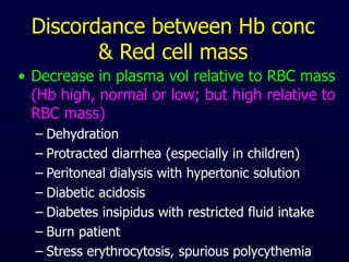
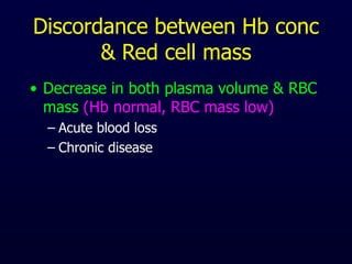
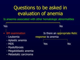
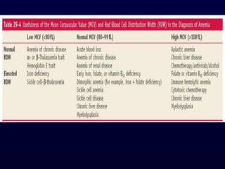
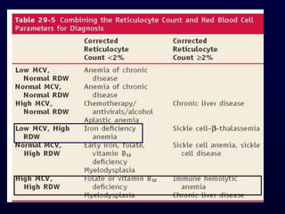
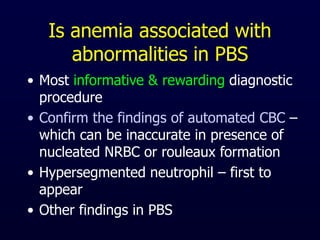
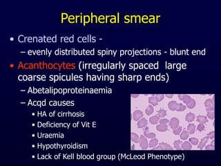
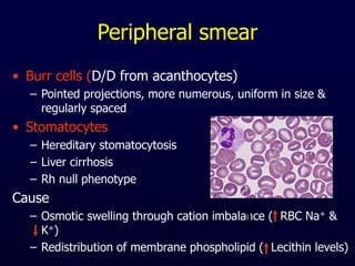
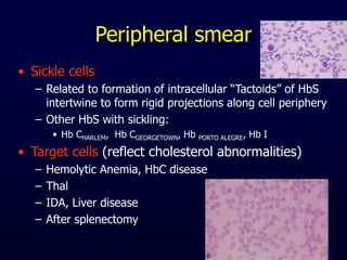
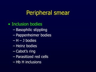
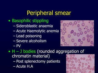
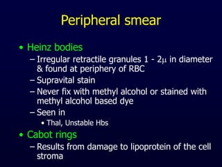
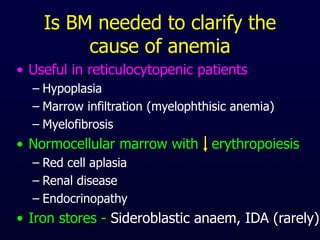
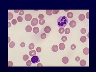
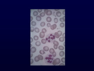
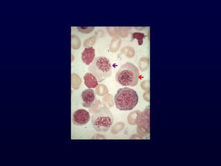
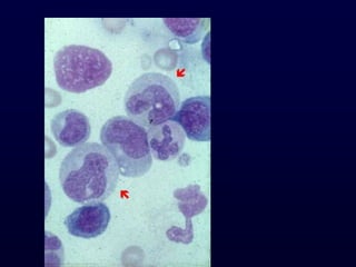
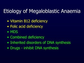
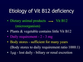
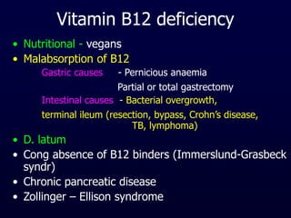
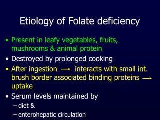
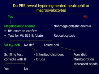
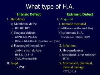
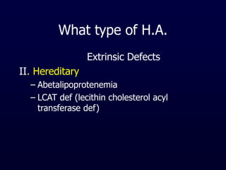
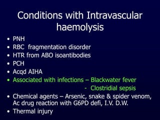
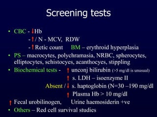
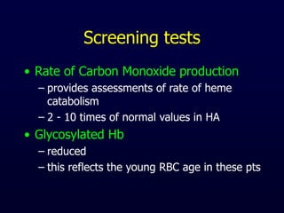
Classification &
Pathogenesis of
Anemia
Anemia
• Definition
• History
– Age
• normal values - adult, children
• In < 5 yrs - Hb < 11 g/dl Anemia (WHO crit)
• Girls (> 6yrs) + Boys (6 - 14 yrs) - Hb < 12 g/dl
• Boys (>15yrs) - Hb < 13 g/dl Anemia
• Elderly (low values - decrease in androgen secretion,
age related in stem cell proliferation)
• Cause of difference of values of Hb of children &
adults
Anemia
– Diet
– Drugs
– Race - Blacks - low values by 0.5 - 0.6 g/dl
– Change in stool habits
– Alcohol abuse
– Travel to malarious zone
Anaemia
• History
– Menstrual flow - > 7 days, clots, > 12 pads
– No. of pregnancy & abortions
– Fever - infection / neoplasm / collagen dis
– Skin
• Hyperpigmentation - Faconi’s anemia
• Bruises, ecchymosis, petechiae - PFD, liver disease
• Carotenemia - IDA in infants
• Jaundice - HA / Hepatitis
• Cavernous haemangioma - MAHA
Anaemia
• History
– Facies - haemolytic - thal
– Eyes
• Icterus
• Microcornea - Fanconi’s anemia
• Microaneurysmal retinal vessels - S & C Hbpathies
• Cataract - Galactosaemia
• Oedema of eyelid - Inf mononucleosis
• Blindness - Osteopetrosis
– Mouth - glossitis, angular stomatitis
Anaemia
• History
– Hands
• Triphalangeal thumb - Red cell aplasia
• Hypoplasia of thenar eminus - Fanconi’s anaemia
• Spoon nails - IDA
– Spleen enlargement
– Sternal tenderness
– Occult blood in stool
– Urine examination -
• haematuria, haemoglobinuria
Clinical effects of Anemia
• CVS & Pulmonary features of Anemia
– Depends on :-
• rate of reduction of O2 carrying capacity
• compensatory mechanism
• features of underlying disorder
– If anemia develops rapidly
• shortness of breath, tachycardia, dizziness/
faintness, postural hypotension, extreme fatigue
– In chronic anemias
• moderate dysponea or palpitation present
• some pts - CCF, Angina, intermittent claudication
Clinical effects of Anemia
• CVS & Pulmonary features of Anemia
– Heart murmur - systolic - pulmonic area
• Pallor -
– D/D from
• vasoconstriction of peripheral vessels
• pigmentation of skin
• myxedema
– Pallor + icterus
– Pallor + petechiae/ecchymosis = BM failure
Clinical effects of Anemia
• Skin & Mucosal changes
– thinning, loss of luster, early graying of hairs
– nails - loss of luster, brittle, concave
– Chronic leg ulcer - SCD
– Symmetric dermatitis
– Fissure at angle of mouth, glossitis
Clinical effects of Anemia
• Neuromuscular features
– headache, vertigo, tinnitus, faintness, scotoma
– lack of mental concentration, drowsiness
– restlessness & muscular weakness
– paresthesias
• Eye findings
– 20% pts - flame shaped hge, hard exudates,
cotton - wool spots
Clinical effects of Anemia
• GI changes
– Manifestations of underlying disorder
• hiatus hernia, DU, Gastric carcinoma
– Consequence of anemic conditions
• glossitis, atrophy of papillae of tongue
• dysphagia (IDA)
Discordance between Hb conc
& Red cell mass
• Increase in plasma vol relative to RBC
mass (Hb disproportionately low)
– Hydremia of pregnancy
– Congestive splenomegaly
– Recumbency (vs. upright)
Discordance between Hb conc
& Red cell mass
• Decrease in plasma vol relative to RBC mass
(Hb high, normal or low; but high relative to
RBC mass)
– Dehydration
– Protracted diarrhea (especially in children)
– Peritoneal dialysis with hypertonic solution
– Diabetic acidosis
– Diabetes insipidus with restricted fluid intake
– Burn patient
– Stress erythrocytosis, spurious polycythemia
Discordance between Hb conc
& Red cell mass
• Decrease in both plasma volume & RBC
mass (Hb normal, RBC mass low)
– Acute blood loss
– Chronic disease
Questions to be asked in
evaluation of anemia
Is anaemia associated with other hematologic abnormalities
Yes No
• BM examination Is there an appropriate Retic
- Leukemia response to anemia
- Aplastic anemia
- MDS Yes No
- Myelofibrosis
- Megaloblastic anemia
- Metastatic carcinoma
Questions to be asked-------
Yes No
• Is there Hemolysis? - Decrease in red cell prec
( Bilirubin, LDH, Haptoglobin - Ineffective erythropoiesis
Haemosidnuria)
What are red cell indices
Yes No
• Evaluate for cause Evaluate for
of hemolysis hemorrhagic cause
MCV (>100) MCV (80-100) MCV (<80)
- Megaloblastic A - ACD, PRCA - IDA, Thal, HbE
Reticuloctye count
• Corrected retic count =
– % retic x pts Hct / 45
• Reticulocyte index = Corrected retic x 0.5
(corrected for extended period of presence
in the circulation)
• Absolute retic count =
– % retic x RBC count / L3
– Normal = 25 to 75 x 109 / L
Reticulocyte count
• Corrected retic count < 2% or
• Absolute reticulocyte count < 100,000/l
Hypoproliferative anaemias
• Values above these –
– associated with either an appropriate
response to blood loss or with hemolytic
anemias
MCV & RDW
• MCV – fl, average cell size
• RDW –
– the SD of red blood cell volume divided by the
mean volume
– Reflects the variation in cell size (anisocytosis)
in the population of red blood cells
– upper limit 14.6% (12.4 - 14.6%) - CV
– Increased in
• IDA (even before MCV falls)
• Megaloblastic anaemia
Red cell Indices
• MCV = Hct (%) / RBC count (x1012/L) x 100
normal = 80 - 100 fl
• MCHC = Hb (g/dl) / Hct (%) x 100
normal = 31 - 36%
• MCH = Hb (g/dl) / RBC count (x1012/L) x 10
normal = 28 - 32 pg
• Hematological rule of THREE
Is anemia associated with
abnormalities in PBS
• Most informative & rewarding diagnostic
procedure
• Confirm the findings of automated CBC –
which can be inaccurate in presence of
nucleated NRBC or rouleaux formation
• Hypersegmented neutrophil – first to
appear
• Other findings in PBS
Peripheral smear
• Crenated red cells -
– evenly distributed spiny projections - blunt end
• Acanthocytes (irregularly spaced large
coarse spicules having sharp ends)
– Abetalipoproteinaemia
– Acqd causes
• HA of cirrhosis
• Deficiency of Vit E
• Uraemia
• Hypothyroidism
• Lack of Kell blood group (McLeod Phenotype)
Peripheral smear
• Burr cells (D/D from acanthocytes)
– Pointed projections, more numerous, uniform in size &
regularly spaced
• Stomatocytes
– Hereditary stomatocytosis
– Liver cirrhosis
– Rh null phenotype
Cause
– Osmotic swelling through cation imbalance ( RBC Na+ &
K+)
– Redistribution of membrane phospholipid ( Lecithin levels)
Peripheral smear
• Sickle cells
– Related to formation of intracellular “Tactoids” of HbS
intertwine to form rigid projections along cell periphery
– Other HbS with sickling:
• Hb CHARLEM, Hb CGEORGETOWN, Hb PORTO ALEGRE, Hb I
• Target cells (reflect cholesterol abnormalities)
– Hemolytic Anemia, HbC disease
– Thal
– IDA, Liver disease
– After splenectomy
Peripheral smear
• Inclusion bodies
– Basophilic stippling
– Pappenheimer bodies
– H – J bodies
– Heinz bodies
– Cabot’s ring
– Parasitized red cells
– Hb H inclusions
Peripheral smear
• Basophilic stippling
– Sideroblastic anaemia
– Acute Haemolytic anemia
– Lead poisoning
– Severe alcoholism
– PV
• H – J bodies (rounded aggregation of
chromatin material)
– Post splenectomy patients
– Acute H.A
Peripheral smear
• Heinz bodies
– Irregular retractile granules 1 - 2 in diameter
& found at periphery of RBC
– Supravital stain
– Never fix with methyl alcohol or stained with
methyl alcohol based dye
– Seen in
• Thal, Unstable Hbs
• Cabot rings
– Results from damage to lipoprotein of the cell
stroma
Is BM needed to clarify the
cause of anemia
• Useful in reticulocytopenic patients
– Hypoplasia
– Marrow infiltration (myelophthisic anemia)
– Myelofibrosis
• Normocellular marrow with erythropoiesis
– Red cell aplasia
– Renal disease
– Endocrinopathy
• Iron stores - Sideroblastic anaem, IDA (rarely)
Is BM needed to clarify the
cause of anemia
• Low retic count with normal marrow
erythropoiesis - ineffective erythropoiesis
– Megaloblastic anemia
– Thal
– Sideroblastic anemia
Macrocytic anemia
• Common finding
• 1.7% - 3.6% pts.- MCV is in absence of
anaemia
• Mild macrocytosis - common & may
remain unexplained
• This finding should not be ignored
• This could be imp. clue to underlying
disorder
Macrocytic Anaemia
• Megaloblastic macrocytic anaemia
• Normoblastic macrocytic anaemia
Normoblastic macrocytic anemia
(MCV - 100 - 110 fl)
• Pathology in RBC membrane lipids
Causes
• Macrocytosis Common -
Haemolytic anaemia
Post - haemorrhagic anaemia
• Macrocytosis occasional
Liver disease, CDA I & III,
Hypothyroidism, Alcohol,
Cytotoxic drugs, Leukemia's, MDS
Myelophthisic anaemia, Aplastic anaemia
Macrocytic anaemia & liver
disease
• Cause : Multi factorial -
– Intravascular dilution due to hypervoluemia
– Impaired ability of marrow to respond
– Severe HA (spur cell anaemia)
• Characterized by
– thin macrocytes (target cells)
– surface area without increase in vol
– memb lipids - esp cholesterol & phospholipids
Megaloblast
• Ehrlich - 1880 - first described
• Minot & Murphy - 1926 - disease could be
reversed by eating liver - Nobel prize
• Castle - 1929 -Gastric juice - IF which
binds with extrinsic factor in meat
Megaloblastic Anaemia
• Impaired DNA synthesis &
• Distinctive morphologic changes in
blood and bone marrow
Etiology of Megaloblastic Anaemia
• Vitamin B12 deficiency
• Folic acid deficiency
• MDS
• Combined deficiency
• Inherited disorders of DNA synthesis
• Drugs - inhibit DNA synthesis
Etiology of Vit B12 deficiency
• Dietary animal products Vit B12
(microorganism)
• Plants & vegetable contains little Vit B12
• Daily requirement - 2 - 3 mg
• Body stores - sufficient for many years
(Body stores to daily requirement ratio 1000:1)
• 1g - lost daily - biliary or renal excretion
Vitamin B12 deficiency
• Nutritional - vegans
• Malabsorption of B12
– Gastric causes - Pernicious anaemia
Partial or total gastrectomy
– Intestinal causes - Bacterial overgrowth,
terminal ileum (resection, bypass, Crohn’s disease,
TB, lymphoma)
• D. latum
• Cong absence of B12 binders (Immerslund-Grasbeck
syndr)
• Chronic pancreatic disease
• Zollinger – Ellison syndrome
Etiology of Folate deficiency
• Present in leafy vegetables, fruits,
mushrooms & animal protein
• Destroyed by prolonged cooking
• After ingestion interacts with small int.
brush border associated binding proteins
uptake
• Serum levels maintained by
– diet &
– enterohepatic circulation
Folic acid deficiency
• Poor intake - extremes of age, alcoholics
• Increased demand - pregnancy & lactation,
Hemolysis, Exfoliative dermatitis, Rapidly
growing malignancy
• Malabsorption - Sprue, Crohn’s disease,
Short bowel syndrome, Amyloidosis
Inherited disorders of DNA
synthesis
• Orotic aciduria
• Lesch – Nyhan syndrome
• Thiamine responsive megaloblastic anaemia
• MTHFR
• Formiminotransferase
• Dihydrofolate reductase
• Transcobalamin II deficiency
Other causes
• Myelodysplasia & Erythroleukemia
• Medications
– Folate antagonists (e.g. methotrexate)
– Purine antagonists (e.g. 6 – mercaptopurine)
– Pyrimidine antagonists (e.g. cytosine arabinoside)
– Alkylating agents (e.g. cyclophosphamide)
– Zidovudine , OC, Nitrous oxide, Arsenic
– Dihydrofolate reductase inhibitors
(sulfa drugs, methotrexate)
• Toxins
– Nitrous oxide, Arsenic, Chlordane
• CDA
Do PBS reveal hypersegmented neutrophil or
macroovaloctytes
Yes No
Megaloblastic anemia Nonmegaloblastic anemia
• BM exam to confirm
• Test for Vit B12 & folate Reticulocytosis
Vit B12 defi No defi Folate defi
Schilling test - Inherited disorders Poor diet
corrects with IF - Drugs Malabsorption
Increased needs
Yes No
Reticulocytosis
Increased Normal or decreased
Hemolytic anemia Alcohol toxicity
Hemorrhagic anemia Hypothroidism
Liver disease
If above negative, get BM exam
MDS, Red cell aplasia
Acqd sideroblastic anemia
Microcytic anemia
• Disorders of Iron metabolism
– IDA, ACD
• Disorders of globin synthesis
– Alpha and beta thal
– Hb E syndromes
– Hb C syndromes
– Unstable Hbs
• Sideroblastic anemia
• Lead intoxication (usually normocytic)
Approach to Microcytic anemia
Reticulocytosis
Low / Normal Increased
s. iron s. iron N serum iron s. iron
TIBC N / TIBC Normal TIBC Normal TIBC
Ferritin N or Ferritin Normal ferritin Ferritin
IDA Increased Hb electrophoresis BM + iron stain
- ESR, CRP
ACD Thal Sideroblastic an
Reticulocytosis
- Review PBS for abnormal morphology
- Lab test for increased red cell destruction
- obtain Hb studies
Homozygous beta thal
Hemolytic elliptocytosis
Hereditary pyropokilocytosis
Classification of Normocytic anemia
• Anemia associated with appropriately
increased erythrocyte production
– Post hemorrhagic anemia
– Hemolytic anemia
• Decreased erythropoietin secretion
– Impaired source - Renal or hepatic
– Reduced stimulus (decreased tissue needs)
• Anemia of endocrine deficiency
– PCM, ACD
Classification of Normocytic anemia
• Anemia with impaired marrow response
– BM hypoplasia
• Red blood cell aplasia
• Aplastic anemia
– BM infiltrative disorders
– MDS
– CDA II
– IDA - early
Approach to Normocytic anemia
Reticulocytosis
Increased RBC production N / decreased RBC production
H/O jaundice, splenomegaly
Presence of PBS abnormality
Elevated bilirubin or LDH
Yes No
Hemolysis Hemorrhogic abnormalities
Normal / decreased RBC production
- Serum chemistries to screen for S. iron
renal, hepatic and endocrine disease
- Consider EPO levels, thyroid studies
Positive Negative N / high Low
Anemia of BM aspirate / biopsy
- renal dis
- liver dis - Infiltrative disorders ACD
- endocrine - Red cell aplasia Early IDA
failure - MDS, CDA - II
Diagnostic approach to HA
• Anaemia - RBC destruction
RBC production OR BOTH
• BM – normally – increased production 6 – 8
times normal whenever destruction
• H.A. - destruction inspite of production
Clinical History
• Pallor, jaundice
• Passage of yellow / dark urine
• H/O gallstones, leg ulcers, skeletal abnormalities
• Splenomegaly, Hepatomegaly, aplastic crises
Family History (since birth/early childhood)
Yes No
Heriditary HA H/O – infection
- fever, drugs,
(ACQUIRED H.A.)
Intravascular haemolysis
What type of H.A.
Intrisic Defect Extrinsic Defect
1. Hereditary I. Acquired
a) Membrane defect 1. Immune mediated
– HS, HE, HPP a) AIHA (warm Abs, cold Abs)
b) Enzyme defects b)Isoimmune H.A.
– G6PD defi, PK defi Transfusion related, HDN
– Others- Glutathione reductase defi (rare)
c) Haemoglobinopathies / 2. Infections
globin chain defects 3. Hypersplenism
– Hb SS, Hb S -  4. Sec to Renal / Liver pathology
– Thal, Abnormal Hb / HTN
II. Acqd. 5. Mechanical, chemical,
- PNH thermal damage
- TTP, HUS
What type of H.A.
Extrinsic Defects
II. Hereditary
– Abetalipoprotenemia
– LCAT def (lecithin cholesterol acyl
transferase def)
Conditions with Intravascular
haemolysis
• PNH
• RBC fragmentation disorder
• HTR from ABO isoantibodies
• PCH
• Acqd AIHA
• Associated with infections – Blackwater fever
- Clostridial sepsis
• Chemical agents – Arsenic, snake & spider venom,
Ac drug reaction with G6PD defi, I.V. D.W.
• Thermal injury
Screening tests
• CBC - Hb
- / N - MCV, RDW
- Retic count BM – erythroid hyperplasia
• PS – macrocytes, polychramasia, NRBC, spherocytes,
elliptocytes, schistocyes, acanthocyes, stippling
• Biochemical tests - unconj bilirubin (>5 mg/dl is unusual)
s. LDH – isoenzyme II
Absent / s. haptoglobin (N=30 –190 mg/dl
Plasma Hb > 10 mg/dl
Fecal urobilinogen, Urine haemosiderin +ve
• Others – Red cell survival studies
Screening tests
• Rate of Carbon Monoxide production
– provides assessments of rate of heme
catabolism
– 2 - 10 times of normal values in HA
• Glycosylated Hb
– reduced
– this reflects the young RBC age in these pts
Features of I.V. haemolysis
• / absent Haptoglobin*
• Haemoglobinaemia (N < 1 mg/dl)
• Haemoglobinuria
• Haemosidinuria
• Methemalbuminemia – Schumm test
• Hemopexin
*Haptoglobin – acute phase reactant
- in inflam disorder - R.A., SLE
- neoplastic cond, & infections
Signs of Accelerated erythropoiesis
• Reticulocytosis
• Red cell creatine
– levels in young RBC - 6 to 8 times higher
– elevation persists for 20 days of RBC life span
compared to retic maturation time of 1 to 3
days
• Morphologic findings in PBS & BM
• Ferrokinetic studies
– increased plasma iron turn over - 2 to 8 times
Lab test useful in the D/D of
Hemolysis
• Specific morphologic abnormalities
• DAT
• Osmotic fragility test
• Hemolytic disorders with Heinz bodies
– G6PD defi (acute hemolytic episode)
– Unstable Hb disease
– Thal
– Chemical injury
Specific tests
PS Spherocytes (+)
+ve - ve
– AIHA (Warm Ab, Cold Ag, PCH) OF
– ABO & Rh incompatibility
– Drug induced HA -VE +VE
Incubated
Enzymopathies OFT
G6PD def PK def s/o HS
Heinz bodies confirm by
Enzyme screening test - Family studies
Enzyme assay - Memb protein anal
Autohaemolysis test
Coombs test
Specific tests
PS – Microcytic anaemia
Reticulocytosis - mild / N
Refractory anaemia
Leucopenia
Ham’s test
+ve -ve
Sucrose lysis test Other Ref anemia
CDA
PNH (confirm by CD55/59)
Autohemolysis test
• Measures spontaneous haemolysis of RBC when
incubated at 370C for 48 h in sterile conditions
• N= 0.2 – 2%
• With addition of Glucose = 0 – 0.9%
• With addition of ATP = 0 – 0.8%
• Type 1: slightly to moderately increased
partially corrected with glucose
seen in – G6PD def, HE
(Disorders of Pentose Phosphate Pathway)
Autohemolysis test
• Type II – autohaemolysis greatly increased
corrected with ATP & not with Glucose
seen in PK def
(disorders of glycolytic pathway)
• Type III – autohaemolyis greatly increased
correction with ATP or Glucose
seen in HS (membrane defect)
• Comments- neither specific nor sensitive
- But when positive – differentiate
membrane abnormality from enzyme defects
Conditions sometimes
mistaken for H.A.
• Associated with anemia & reticulocytosis
– Hge, Recovery from iron, folate or vit B12 def
– Recovery from marrow failure
• Associated with jaundice & anemia
– Ineffective erythropoiesis (intramedullary haemolysis)
– Bleeding into body cavity or tissue
• Associated with jaundice without anemia
– Defective bilirubin conjugation
– Crigler – Najar syndrome
– Gilbert syndrome
• Marrow invasion (MF, metastatic disease)
• Myoglobinuria
Approach to Haemoglobinopathies
& Thalassemia
CBC + PS ( Hb, micro – hypo)
MCV (normal) MCV (<77fl)
MCH (>27pg) MCH (<27pg)
Hb electrophoresis
Abn band Hb F Hb A2
Sickling test Quantitation Symptoms + Thal maj
Assymptomatic  thal / HPFH
+ve Hb S (HPLC for subclassfication)
- ve Hb D Punjab / Rare Hb variant (HPLC)
Approach to Haemoglobinopathies
& Thalassemia
Hb A2
Normal Border line Raised
IDA  Thal > 3.5% >10%
 thal IDA but <10%
Normal – A2  Thal
Repeat after IDA  Thal HbE /
correction triat variant-HPLC

Anemia classification & pathogenesis.ppt

  • 1.
  • 2.
    Anemia • Definition • History –Age • normal values - adult, children • In < 5 yrs - Hb < 11 g/dl Anemia (WHO crit) • Girls (> 6yrs) + Boys (6 - 14 yrs) - Hb < 12 g/dl • Boys (>15yrs) - Hb < 13 g/dl Anemia • Elderly (low values - decrease in androgen secretion, age related in stem cell proliferation) • Cause of difference of values of Hb of children & adults
  • 3.
    Anemia – Diet – Drugs –Race - Blacks - low values by 0.5 - 0.6 g/dl – Change in stool habits – Alcohol abuse – Travel to malarious zone
  • 4.
    Anaemia • History – Menstrualflow - > 7 days, clots, > 12 pads – No. of pregnancy & abortions – Fever - infection / neoplasm / collagen dis – Skin • Hyperpigmentation - Faconi’s anemia • Bruises, ecchymosis, petechiae - PFD, liver disease • Carotenemia - IDA in infants • Jaundice - HA / Hepatitis • Cavernous haemangioma - MAHA
  • 5.
    Anaemia • History – Facies- haemolytic - thal – Eyes • Icterus • Microcornea - Fanconi’s anemia • Microaneurysmal retinal vessels - S & C Hbpathies • Cataract - Galactosaemia • Oedema of eyelid - Inf mononucleosis • Blindness - Osteopetrosis – Mouth - glossitis, angular stomatitis
  • 6.
    Anaemia • History – Hands •Triphalangeal thumb - Red cell aplasia • Hypoplasia of thenar eminus - Fanconi’s anaemia • Spoon nails - IDA – Spleen enlargement – Sternal tenderness – Occult blood in stool – Urine examination - • haematuria, haemoglobinuria
  • 7.
    Clinical effects ofAnemia • CVS & Pulmonary features of Anemia – Depends on :- • rate of reduction of O2 carrying capacity • compensatory mechanism • features of underlying disorder – If anemia develops rapidly • shortness of breath, tachycardia, dizziness/ faintness, postural hypotension, extreme fatigue – In chronic anemias • moderate dysponea or palpitation present • some pts - CCF, Angina, intermittent claudication
  • 8.
    Clinical effects ofAnemia • CVS & Pulmonary features of Anemia – Heart murmur - systolic - pulmonic area • Pallor - – D/D from • vasoconstriction of peripheral vessels • pigmentation of skin • myxedema – Pallor + icterus – Pallor + petechiae/ecchymosis = BM failure
  • 9.
    Clinical effects ofAnemia • Skin & Mucosal changes – thinning, loss of luster, early graying of hairs – nails - loss of luster, brittle, concave – Chronic leg ulcer - SCD – Symmetric dermatitis – Fissure at angle of mouth, glossitis
  • 10.
    Clinical effects ofAnemia • Neuromuscular features – headache, vertigo, tinnitus, faintness, scotoma – lack of mental concentration, drowsiness – restlessness & muscular weakness – paresthesias • Eye findings – 20% pts - flame shaped hge, hard exudates, cotton - wool spots
  • 11.
    Clinical effects ofAnemia • GI changes – Manifestations of underlying disorder • hiatus hernia, DU, Gastric carcinoma – Consequence of anemic conditions • glossitis, atrophy of papillae of tongue • dysphagia (IDA)
  • 13.
    Discordance between Hbconc & Red cell mass • Increase in plasma vol relative to RBC mass (Hb disproportionately low) – Hydremia of pregnancy – Congestive splenomegaly – Recumbency (vs. upright)
  • 14.
    Discordance between Hbconc & Red cell mass • Decrease in plasma vol relative to RBC mass (Hb high, normal or low; but high relative to RBC mass) – Dehydration – Protracted diarrhea (especially in children) – Peritoneal dialysis with hypertonic solution – Diabetic acidosis – Diabetes insipidus with restricted fluid intake – Burn patient – Stress erythrocytosis, spurious polycythemia
  • 15.
    Discordance between Hbconc & Red cell mass • Decrease in both plasma volume & RBC mass (Hb normal, RBC mass low) – Acute blood loss – Chronic disease
  • 16.
    Questions to beasked in evaluation of anemia Is anaemia associated with other hematologic abnormalities Yes No • BM examination Is there an appropriate Retic - Leukemia response to anemia - Aplastic anemia - MDS Yes No - Myelofibrosis - Megaloblastic anemia - Metastatic carcinoma
  • 17.
    Questions to beasked------- Yes No • Is there Hemolysis? - Decrease in red cell prec ( Bilirubin, LDH, Haptoglobin - Ineffective erythropoiesis Haemosidnuria) What are red cell indices Yes No • Evaluate for cause Evaluate for of hemolysis hemorrhagic cause MCV (>100) MCV (80-100) MCV (<80) - Megaloblastic A - ACD, PRCA - IDA, Thal, HbE
  • 18.
    Reticuloctye count • Correctedretic count = – % retic x pts Hct / 45 • Reticulocyte index = Corrected retic x 0.5 (corrected for extended period of presence in the circulation) • Absolute retic count = – % retic x RBC count / L3 – Normal = 25 to 75 x 109 / L
  • 19.
    Reticulocyte count • Correctedretic count < 2% or • Absolute reticulocyte count < 100,000/l Hypoproliferative anaemias • Values above these – – associated with either an appropriate response to blood loss or with hemolytic anemias
  • 21.
    MCV & RDW •MCV – fl, average cell size • RDW – – the SD of red blood cell volume divided by the mean volume – Reflects the variation in cell size (anisocytosis) in the population of red blood cells – upper limit 14.6% (12.4 - 14.6%) - CV – Increased in • IDA (even before MCV falls) • Megaloblastic anaemia
  • 22.
    Red cell Indices •MCV = Hct (%) / RBC count (x1012/L) x 100 normal = 80 - 100 fl • MCHC = Hb (g/dl) / Hct (%) x 100 normal = 31 - 36% • MCH = Hb (g/dl) / RBC count (x1012/L) x 10 normal = 28 - 32 pg • Hematological rule of THREE
  • 25.
    Is anemia associatedwith abnormalities in PBS • Most informative & rewarding diagnostic procedure • Confirm the findings of automated CBC – which can be inaccurate in presence of nucleated NRBC or rouleaux formation • Hypersegmented neutrophil – first to appear • Other findings in PBS
  • 26.
    Peripheral smear • Crenatedred cells - – evenly distributed spiny projections - blunt end • Acanthocytes (irregularly spaced large coarse spicules having sharp ends) – Abetalipoproteinaemia – Acqd causes • HA of cirrhosis • Deficiency of Vit E • Uraemia • Hypothyroidism • Lack of Kell blood group (McLeod Phenotype)
  • 27.
    Peripheral smear • Burrcells (D/D from acanthocytes) – Pointed projections, more numerous, uniform in size & regularly spaced • Stomatocytes – Hereditary stomatocytosis – Liver cirrhosis – Rh null phenotype Cause – Osmotic swelling through cation imbalance ( RBC Na+ & K+) – Redistribution of membrane phospholipid ( Lecithin levels)
  • 28.
    Peripheral smear • Sicklecells – Related to formation of intracellular “Tactoids” of HbS intertwine to form rigid projections along cell periphery – Other HbS with sickling: • Hb CHARLEM, Hb CGEORGETOWN, Hb PORTO ALEGRE, Hb I • Target cells (reflect cholesterol abnormalities) – Hemolytic Anemia, HbC disease – Thal – IDA, Liver disease – After splenectomy
  • 29.
    Peripheral smear • Inclusionbodies – Basophilic stippling – Pappenheimer bodies – H – J bodies – Heinz bodies – Cabot’s ring – Parasitized red cells – Hb H inclusions
  • 30.
    Peripheral smear • Basophilicstippling – Sideroblastic anaemia – Acute Haemolytic anemia – Lead poisoning – Severe alcoholism – PV • H – J bodies (rounded aggregation of chromatin material) – Post splenectomy patients – Acute H.A
  • 31.
    Peripheral smear • Heinzbodies – Irregular retractile granules 1 - 2 in diameter & found at periphery of RBC – Supravital stain – Never fix with methyl alcohol or stained with methyl alcohol based dye – Seen in • Thal, Unstable Hbs • Cabot rings – Results from damage to lipoprotein of the cell stroma
  • 32.
    Is BM neededto clarify the cause of anemia • Useful in reticulocytopenic patients – Hypoplasia – Marrow infiltration (myelophthisic anemia) – Myelofibrosis • Normocellular marrow with erythropoiesis – Red cell aplasia – Renal disease – Endocrinopathy • Iron stores - Sideroblastic anaem, IDA (rarely)
  • 33.
    Is BM neededto clarify the cause of anemia • Low retic count with normal marrow erythropoiesis - ineffective erythropoiesis – Megaloblastic anemia – Thal – Sideroblastic anemia
  • 34.
    Macrocytic anemia • Commonfinding • 1.7% - 3.6% pts.- MCV is in absence of anaemia • Mild macrocytosis - common & may remain unexplained • This finding should not be ignored • This could be imp. clue to underlying disorder
  • 35.
    Macrocytic Anaemia • Megaloblasticmacrocytic anaemia • Normoblastic macrocytic anaemia
  • 36.
    Normoblastic macrocytic anemia (MCV- 100 - 110 fl) • Pathology in RBC membrane lipids Causes • Macrocytosis Common - Haemolytic anaemia Post - haemorrhagic anaemia • Macrocytosis occasional Liver disease, CDA I & III, Hypothyroidism, Alcohol, Cytotoxic drugs, Leukemia's, MDS Myelophthisic anaemia, Aplastic anaemia
  • 37.
    Macrocytic anaemia &liver disease • Cause : Multi factorial - – Intravascular dilution due to hypervoluemia – Impaired ability of marrow to respond – Severe HA (spur cell anaemia) • Characterized by – thin macrocytes (target cells) – surface area without increase in vol – memb lipids - esp cholesterol & phospholipids
  • 38.
    Megaloblast • Ehrlich -1880 - first described • Minot & Murphy - 1926 - disease could be reversed by eating liver - Nobel prize • Castle - 1929 -Gastric juice - IF which binds with extrinsic factor in meat
  • 39.
    Megaloblastic Anaemia • ImpairedDNA synthesis & • Distinctive morphologic changes in blood and bone marrow
  • 45.
    Etiology of MegaloblasticAnaemia • Vitamin B12 deficiency • Folic acid deficiency • MDS • Combined deficiency • Inherited disorders of DNA synthesis • Drugs - inhibit DNA synthesis
  • 46.
    Etiology of VitB12 deficiency • Dietary animal products Vit B12 (microorganism) • Plants & vegetable contains little Vit B12 • Daily requirement - 2 - 3 mg • Body stores - sufficient for many years (Body stores to daily requirement ratio 1000:1) • 1g - lost daily - biliary or renal excretion
  • 47.
    Vitamin B12 deficiency •Nutritional - vegans • Malabsorption of B12 – Gastric causes - Pernicious anaemia Partial or total gastrectomy – Intestinal causes - Bacterial overgrowth, terminal ileum (resection, bypass, Crohn’s disease, TB, lymphoma) • D. latum • Cong absence of B12 binders (Immerslund-Grasbeck syndr) • Chronic pancreatic disease • Zollinger – Ellison syndrome
  • 48.
    Etiology of Folatedeficiency • Present in leafy vegetables, fruits, mushrooms & animal protein • Destroyed by prolonged cooking • After ingestion interacts with small int. brush border associated binding proteins uptake • Serum levels maintained by – diet & – enterohepatic circulation
  • 49.
    Folic acid deficiency •Poor intake - extremes of age, alcoholics • Increased demand - pregnancy & lactation, Hemolysis, Exfoliative dermatitis, Rapidly growing malignancy • Malabsorption - Sprue, Crohn’s disease, Short bowel syndrome, Amyloidosis
  • 50.
    Inherited disorders ofDNA synthesis • Orotic aciduria • Lesch – Nyhan syndrome • Thiamine responsive megaloblastic anaemia • MTHFR • Formiminotransferase • Dihydrofolate reductase • Transcobalamin II deficiency
  • 51.
    Other causes • Myelodysplasia& Erythroleukemia • Medications – Folate antagonists (e.g. methotrexate) – Purine antagonists (e.g. 6 – mercaptopurine) – Pyrimidine antagonists (e.g. cytosine arabinoside) – Alkylating agents (e.g. cyclophosphamide) – Zidovudine , OC, Nitrous oxide, Arsenic – Dihydrofolate reductase inhibitors (sulfa drugs, methotrexate) • Toxins – Nitrous oxide, Arsenic, Chlordane • CDA
  • 52.
    Do PBS revealhypersegmented neutrophil or macroovaloctytes Yes No Megaloblastic anemia Nonmegaloblastic anemia • BM exam to confirm • Test for Vit B12 & folate Reticulocytosis Vit B12 defi No defi Folate defi Schilling test - Inherited disorders Poor diet corrects with IF - Drugs Malabsorption Increased needs Yes No
  • 53.
    Reticulocytosis Increased Normal ordecreased Hemolytic anemia Alcohol toxicity Hemorrhagic anemia Hypothroidism Liver disease If above negative, get BM exam MDS, Red cell aplasia Acqd sideroblastic anemia
  • 54.
    Microcytic anemia • Disordersof Iron metabolism – IDA, ACD • Disorders of globin synthesis – Alpha and beta thal – Hb E syndromes – Hb C syndromes – Unstable Hbs • Sideroblastic anemia • Lead intoxication (usually normocytic)
  • 55.
    Approach to Microcyticanemia Reticulocytosis Low / Normal Increased s. iron s. iron N serum iron s. iron TIBC N / TIBC Normal TIBC Normal TIBC Ferritin N or Ferritin Normal ferritin Ferritin IDA Increased Hb electrophoresis BM + iron stain - ESR, CRP ACD Thal Sideroblastic an
  • 56.
    Reticulocytosis - Review PBSfor abnormal morphology - Lab test for increased red cell destruction - obtain Hb studies Homozygous beta thal Hemolytic elliptocytosis Hereditary pyropokilocytosis
  • 57.
    Classification of Normocyticanemia • Anemia associated with appropriately increased erythrocyte production – Post hemorrhagic anemia – Hemolytic anemia • Decreased erythropoietin secretion – Impaired source - Renal or hepatic – Reduced stimulus (decreased tissue needs) • Anemia of endocrine deficiency – PCM, ACD
  • 58.
    Classification of Normocyticanemia • Anemia with impaired marrow response – BM hypoplasia • Red blood cell aplasia • Aplastic anemia – BM infiltrative disorders – MDS – CDA II – IDA - early
  • 59.
    Approach to Normocyticanemia Reticulocytosis Increased RBC production N / decreased RBC production H/O jaundice, splenomegaly Presence of PBS abnormality Elevated bilirubin or LDH Yes No Hemolysis Hemorrhogic abnormalities
  • 60.
    Normal / decreasedRBC production - Serum chemistries to screen for S. iron renal, hepatic and endocrine disease - Consider EPO levels, thyroid studies Positive Negative N / high Low Anemia of BM aspirate / biopsy - renal dis - liver dis - Infiltrative disorders ACD - endocrine - Red cell aplasia Early IDA failure - MDS, CDA - II
  • 61.
    Diagnostic approach toHA • Anaemia - RBC destruction RBC production OR BOTH • BM – normally – increased production 6 – 8 times normal whenever destruction • H.A. - destruction inspite of production
  • 62.
    Clinical History • Pallor,jaundice • Passage of yellow / dark urine • H/O gallstones, leg ulcers, skeletal abnormalities • Splenomegaly, Hepatomegaly, aplastic crises Family History (since birth/early childhood) Yes No Heriditary HA H/O – infection - fever, drugs, (ACQUIRED H.A.)
  • 63.
  • 64.
    What type ofH.A. Intrisic Defect Extrinsic Defect 1. Hereditary I. Acquired a) Membrane defect 1. Immune mediated – HS, HE, HPP a) AIHA (warm Abs, cold Abs) b) Enzyme defects b)Isoimmune H.A. – G6PD defi, PK defi Transfusion related, HDN – Others- Glutathione reductase defi (rare) c) Haemoglobinopathies / 2. Infections globin chain defects 3. Hypersplenism – Hb SS, Hb S -  4. Sec to Renal / Liver pathology – Thal, Abnormal Hb / HTN II. Acqd. 5. Mechanical, chemical, - PNH thermal damage - TTP, HUS
  • 65.
    What type ofH.A. Extrinsic Defects II. Hereditary – Abetalipoprotenemia – LCAT def (lecithin cholesterol acyl transferase def)
  • 66.
    Conditions with Intravascular haemolysis •PNH • RBC fragmentation disorder • HTR from ABO isoantibodies • PCH • Acqd AIHA • Associated with infections – Blackwater fever - Clostridial sepsis • Chemical agents – Arsenic, snake & spider venom, Ac drug reaction with G6PD defi, I.V. D.W. • Thermal injury
  • 67.
    Screening tests • CBC- Hb - / N - MCV, RDW - Retic count BM – erythroid hyperplasia • PS – macrocytes, polychramasia, NRBC, spherocytes, elliptocytes, schistocyes, acanthocyes, stippling • Biochemical tests - unconj bilirubin (>5 mg/dl is unusual) s. LDH – isoenzyme II Absent / s. haptoglobin (N=30 –190 mg/dl Plasma Hb > 10 mg/dl Fecal urobilinogen, Urine haemosiderin +ve • Others – Red cell survival studies
  • 68.
    Screening tests • Rateof Carbon Monoxide production – provides assessments of rate of heme catabolism – 2 - 10 times of normal values in HA • Glycosylated Hb – reduced – this reflects the young RBC age in these pts
  • 69.
    Features of I.V.haemolysis • / absent Haptoglobin* • Haemoglobinaemia (N < 1 mg/dl) • Haemoglobinuria • Haemosidinuria • Methemalbuminemia – Schumm test • Hemopexin *Haptoglobin – acute phase reactant - in inflam disorder - R.A., SLE - neoplastic cond, & infections
  • 70.
    Signs of Acceleratederythropoiesis • Reticulocytosis • Red cell creatine – levels in young RBC - 6 to 8 times higher – elevation persists for 20 days of RBC life span compared to retic maturation time of 1 to 3 days • Morphologic findings in PBS & BM • Ferrokinetic studies – increased plasma iron turn over - 2 to 8 times
  • 71.
    Lab test usefulin the D/D of Hemolysis • Specific morphologic abnormalities • DAT • Osmotic fragility test • Hemolytic disorders with Heinz bodies – G6PD defi (acute hemolytic episode) – Unstable Hb disease – Thal – Chemical injury
  • 72.
    Specific tests PS Spherocytes(+) +ve - ve – AIHA (Warm Ab, Cold Ag, PCH) OF – ABO & Rh incompatibility – Drug induced HA -VE +VE Incubated Enzymopathies OFT G6PD def PK def s/o HS Heinz bodies confirm by Enzyme screening test - Family studies Enzyme assay - Memb protein anal Autohaemolysis test Coombs test
  • 73.
    Specific tests PS –Microcytic anaemia Reticulocytosis - mild / N Refractory anaemia Leucopenia Ham’s test +ve -ve Sucrose lysis test Other Ref anemia CDA PNH (confirm by CD55/59)
  • 74.
    Autohemolysis test • Measuresspontaneous haemolysis of RBC when incubated at 370C for 48 h in sterile conditions • N= 0.2 – 2% • With addition of Glucose = 0 – 0.9% • With addition of ATP = 0 – 0.8% • Type 1: slightly to moderately increased partially corrected with glucose seen in – G6PD def, HE (Disorders of Pentose Phosphate Pathway)
  • 75.
    Autohemolysis test • TypeII – autohaemolysis greatly increased corrected with ATP & not with Glucose seen in PK def (disorders of glycolytic pathway) • Type III – autohaemolyis greatly increased correction with ATP or Glucose seen in HS (membrane defect) • Comments- neither specific nor sensitive - But when positive – differentiate membrane abnormality from enzyme defects
  • 76.
    Conditions sometimes mistaken forH.A. • Associated with anemia & reticulocytosis – Hge, Recovery from iron, folate or vit B12 def – Recovery from marrow failure • Associated with jaundice & anemia – Ineffective erythropoiesis (intramedullary haemolysis) – Bleeding into body cavity or tissue • Associated with jaundice without anemia – Defective bilirubin conjugation – Crigler – Najar syndrome – Gilbert syndrome • Marrow invasion (MF, metastatic disease) • Myoglobinuria
  • 77.
    Approach to Haemoglobinopathies &Thalassemia CBC + PS ( Hb, micro – hypo) MCV (normal) MCV (<77fl) MCH (>27pg) MCH (<27pg) Hb electrophoresis Abn band Hb F Hb A2 Sickling test Quantitation Symptoms + Thal maj Assymptomatic  thal / HPFH +ve Hb S (HPLC for subclassfication) - ve Hb D Punjab / Rare Hb variant (HPLC)
  • 78.
    Approach to Haemoglobinopathies &Thalassemia Hb A2 Normal Border line Raised IDA  Thal > 3.5% >10%  thal IDA but <10% Normal – A2  Thal Repeat after IDA  Thal HbE / correction triat variant-HPLC