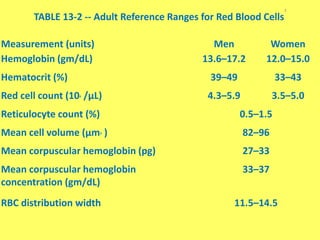
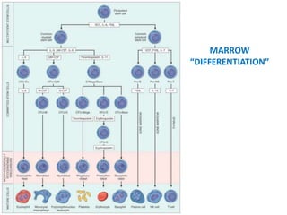
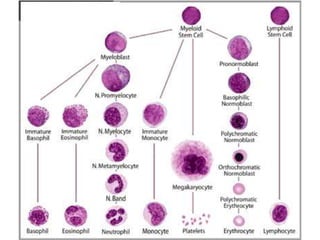
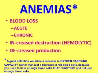
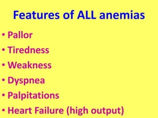
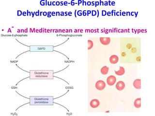
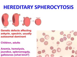
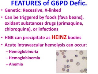
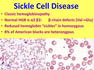
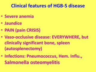
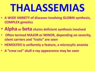
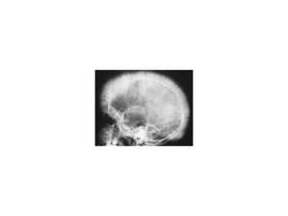
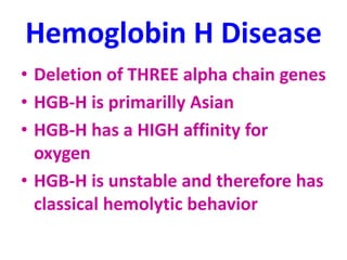
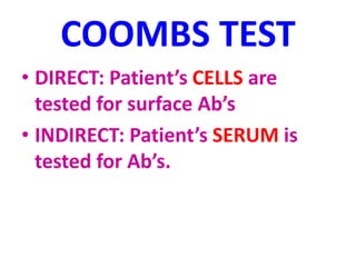
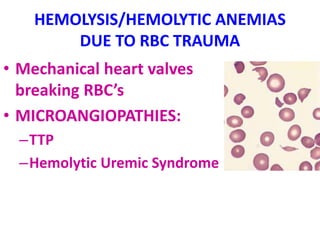
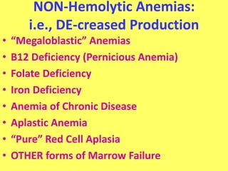
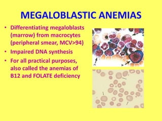
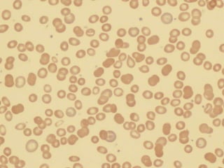
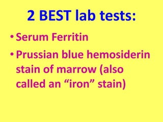
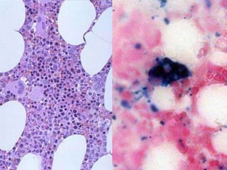
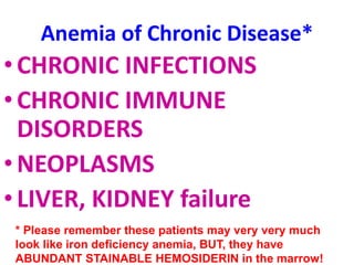
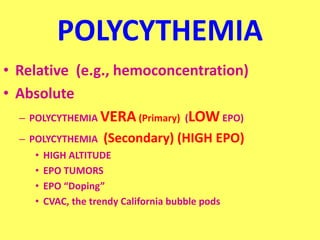
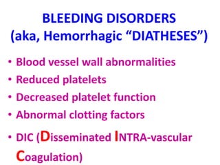
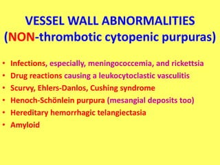
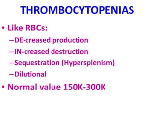
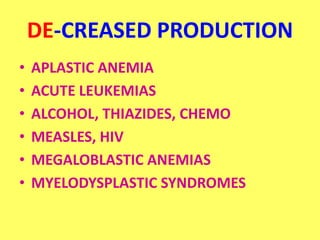
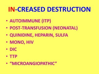
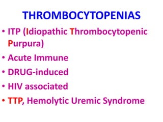
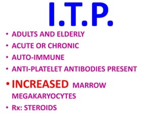
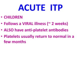
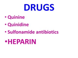
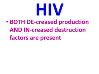
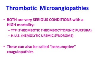
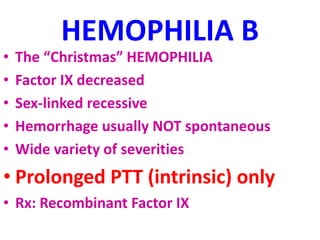
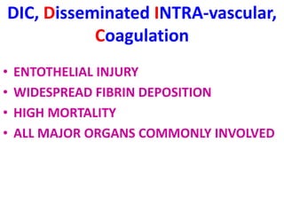
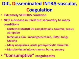
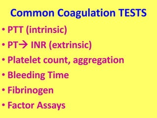
RBC and BLEEDING DISORDERS
RBC and Bleeding Disorders
• NORMAL
– Anatomy, histology
– Development
– Physiology
• ANEMIAS
– Blood loss: acute, chronic
– Hemolytic
– Diminished erythropoesis
• POLYCYTHEMIA
• BLEEDING DISORDERS
TABLE 13-2 -- Adult Reference Ranges for Red Blood Cells
*
Measurement (units) Men Women
Hemoglobin (gm/dL) 13.6–17.2 12.0–15.0
Hematocrit (%) 39–49 33–43
Red cell count (106 /µL) 4.3–5.9 3.5–5.0
Reticulocyte count (%) 0.5–1.5
Mean cell volume (µm3 ) 82–96
Mean corpuscular hemoglobin (pg) 27–33
Mean corpuscular hemoglobin
concentration (gm/dL)
33–37
RBC distribution width 11.5–14.5
WHERE is MARROW?
• Yolk Sac: very early embryo
• Liver, Spleen: NEWBORN
• BONE
– CHILDHOOD: AXIAL SKELETON & APPENDICULAR
SKELETON BOTH HAVE RED (active) MARROW
– ADULT: AXIAL SKELETON RED MARROW,
APPENDICULAR SKELETON YELLOW MARROW
MARROW FEATURES
• CELLULARITY
• MEGAKARYOCYTES
• M:E RATIO
• MYELOID MATURATION
• ERYTHROID MATURATION
• LYMPHS, PLASMA CELLS
• STORAGE IRON, i.e., HEMOSIDERIN
• “FOREIGN CELLS”
MARROW
“DIFFERENTIATION”
ANEMIAS*
• BLOOD LOSS
–ACUTE
–CHRONIC
• IN-creased destruction (HEMOLYTIC)
• DE-creased production
*A good definition would be a decrease in OXYGEN CARRYING
CAPACITY, rather than just a decrease in red blood cells, because
you need to have enough blood cells THAT FUNCTION, and not just
enough blood cells.
Features of ALL anemias
• Pallor
• Tiredness
• Weakness
• Dyspnea
• Palpitations
• Heart Failure (high output)
Blood Loss
Acute: trauma
Chronic: lesions of gastrointestinal tract,
gynecologic disturbances. The features of
chronic blood loss anemia are the same as iron
deficiency anemia, and is defined as a situation
in which the production cannot keep up with
the loss
HEMOLYTIC
• HEREDITARY
– MEMBRANE disorders: e.g., spherocytosis
– ENZYME disorders: e.g., G6PD deficciency
– HGB disorders (hemoglobinopathies)
• ACQUIRED
– MEMBRANE disorders (PNH)
– ANTIBODY MEDIATED, transfusion or autoantibodies
– MECHANICAL TRAUMA
– INFECTIONS
– DRUGS, TOXINS, HYPERSPLENISM
IMPAIRED PRODUCTION
• Disturbance of proliferation and differentiation of
stem cells: aplastic anemias, pure RBC aplasia,
renal failure
• Disturbance of proliferation and maturation of
erythroblasts
• Defective DNA synthesis: (Megaloblastic)
• Defective heme synthesis: (Fe)
• Deficient globin synthesis: (Thalassemias)
Glucose-6-Phosphate
Dehydrogenase (G6PD) Deficiency
• A
- and Mediterranean are most significant types
MODIFIERS
• MCV, microcytosis, macrocytosis
• MCH
• MCHC, hypochromic
• RDW, anisocytosis
HEMOLYTIC ANEMIAS
• Life span LESS than 120 days
• Marrow hyperplasia (M:E), EPO+
• Increased catabolic products, e.g.,
bilirubin, serum HGB, hemosiderin
• Decreased haptoglobin
HEMOLYSIS
• INTRA-vascular (vessels)
• EXTRA-vascular (spleen)
M:E Ratio normally 3:1
HEREDITARY SPHEROCYTOSIS
Genetic defects affecting
ankyrin, spectrin, usually
autosomal dominant
Children, adults
Anemia, hemolysis,
jaundice, splenomegaly,
gallstones (what kind?)
FEATURES of G6PD Defic.
• Genetic: Recessive, X-linked
• Can be triggered by foods (fava beans),
oxidant substances drugs (primaquine,
chloroquine), or infections
• HGB can precipitate as HEINZ bodies
• Acute intravascular hemolysis can occur:
–Hemoglobinuria
–Hemoglobinemia
–Anemia
Sickle Cell Disease
• Classic hemoglobinopathy
• Normal HGB is α2 β2: β-chain defects (Val->Glu)
• Reduced hemoglobin “sickles” in homozygous
• 8% of American blacks are heterozygous
Clinical features of HGB-S disease
• Severe anemia
• Jaundice
• PAIN (pain CRISIS)
• Vaso-occlusive disease: EVERYWHERE, but
clinically significant bone, spleen
(autosplenectomy)
• Infections: Pneumococcus, Hem. Influ.,
Salmonella osteomyelitis
THALASSEMIAS
• A WIDE VARIETY of diseases involving GLOBIN synthesis,
COMPLEX genetics
• Alpha or beta chains deficient synthesis involved
• Often termed MAJOR or MINOR, depending on severity,
silent carriers and “traits” are seen
• HEMOLYSIS is uniformly a feature, a microcytic anemia
• A “crew cut” skull x-ray appearance may be seen
Hemoglobin H Disease
• Deletion of THREE alpha chain genes
• HGB-H is primarilly Asian
• HGB-H has a HIGH affinity for
oxygen
• HGB-H is unstable and therefore has
classical hemolytic behavior
HYDROPS FETALIS
• FOUR alpha chain genes are deleted, so this is
the MOST SEVERE form of thalassemia
• Many/most never make it to term
• Children born will have a SEVERE hemolytic
anemia as in the erythroblastosis fetalis of Rh
disease:
– Pallor (as in all anemias)
– Edema (hence the name “hydrops”)
– Massive hepatosplenomegaly (hemolysis)
Paroxysmal Nocturnal
Hemoglobinuria (PNH)
• ACQUIRED, NOT INHERITED like all the previous
hemolytic anemias were
• ACQUIRED mutations in phosphatidylinositol
glycan A (PIGA)
• It is “P” and “N” only 25% of the time
GlycosylphosPhatidylInositol
Immunohemolytic Anemia
• All of these have the presence of antibodies
and/or compliment present on RBC surfaces
• NOT all are AUTOimmune, some are caused
by drugs
• Antibodies can be
–WARM (IgG)
–COLD AGGLUTININ (IgM)
–COLD HEMOLYSIN (paroxysmal) (IgG)
IMMUNOHEMOLYTIC ANEMIAS
• WARM (IgG), will NOT hemolyze at room temp
– Primary Idiopathic (most common)
– Secondary (Tumors, especially leuk/lymph, drugs)
• COLD AGGLUTININS: (IgM), WILL hemolyze at
room temp
– Mycoplasma pneumoniae, HIV, mononucleosis
• COLD HEMOLYSINS: (IgG) Cold Paroxysmal
Hemoglobinuria, hemo-LYSIS in body, ALSO often
follows mycoplasma pneumoniae
COOMBS TEST
• DIRECT: Patient’s CELLS are
tested for surface Ab’s
• INDIRECT: Patient’s SERUM is
tested for Ab’s.
HEMOLYSIS/HEMOLYTIC ANEMIAS
DUE TO RBC TRAUMA
• Mechanical heart valves
breaking RBC’s
• MICROANGIOPATHIES:
–TTP
–Hemolytic Uremic Syndrome
NON-Hemolytic Anemias:
i.e., DE-creased Production
• “Megaloblastic” Anemias
• B12 Deficiency (Pernicious Anemia)
• Folate Deficiency
• Iron Deficiency
• Anemia of Chronic Disease
• Aplastic Anemia
• “Pure” Red Cell Aplasia
• OTHER forms of Marrow Failure
MEGALOBLASTIC ANEMIAS
• Differentiating megaloblasts
(marrow) from macrocytes
(peripheral smear, MCV>94)
• Impaired DNA synthesis
• For all practical purposes,
also called the anemias of
B12 and FOLATE deficiency
Vit-B12 Physiology
• Oral ingestion
• Combines with INTRINSIC FACTOR in the
gastric mucosa
• Absorbed in the terminal ileum
• DEFECTS at ANY of these sites can
produce a MEGALOBLASTIC anemia
Decreased intake
Inadequate diet, vegetarianism
Impaired absorption
Intrinsic factor deficiency
Pernicious anemia
Gastrectomy
Malabsorption states
Diffuse intestinal disease, e.g., lymphoma, systemic sclerosis
Ileal resection, ileitis
Competitive parasitic uptake
Fish tapeworm infestation
Bacterial overgrowth in blind loops and diverticula of bowel
Increased requirement
Pregnancy, hyperthyroidism, disseminated cancer
Please remember that ALL
megaloblastic anemias are also
MACROCYTIC (MCV>94 or
MCV~100), and that not only are
the RBC’s BIG, but so are the
neutrophils, and neutrophilic
precursors in the bone marrow
too, and even more so,
HYPERSEGMENTED!!!
PERNICIOUS ANEMIA
• MEGALOBLASTIC anemia
• LEUKOPENIA and HYPERSEGS
• JAUNDICE
• NEUROLOGIC posterolateral spinal tracts
• ACHLORHYDRIA
• Can’t absorb B12
• LOW serum B12
• Flunk Schilling test, i.e., can’t absorb B12,
using a radioactive tracer
FOLATE DEFICIENCY
MEGALOBLASTIC AMEMIAS
• Decreased Intake: diet, etoh-ism, infancy
• Impaired Absorption: intestinal disease
• DRUGS: anticonvulsants, BCPs, CHEMO
• Increased Loss: Hemodialysis
• Increased Requirement: Pregnancy, infancy
• Impaired Usage
Fe Deficiency Anemia
• Due to increased loss or decreased
ingestion, almost always, in USA,
nowadays, increased loss is the reason
• Microcytic (low MCV), Hypochromic
(low MCHC)
• THE ONLY WAY WE CAN LOSE IRON IS BY
LOSING BLOOD
Fe
Transferrin
Ferritin (GREAT test)
Hemosiderin
Clinical Fe-Defic-Anemia
• Adult men: GI Blood Loss
• PRE menopausal women:
menorrhagia
• POST menopausal women: GI Blood
Loss
2 BEST lab tests:
•Serum Ferritin
•Prussian blue hemosiderin
stain of marrow (also
called an “iron” stain)
Anemia of Chronic Disease*
•CHRONIC INFECTIONS
•CHRONIC IMMUNE
DISORDERS
•NEOPLASMS
•LIVER, KIDNEY failure
* Please remember these patients may very very much
look like iron deficiency anemia, BUT, they have
ABUNDANT STAINABLE HEMOSIDERIN in the marrow!
APLASTIC ANEMIAS
• ALMOST ALWAYS involve platelet and
WBC suppression as well
• Some are idiopathic, but MOST are
related to drugs, radiation
• FANCONI’s ANEMIA is the only one that
is inherited, and NOT acquired
• Act at STEM CELL level, except for “pure”
red cell aplasia
APLASTIC ANEMIAS
APLASTIC ANEMIAS
• CHLORAMPHENICOL
• OTHER ANTIBIOTICS
• CHEMO
• INSECTICIDES
• VIRUSES
–EBV
–HEPATITIS
–VZ
MYELOPHTHISIC ANEMIAS
• Are anemias caused by metastatic
tumor cells replacing the bone
marrow extensively
POLYCYTHEMIA
• Relative (e.g., hemoconcentration)
• Absolute
– POLYCYTHEMIA VERA(Primary) (LOW EPO)
– POLYCYTHEMIA (Secondary) (HIGH EPO)
• HIGH ALTITUDE
• EPO TUMORS
• EPO “Doping”
• CVAC, the trendy California bubble pods
P. VERA
•A “myeloproliferative”
disease
•ALL cell lines are increased,
not just RBCs
BLEEDING DISORDERS
(aka, Hemorrhagic “DIATHESES”)
• Blood vessel wall abnormalities
• Reduced platelets
• Decreased platelet function
• Abnormal clotting factors
• DIC (Disseminated INTRA-vascular
Coagulation)
VESSEL WALL ABNORMALITIES
(NON-thrombotic cytopenic purpuras)
• Infections, especially, meningococcemia, and rickettsia
• Drug reactions causing a leukocytoclastic vasculitis
• Scurvy, Ehlers-Danlos, Cushing syndrome
• Henoch-Schönlein purpura (mesangial deposits too)
• Hereditary hemorrhagic telangiectasia
• Amyloid
THROMBOCYTOPENIAS
• Like RBCs:
–DE-creased production
–IN-creased destruction
–Sequestration (Hypersplenism)
–Dilutional
• Normal value 150K-300K
DE-CREASED PRODUCTION
• APLASTIC ANEMIA
• ACUTE LEUKEMIAS
• ALCOHOL, THIAZIDES, CHEMO
• MEASLES, HIV
• MEGALOBLASTIC ANEMIAS
• MYELODYSPLASTIC SYNDROMES
IN-CREASED DESTRUCTION
• AUTOIMMUNE (ITP)
• POST-TRANSFUSION (NEONATAL)
• QUINIDINE, HEPARIN, SULFA
• MONO, HIV
• DIC
• TTP
• “MICROANGIOPATHIC”
THROMBOCYTOPENIAS
• ITP (Idiopathic Thrombocytopenic
Purpura)
• Acute Immune
• DRUG-induced
• HIV associated
• TTP, Hemolytic Uremic Syndrome
I.T.P.
• ADULTS AND ELDERLY
• ACUTE OR CHRONIC
• AUTO-IMMUNE
• ANTI-PLATELET ANTIBODIES PRESENT
•INCREASED MARROW
MEGAKARYOCYTES
• Rx: STEROIDS
ACUTE ITP
• CHILDREN
• Follows a VIRAL illness (~ 2 weeks)
• ALSO have anti-platelet antibodies
• Platelets usually return to normal in a
few months
DRUGS
• Quinine
• Quinidine
• Sulfonamide antibiotics
•HEPARIN
HIV
• BOTH DE-creased production
AND IN-creased destruction
factors are present
Thrombotic Microangiopathies
• BOTH are very SERIOUS CONDITIONS with a
HIGH mortality:
– TTP (THROMBOTIC THROMBOCYTOPENIC PURPURA)
– H.U.S. (HEMOLYTIC UREMIC SYNDROME)
• These can also be called “consumptive”
coagulopathies
“QUALITATIVE” platelet disorders
• Mostly congenital (genetic):
–Bernard-Soulier syndrome (Glycoprotein-1-
b deficiency)
–Glanzmann’s thrombasthenia (Glyc.-IIB/IIIA
deficiency)
–Storage pool disorders, i.e., platelets mis-
function AFTER they degranulate
• ACQUIRED: ASPIRIN, ASPIRIN, ASPIRIN
BLEEDING DISORDERS due to
CLOTTING FACTOR DEFICIENCIES
• NOT spontaneous, but following surgery or trauma
• ALL factor deficiencies are possible
• Factor VIII and IX both are the classic X-linked
recessive hemophilias, A and B, respectively
• ACQUIRED disorders often due to Vitamin-K
deficiencies
• von Willebrand disease the most common, 1%
von Willebrand Disease
• 1% prevalence, most common bleeding disorder
• Spontaneous and wound bleeding
• Usually autosomal dominant
• Gazillions of variants, genetics even more complex
• Prolonged BLEEDING TIME, NL platelet count
• vWF is von Willebrand Factor, which complexes with
Factor VIII, it is the von Willebrand Factor which is
defective in von Willebrand disease
• Usually BOTH platelet and FactorVIII-vWF
disorders are present
HEMOPHILIA A
• The “classic” HEMOPHILIA
• Factor VIII decreased
• Co-factor of Factor IX to activate Factor X
• Sex-linked recessive
• Hemorrhage usually NOT spontaneous
• Wide variety of severities
• Prolonged PTT (intrinsic) only
• Rx: Recombinant Factor VIII
HEMOPHILIA B
• The “Christmas” HEMOPHILIA
• Factor IX decreased
• Sex-linked recessive
• Hemorrhage usually NOT spontaneous
• Wide variety of severities
• Prolonged PTT (intrinsic) only
• Rx: Recombinant Factor IX
DIC, Disseminated INTRA-vascular,
Coagulation
• ENTOTHELIAL INJURY
• WIDESPREAD FIBRIN DEPOSITION
• HIGH MORTALITY
• ALL MAJOR ORGANS COMMONLY INVOLVED
DIC, Disseminated INTRA-vascular,
Coagulation
• Extremely SERIOUS condition
• NOT a disease in itself but secondary to many
conditions
– Obstetric: MAJOR OB complications, toxemia, sepsis,
abruption
– Infections: Gm-, meningococcemia, RMSF, fungi,
Malaria
– Many neoplasms, acute promyelocytic leukemia
– Massive tissue injury: trauma, burns, surgery
• “Consumptive” coagulopathy
Common Coagulation TESTS
• PTT (intrinsic)
• PT INR (extrinsic)
• Platelet count, aggregation
• Bleeding Time
• Fibrinogen
• Factor Assays

Red blood cell and its bleeding disorders

  • 1.
  • 2.
    RBC and BleedingDisorders • NORMAL – Anatomy, histology – Development – Physiology • ANEMIAS – Blood loss: acute, chronic – Hemolytic – Diminished erythropoesis • POLYCYTHEMIA • BLEEDING DISORDERS
  • 6.
    TABLE 13-2 --Adult Reference Ranges for Red Blood Cells * Measurement (units) Men Women Hemoglobin (gm/dL) 13.6–17.2 12.0–15.0 Hematocrit (%) 39–49 33–43 Red cell count (106 /µL) 4.3–5.9 3.5–5.0 Reticulocyte count (%) 0.5–1.5 Mean cell volume (µm3 ) 82–96 Mean corpuscular hemoglobin (pg) 27–33 Mean corpuscular hemoglobin concentration (gm/dL) 33–37 RBC distribution width 11.5–14.5
  • 8.
    WHERE is MARROW? •Yolk Sac: very early embryo • Liver, Spleen: NEWBORN • BONE – CHILDHOOD: AXIAL SKELETON & APPENDICULAR SKELETON BOTH HAVE RED (active) MARROW – ADULT: AXIAL SKELETON RED MARROW, APPENDICULAR SKELETON YELLOW MARROW
  • 9.
    MARROW FEATURES • CELLULARITY •MEGAKARYOCYTES • M:E RATIO • MYELOID MATURATION • ERYTHROID MATURATION • LYMPHS, PLASMA CELLS • STORAGE IRON, i.e., HEMOSIDERIN • “FOREIGN CELLS”
  • 10.
  • 12.
    ANEMIAS* • BLOOD LOSS –ACUTE –CHRONIC •IN-creased destruction (HEMOLYTIC) • DE-creased production *A good definition would be a decrease in OXYGEN CARRYING CAPACITY, rather than just a decrease in red blood cells, because you need to have enough blood cells THAT FUNCTION, and not just enough blood cells.
  • 13.
    Features of ALLanemias • Pallor • Tiredness • Weakness • Dyspnea • Palpitations • Heart Failure (high output)
  • 14.
    Blood Loss Acute: trauma Chronic:lesions of gastrointestinal tract, gynecologic disturbances. The features of chronic blood loss anemia are the same as iron deficiency anemia, and is defined as a situation in which the production cannot keep up with the loss
  • 15.
    HEMOLYTIC • HEREDITARY – MEMBRANEdisorders: e.g., spherocytosis – ENZYME disorders: e.g., G6PD deficciency – HGB disorders (hemoglobinopathies) • ACQUIRED – MEMBRANE disorders (PNH) – ANTIBODY MEDIATED, transfusion or autoantibodies – MECHANICAL TRAUMA – INFECTIONS – DRUGS, TOXINS, HYPERSPLENISM
  • 16.
    IMPAIRED PRODUCTION • Disturbanceof proliferation and differentiation of stem cells: aplastic anemias, pure RBC aplasia, renal failure • Disturbance of proliferation and maturation of erythroblasts • Defective DNA synthesis: (Megaloblastic) • Defective heme synthesis: (Fe) • Deficient globin synthesis: (Thalassemias)
  • 17.
    Glucose-6-Phosphate Dehydrogenase (G6PD) Deficiency •A - and Mediterranean are most significant types
  • 18.
    MODIFIERS • MCV, microcytosis,macrocytosis • MCH • MCHC, hypochromic • RDW, anisocytosis
  • 19.
    HEMOLYTIC ANEMIAS • Lifespan LESS than 120 days • Marrow hyperplasia (M:E), EPO+ • Increased catabolic products, e.g., bilirubin, serum HGB, hemosiderin • Decreased haptoglobin
  • 20.
  • 21.
  • 22.
    HEREDITARY SPHEROCYTOSIS Genetic defectsaffecting ankyrin, spectrin, usually autosomal dominant Children, adults Anemia, hemolysis, jaundice, splenomegaly, gallstones (what kind?)
  • 23.
    FEATURES of G6PDDefic. • Genetic: Recessive, X-linked • Can be triggered by foods (fava beans), oxidant substances drugs (primaquine, chloroquine), or infections • HGB can precipitate as HEINZ bodies • Acute intravascular hemolysis can occur: –Hemoglobinuria –Hemoglobinemia –Anemia
  • 24.
    Sickle Cell Disease •Classic hemoglobinopathy • Normal HGB is α2 β2: β-chain defects (Val->Glu) • Reduced hemoglobin “sickles” in homozygous • 8% of American blacks are heterozygous
  • 25.
    Clinical features ofHGB-S disease • Severe anemia • Jaundice • PAIN (pain CRISIS) • Vaso-occlusive disease: EVERYWHERE, but clinically significant bone, spleen (autosplenectomy) • Infections: Pneumococcus, Hem. Influ., Salmonella osteomyelitis
  • 27.
    THALASSEMIAS • A WIDEVARIETY of diseases involving GLOBIN synthesis, COMPLEX genetics • Alpha or beta chains deficient synthesis involved • Often termed MAJOR or MINOR, depending on severity, silent carriers and “traits” are seen • HEMOLYSIS is uniformly a feature, a microcytic anemia • A “crew cut” skull x-ray appearance may be seen
  • 29.
    Hemoglobin H Disease •Deletion of THREE alpha chain genes • HGB-H is primarilly Asian • HGB-H has a HIGH affinity for oxygen • HGB-H is unstable and therefore has classical hemolytic behavior
  • 30.
    HYDROPS FETALIS • FOURalpha chain genes are deleted, so this is the MOST SEVERE form of thalassemia • Many/most never make it to term • Children born will have a SEVERE hemolytic anemia as in the erythroblastosis fetalis of Rh disease: – Pallor (as in all anemias) – Edema (hence the name “hydrops”) – Massive hepatosplenomegaly (hemolysis)
  • 31.
    Paroxysmal Nocturnal Hemoglobinuria (PNH) •ACQUIRED, NOT INHERITED like all the previous hemolytic anemias were • ACQUIRED mutations in phosphatidylinositol glycan A (PIGA) • It is “P” and “N” only 25% of the time GlycosylphosPhatidylInositol
  • 32.
    Immunohemolytic Anemia • Allof these have the presence of antibodies and/or compliment present on RBC surfaces • NOT all are AUTOimmune, some are caused by drugs • Antibodies can be –WARM (IgG) –COLD AGGLUTININ (IgM) –COLD HEMOLYSIN (paroxysmal) (IgG)
  • 33.
    IMMUNOHEMOLYTIC ANEMIAS • WARM(IgG), will NOT hemolyze at room temp – Primary Idiopathic (most common) – Secondary (Tumors, especially leuk/lymph, drugs) • COLD AGGLUTININS: (IgM), WILL hemolyze at room temp – Mycoplasma pneumoniae, HIV, mononucleosis • COLD HEMOLYSINS: (IgG) Cold Paroxysmal Hemoglobinuria, hemo-LYSIS in body, ALSO often follows mycoplasma pneumoniae
  • 34.
    COOMBS TEST • DIRECT:Patient’s CELLS are tested for surface Ab’s • INDIRECT: Patient’s SERUM is tested for Ab’s.
  • 35.
    HEMOLYSIS/HEMOLYTIC ANEMIAS DUE TORBC TRAUMA • Mechanical heart valves breaking RBC’s • MICROANGIOPATHIES: –TTP –Hemolytic Uremic Syndrome
  • 36.
    NON-Hemolytic Anemias: i.e., DE-creasedProduction • “Megaloblastic” Anemias • B12 Deficiency (Pernicious Anemia) • Folate Deficiency • Iron Deficiency • Anemia of Chronic Disease • Aplastic Anemia • “Pure” Red Cell Aplasia • OTHER forms of Marrow Failure
  • 37.
    MEGALOBLASTIC ANEMIAS • Differentiatingmegaloblasts (marrow) from macrocytes (peripheral smear, MCV>94) • Impaired DNA synthesis • For all practical purposes, also called the anemias of B12 and FOLATE deficiency
  • 38.
    Vit-B12 Physiology • Oralingestion • Combines with INTRINSIC FACTOR in the gastric mucosa • Absorbed in the terminal ileum • DEFECTS at ANY of these sites can produce a MEGALOBLASTIC anemia
  • 39.
    Decreased intake Inadequate diet,vegetarianism Impaired absorption Intrinsic factor deficiency Pernicious anemia Gastrectomy Malabsorption states Diffuse intestinal disease, e.g., lymphoma, systemic sclerosis Ileal resection, ileitis Competitive parasitic uptake Fish tapeworm infestation Bacterial overgrowth in blind loops and diverticula of bowel Increased requirement Pregnancy, hyperthyroidism, disseminated cancer
  • 40.
    Please remember thatALL megaloblastic anemias are also MACROCYTIC (MCV>94 or MCV~100), and that not only are the RBC’s BIG, but so are the neutrophils, and neutrophilic precursors in the bone marrow too, and even more so, HYPERSEGMENTED!!!
  • 41.
    PERNICIOUS ANEMIA • MEGALOBLASTICanemia • LEUKOPENIA and HYPERSEGS • JAUNDICE • NEUROLOGIC posterolateral spinal tracts • ACHLORHYDRIA • Can’t absorb B12 • LOW serum B12 • Flunk Schilling test, i.e., can’t absorb B12, using a radioactive tracer
  • 42.
    FOLATE DEFICIENCY MEGALOBLASTIC AMEMIAS •Decreased Intake: diet, etoh-ism, infancy • Impaired Absorption: intestinal disease • DRUGS: anticonvulsants, BCPs, CHEMO • Increased Loss: Hemodialysis • Increased Requirement: Pregnancy, infancy • Impaired Usage
  • 43.
    Fe Deficiency Anemia •Due to increased loss or decreased ingestion, almost always, in USA, nowadays, increased loss is the reason • Microcytic (low MCV), Hypochromic (low MCHC) • THE ONLY WAY WE CAN LOSE IRON IS BY LOSING BLOOD
  • 44.
  • 45.
    Clinical Fe-Defic-Anemia • Adultmen: GI Blood Loss • PRE menopausal women: menorrhagia • POST menopausal women: GI Blood Loss
  • 47.
    2 BEST labtests: •Serum Ferritin •Prussian blue hemosiderin stain of marrow (also called an “iron” stain)
  • 49.
    Anemia of ChronicDisease* •CHRONIC INFECTIONS •CHRONIC IMMUNE DISORDERS •NEOPLASMS •LIVER, KIDNEY failure * Please remember these patients may very very much look like iron deficiency anemia, BUT, they have ABUNDANT STAINABLE HEMOSIDERIN in the marrow!
  • 50.
    APLASTIC ANEMIAS • ALMOSTALWAYS involve platelet and WBC suppression as well • Some are idiopathic, but MOST are related to drugs, radiation • FANCONI’s ANEMIA is the only one that is inherited, and NOT acquired • Act at STEM CELL level, except for “pure” red cell aplasia
  • 51.
  • 52.
    APLASTIC ANEMIAS • CHLORAMPHENICOL •OTHER ANTIBIOTICS • CHEMO • INSECTICIDES • VIRUSES –EBV –HEPATITIS –VZ
  • 53.
    MYELOPHTHISIC ANEMIAS • Areanemias caused by metastatic tumor cells replacing the bone marrow extensively
  • 54.
    POLYCYTHEMIA • Relative (e.g.,hemoconcentration) • Absolute – POLYCYTHEMIA VERA(Primary) (LOW EPO) – POLYCYTHEMIA (Secondary) (HIGH EPO) • HIGH ALTITUDE • EPO TUMORS • EPO “Doping” • CVAC, the trendy California bubble pods
  • 55.
    P. VERA •A “myeloproliferative” disease •ALLcell lines are increased, not just RBCs
  • 56.
    BLEEDING DISORDERS (aka, Hemorrhagic“DIATHESES”) • Blood vessel wall abnormalities • Reduced platelets • Decreased platelet function • Abnormal clotting factors • DIC (Disseminated INTRA-vascular Coagulation)
  • 57.
    VESSEL WALL ABNORMALITIES (NON-thromboticcytopenic purpuras) • Infections, especially, meningococcemia, and rickettsia • Drug reactions causing a leukocytoclastic vasculitis • Scurvy, Ehlers-Danlos, Cushing syndrome • Henoch-Schönlein purpura (mesangial deposits too) • Hereditary hemorrhagic telangiectasia • Amyloid
  • 58.
    THROMBOCYTOPENIAS • Like RBCs: –DE-creasedproduction –IN-creased destruction –Sequestration (Hypersplenism) –Dilutional • Normal value 150K-300K
  • 59.
    DE-CREASED PRODUCTION • APLASTICANEMIA • ACUTE LEUKEMIAS • ALCOHOL, THIAZIDES, CHEMO • MEASLES, HIV • MEGALOBLASTIC ANEMIAS • MYELODYSPLASTIC SYNDROMES
  • 60.
    IN-CREASED DESTRUCTION • AUTOIMMUNE(ITP) • POST-TRANSFUSION (NEONATAL) • QUINIDINE, HEPARIN, SULFA • MONO, HIV • DIC • TTP • “MICROANGIOPATHIC”
  • 61.
    THROMBOCYTOPENIAS • ITP (IdiopathicThrombocytopenic Purpura) • Acute Immune • DRUG-induced • HIV associated • TTP, Hemolytic Uremic Syndrome
  • 62.
    I.T.P. • ADULTS ANDELDERLY • ACUTE OR CHRONIC • AUTO-IMMUNE • ANTI-PLATELET ANTIBODIES PRESENT •INCREASED MARROW MEGAKARYOCYTES • Rx: STEROIDS
  • 63.
    ACUTE ITP • CHILDREN •Follows a VIRAL illness (~ 2 weeks) • ALSO have anti-platelet antibodies • Platelets usually return to normal in a few months
  • 64.
    DRUGS • Quinine • Quinidine •Sulfonamide antibiotics •HEPARIN
  • 65.
    HIV • BOTH DE-creasedproduction AND IN-creased destruction factors are present
  • 66.
    Thrombotic Microangiopathies • BOTHare very SERIOUS CONDITIONS with a HIGH mortality: – TTP (THROMBOTIC THROMBOCYTOPENIC PURPURA) – H.U.S. (HEMOLYTIC UREMIC SYNDROME) • These can also be called “consumptive” coagulopathies
  • 67.
    “QUALITATIVE” platelet disorders •Mostly congenital (genetic): –Bernard-Soulier syndrome (Glycoprotein-1- b deficiency) –Glanzmann’s thrombasthenia (Glyc.-IIB/IIIA deficiency) –Storage pool disorders, i.e., platelets mis- function AFTER they degranulate • ACQUIRED: ASPIRIN, ASPIRIN, ASPIRIN
  • 68.
    BLEEDING DISORDERS dueto CLOTTING FACTOR DEFICIENCIES • NOT spontaneous, but following surgery or trauma • ALL factor deficiencies are possible • Factor VIII and IX both are the classic X-linked recessive hemophilias, A and B, respectively • ACQUIRED disorders often due to Vitamin-K deficiencies • von Willebrand disease the most common, 1%
  • 69.
    von Willebrand Disease •1% prevalence, most common bleeding disorder • Spontaneous and wound bleeding • Usually autosomal dominant • Gazillions of variants, genetics even more complex • Prolonged BLEEDING TIME, NL platelet count • vWF is von Willebrand Factor, which complexes with Factor VIII, it is the von Willebrand Factor which is defective in von Willebrand disease • Usually BOTH platelet and FactorVIII-vWF disorders are present
  • 70.
    HEMOPHILIA A • The“classic” HEMOPHILIA • Factor VIII decreased • Co-factor of Factor IX to activate Factor X • Sex-linked recessive • Hemorrhage usually NOT spontaneous • Wide variety of severities • Prolonged PTT (intrinsic) only • Rx: Recombinant Factor VIII
  • 71.
    HEMOPHILIA B • The“Christmas” HEMOPHILIA • Factor IX decreased • Sex-linked recessive • Hemorrhage usually NOT spontaneous • Wide variety of severities • Prolonged PTT (intrinsic) only • Rx: Recombinant Factor IX
  • 72.
    DIC, Disseminated INTRA-vascular, Coagulation •ENTOTHELIAL INJURY • WIDESPREAD FIBRIN DEPOSITION • HIGH MORTALITY • ALL MAJOR ORGANS COMMONLY INVOLVED
  • 73.
    DIC, Disseminated INTRA-vascular, Coagulation •Extremely SERIOUS condition • NOT a disease in itself but secondary to many conditions – Obstetric: MAJOR OB complications, toxemia, sepsis, abruption – Infections: Gm-, meningococcemia, RMSF, fungi, Malaria – Many neoplasms, acute promyelocytic leukemia – Massive tissue injury: trauma, burns, surgery • “Consumptive” coagulopathy
  • 74.
    Common Coagulation TESTS •PTT (intrinsic) • PT INR (extrinsic) • Platelet count, aggregation • Bleeding Time • Fibrinogen • Factor Assays

Editor's Notes

  • #4 Classical RBC’s and platelets
  • #5 Classical features of peripheral white cells
  • #6 Erythroblasts (normoblasts) seen in the marrow and/or the peripheral smear.
  • #8 Bone marrow biopsy stained with H&E (left), and smear stained with Giemsa (right). BOTH have special advantages!
  • #10 This is a very intense slide, you may have to write a lot of stuff down and listen well!
  • #12 This is also a very INTENSE slide!
  • #18 G6PD converts glucose-6-phosphate into 6-phosphoglucono-δ-lactone and is the rate-limiting enzyme of the pentose phosphate pathway.
  • #22 The most common reason for a DECREASED M:E ratio with a NORMAL marrow cellularity might be a hemolytic anemia, although a DECREASE in the M cells (myeloid) would also cause this, especially if the cellularity was DE-creased. Please understand this! Please understand the difference between RELATIVE and ABSOLUTE fluctuations of M and E !!!
  • #27 At first the spleen may be enlarged (left) because of HYPERPLENISM due to hemolysis, later it may infarct itself due to small vessel occlussive disease and be quite small (right), perhaps only 1/10 its normal size. A normal adult spleen weighs about 150 grams.
  • #29 Note the “spiculated” appearance of the outer table of the skull due to extreme erythroid hyperplasia!
  • #32 PIGA makes GPI, defective PIGA makes defective or inadequate PGI
  • #35 The Coombs is a routine test used in the workup of just about ALL kinds of hemolytic anemias
  • #37 Anemias of diminished erythropoesis
  • #38 Megaloblasts on top, macrocytes on bottom. What is the difference between a megaloblast and a macrocyte?
  • #45 A great diagram of the iron cycle. Know what transferrin, ferritin, and hemosiderin are!
  • #47 Relate hypochromia, microcytosis, anisocytosis to the Wintrobe indices: Ans: MCHC, MCV, RDW, respectively
  • #49 Golden brown refractile pigment on H&E is HEMOSIDERIN when it stains BLUE by the Prussian Blue method! Any marrow that has stuff staining with Prussian Blue, is NOT an iron deficiency!
  • #51 Fanconi syndrome and Fanconi anemia are two completely different disorders, but named after the same guy. If you understand the cell differentiation concept, why would an aplastic anemia be less likely to involve lymphocytes?
  • #52 The NORMAL adult RED bone marrow in the axial skeleton should be about 50% cells and 50% fat. What is this? Perhaps around 10:90?
  • #53 Does this sound like the usual suspects again?
  • #56 What do you think the most serious consequence might be for a person with increased RBCs and platelets?
  • #59 At what platelet count level does SPONTANEOUS bleeding generally occur? Ans: 20K Platelets normally 150K-300K
  • #62 At what platelet count level does SPONTANEOUS bleeding generally occur? Ans: 20K Platelets normally 150K-300K
  • #63 Any thrombocytopenia of increased destruction should have INCREASED megakaryocytes in the marrow!
  • #70 Von Willebrand disease (vWD) is the most common hereditary coagulation abnormality described in humans, although it can also be acquired as a result of other medical conditions. It arises from a qualitative or quantitative deficiency of von Willebrand factor (vWF), a multimeric protein that is required for plateletadhesion. It is known to affect humans and dogs. There are four types of hereditary vWD. Other factors including ABO blood groups may also play a part in the severity of the condition. The various types of vWD present with varying degrees of bleeding tendency, usually in the form of easy bruising, nosebleeds and bleeding gums. Women may experience heavy menstrual periods and blood loss during childbirth. (Wikipedia)
  • #72 For all practical purposes, the same as Hemophilia A. How to differentiate? Factor assays!
  • #73 What is a “consumptive” coagulopathy? Ans: the platelets and many clotting factors are “consumed”, i.e., used up!