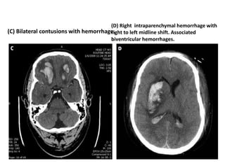
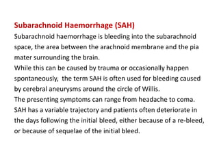
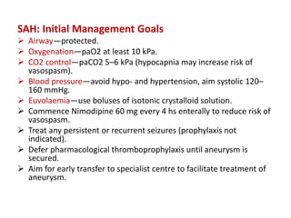
NEURO-ICU
Dr. Rania Gashan
Approach to the Comatose Patient: Initial
Resuscitation and Investigations
Disorders of consciousness may be caused by pathology of the brain
and meninges, or by systemic problems affecting the brain, such as
shock or hypotension, hypoxaemia, intoxication, or metabolic
disturbances including hypoglycaemia or hypercapnia.
Primary brain problems can be structural, such as trauma, stroke, or
intracranial bleeds, or can be caused by infection of the central
nervous system or by seizures or status epilepticus.
Your priority is to look for and treat the potential causes of coma and
the life-threatening consequences of it, and to prevent secondary
brain injury.
Use an ABCDEF approach and consider the effects of coma on airway
patency and airway protection. Measure blood glucose early in all
patients with disordered consciousness.
Intubating early is usually the safest course of action, particularly
of the underlying pathology and expected clinical course are
unclear or before transferring the patient for CT scanning or to a
different unit or hospital.
This will prevent episodes of hypoxaemia, hyper- or hypocapnia
and avoid aspiration of gastric contents. The downside is that
following induction of anaesthesia to facilitate intubation you
can no longer assess neurological status easily, and you should
carefully examine the patient and document your clinical
findings prior to intubation.
Meningism is resistance to neck flexion due to muscle spasm caused
by meningeal irritation. If present, consider starting antibiotics for
suspected bacterial meningitis urgently and before investigations are
complete, particularly in febrile patients. Meningism can also occur in
subarachnoid haemorrhage, where blood causes irritation and
inflammation of the meninges and motor nerve roots.
Assess the respiratory pattern in unconscious patients.
Focal or lateralising signs:
Look for asymmetry of movement and in unconscious patients, assess
muscle tone and check for deviation of the eyes.
Asymmetry or focal deficits suggest a structural cause, and imaging
should be performed as soon as safely possible. If the CT brain is
normal but lateralising signs are present, consider an urgent CT
angiogram to look for stroke.
Take care to avoid periods of hypotension at any stage during
intubation, stabilisation, and transfer, as this can cause secondary brain
injury.
Insert an arterial line early for continuous invasive blood
pressure monitoring, give fluid boluses if required, and consider
early central access and vasopressor support.
Perform arterial blood gas analysis early, in addition to routine
laboratory testing, to identify hypoglycaemia and other metabolic
disturbances that may cause or contribute to coma, and to ensure
adequate oxygenation and normocapnia. Appropriate specific initial
ventilatory and blood pressure targets are considered as part of
general neuroprotective measures below, but in general you should
aim for a mean arterial blood pressure of at least 80 mmHg, ensure
adequate fluid resuscitation, aim for normocapnia and a paO2 of 9–10
kPa.
Pathophysiology of Raised Intracranial Pressure (ICP)
Many types of acute brain injury lead to raised ICP, which in turn
causes reduced blood flow to the brain and can cause brain shift
(herniation) and compression of brain tissue.
It is associated with poor neurological outcomes and increased
mortality.
Intracranial pressure is determined by the combined volume of the
components within the fixed container of the skull:
brain tissue, blood, and cerebrospinal fluid (CSF). It is usually less than
approximately 10–20 mmHg but can spike due to coughing or straining
in health, without causing injury.
Pathologically elevated ICP can be caused by intracranial bleeding or
abscesses, brain swelling due to injured brain tissue, accumulation of
CSF in obstructive hydrocephalus or reduced outflow of blood, such as
in cerebral venous sinus thrombosis
Small increases in intracranial volume can be compensated for
by displacement of CSF or displacement of blood from the
venous sinuses. If the pathology progresses and the increase in
volume exceeds compensatory capacity, ICP increases
exponentially and can rapidly lead to inadequate brain perfusion
and ischaemia, as well as causing herniation .
Brain shift or herniation occurs when a large pressure gradient
squeezes brain tissue across fixed, bony structures.
.
Clinically, this can manifest as pupillary dilatation due to
compression of the third cranial nerve.
This causes compression of the medulla and results in brainstem
dysfunction including the typical description of ‘Cushing’s reflex’,
a combination of hypertension, bradycardia, and abnormal
respiratory pattern.
Without rapid treatment and reduction of the critically elevated
ICP, herniation can result in irreversible brain damage and death
Blood supply to the brain depends on the pressure gradient
between the mean arterial pressure and ICP: cerebral perfusion
pressure (MAP—ICP).
Normally, cerebral blood flow is relatively constant even when
cerebral perfusion pressure (CPP) changes.
Vasoconstirction with increased CPP in order to prevent
hyperaemia, and vasodilatation occurs if the CPP is low so as to
prevent ischaemia.
Autoregulation is usually maintained at a range of CPP between
approximately 60 and 160 mmHg. Below this level of CPP, brain
perfusion will fall precipitously .
Autoregulation is often impaired in acute brain injury, and when
it is completely lost, ICP will follow CPP in a linear fashion as
cerebral blood volume increases with higher arterial blood
pressures.
Several systemic factors also affect cerebral blood flow, most
notably arterial CO2 tension.
Hypercapnia such as due to periods of hypoventilation or airway
obstruction causes cerebral vasodilatation and increased
cerebral blood flow, blood volume, and ICP.
Conversely, hypocapnia causes cerebral vasoconstriction and
reduces blood flow and ICP and blood flow.
Disorders of Consciousness .
Treatment of Raised ICP
Clinical signs of brain herniation such as pupillary dilatation in a
comatose brain-injured patient require immediate treatment to
lower the ICP and avoid irreversible brain damage.
‘Medical’ or non-operative management may be part of a
strategy to control ICP until brain swelling resolves over time, or
it can buy time to arrange urgent surgical treatment of raised ICP.
This may be evacuation of a haematoma, decompressive
craniectomy, or surgical CSF drainage if there is hydrocephalus .
Your aim is to reduce ICP and optimise brain perfusion while
surgical options and a definitive management plan are
considered. Seek expert help immediately.
Management of Critically Elevated ICP/‘Coning’
 Intubate if not already done to protect airway and maintain
oxygenation and normocapnia.
 Elevate head of bed to 30° to improve venous drainage and
reduce ICP.
 Deepen sedation and add/optimise neuromuscular blockade
as well as analgesia, to reduce cerebral metabolic demand and
avoid coughing and straining.
 Elevate MAP to at least 80 mmHg to ensure adequate cerebral
perfusion pressure—use isotonic fluid boluses and
vasopressors, e.g. Noradrenaline.
 A brief period of hyperventilation will reduce cerebral blood
flow through vasoconstriction and thus reduce ICP. This can be
helpful if brainstem herniation is imminent but can cause
prolonged reduction in cerebral blood flow.
 Osmotherapy reduces brain swelling by drawing water from
the brain tissue into the vascular space along an osmotic
gradient (depending on local protocols, give, e.g. 125 mL of
5% Sodium Chloride solution via central venous catheter, or
250 mL of 20% Mannitol intravenously).
 Consider repeat CT imaging and neurosurgical opinion:
Always consider whether surgery or CSF drainage could help
Secondary Brain Injury and Neuroprotective
Measures
The injured brain frequently suffers secondary damage that
happens over time to neurons unharmed by the initial insult.
This is partly driven by an inflammatory response to the initial
injury but is also caused by complications that reduce adequate
brain oxygenation after the initial insult.
Secondary brain injury is therefore at least partly preventable,
and while the damage caused by the initial pathology—be this
trauma, infarction, or a bleed—cannot be reversed, reducing
secondary brain injury can improve functional outcomes.
The mechanisms of avoidable secondary brain injury are
hypotension, hypoxaemia, anaemia, and elevated ICP, which
reduce cerebral perfusion.
All of these result in failure to deliver enough oxygen to the brain.
Additionally, seizures and pyrexia can increase the metabolic and
oxygen demand of brain tissue.
Neuroprotective measures aim to prevent these mechanisms of secondary
injury and apply to brain injuries of any aetiology although targets can be
individualised if needed.
z Oxygenation
Guidelines vary but it is sensible to aim for paO2 > 10 kPa.
z Anaemia
If brain oxygenation is impaired or ICP significantly elevated, aim for
haemoglobin >90 g/L.
z CO2 Control
Avoid hypercapnia due to the associated rise in ICP. Generally,
aim for low normocapnia to reduce ICP, but avoid hypocapnia
in SAH to minimise risk of vasospasm.
z Blood Pressure Control
Avoid hypotension and generally aim for MAP >80 mmHg.
Ensure adequate fluid resuscitation, use vasopressors but diagnose potential
causes of shock: consider bleeding particularly in context of trauma, consider
sepsis and think of myocardial dysfunction in SAH.
Avoid hypertension (aim systolic blood pressure <180 mmHg or
<140 mmHg if significant risk of re-bleeding such as after SAH).
z Sedation
If you think the ICP is elevated, aim for deep sedation to reduce
metabolic demand of the brain. Don’t forget analgesia and
consider neuromuscular blockade if ICP is critically elevated.
z Seizure Control
Treat any observed seizures with anticonvulsants in addition to
sedation. Seizure prophylaxis is not routinely recommended.
z Temperature Control
Treat hyperthermia with antipyretics and active cooling
measures. Diagnose and treat infections, but remember fever is
often due to the brain injury itself.
z Glycaemic Control
Avoid hyper- and hypoglycaemia: use your local protocol.
z General Supportive Measures
Pay attention to evidence-based general Critical Care.
Prevent ventilator-associated pneumonia and pressure sores,
use enteral nutrition, prevent stress ulceration and venous
thrombo-embolism.
Specific Conditions in Neuro-Critical Care
Traumatic Brain Injury (TBI)
Head injuries are a common cause of death and disability, are
often caused by road traffic accidents, and frequently present to
non-specialist centres.
TBI is commonly classified as mild, moderate, and severe based
on the GCS (13–15, 8–13, and <8, respectively).
Outcomes for patients with coma due to head trauma are better
in specialist centres even if no surgical intervention is required,
but you may be involved in managing these patients prior to
transfer. Remember that other parts of the body can be affected
by the trauma too, making the management of these patients
complex and dependent on multiple specialties working together
as a team.
Initial stabilisation will follow the ABCDE approach.
Look for trauma to other organs causing
life-threatening bleeding and maintain stability of
the cervical spine.
Reduced conscious level is a sign of elevated ICP,
particularly if it persists after initial resuscitation.
Seek help and intubate early if the patient
is agitated or does not obey commands.
Arrange a CT scan—this is essential in order to identify
any intracranial bleeding requiring emergency surgery .
Surgery is usually indicated if there is a sizeable extra- or subdural
haematoma causing significant mass effect, but consultation with
your local specialist centre is advised for any patient with significant
or prolonged reduction in conscious level regardless of CT appearance .
Avoid preventable secondary brain injury by paying attention to
neuroprotective measures.
TBI: Initial Management Goals
 Airway—protected (remember C spine precautions); intubate
if in doubt.
 Oxygenation—PaO2 at least 10 kPa.
 CO2 control—paCO2 4.5–5 kPa (hypercapnia will increase
ICP).
 Blood pressure—avoid hypotension, aim MAP >80 mmHg;
insert arterial line.
 Use isotonic crystalloid solution for fluid resuscitation (look
for bleeding from occult trauma to other organs).
 If there are signs of brainstem herniation, elevate head,
deepen sedation, give osmotherapy, and seek expert help
immediately.
 Arrange urgent CT scan and discuss with specialist centre.
Management in specialist centres following transfer will involve
insertion of an ICP bolt in order to provide targeted medical
management of the ICP using the measures outlined above:
sedation, analgesia, neuromuscular blockade, head elevation,
and maintaining CPP above 60 mmHg.
Osmotherapy is used when ICP rises despite this.
A safe target is to keep ICP <20 mmHg.
If these measures fail, then rescue therapies are considered.
These including barbiturate coma or secondary decompressive
craniectomy, where part of the skull is removed, allowing the
swollen brain to expand.
Both are not without controversy and decompressive
craniectomy in particular is associated with a high proportion of
patients surviving with significant disability.
While decompression is effective at preventing death from brain
herniation, it does not undo the extensive damage that has led
to the catastrophic and refractory brain swelling in the first place
In the recovery phase, when the ICP is settling and no longer
dependent on low paCO2 and CPP, interventions can be
gradually de-escalated, including stopping neuromuscular
blockade and reducing sedation.
Recovery can take a long time however and prognostication is
often difficult in the early stages.
(A) Epidural hematoma. (B) Subdural hematoma
(C) Bilateral contusions with hemorrhage.
(D) Right intraparenchymal hemorrhage with
right to left midline shift. Associated
biventricular hemorrhages.
Subarachnoid Haemorrhage (SAH)
Subarachnoid haemorrhage is bleeding into the subarachnoid
space, the area between the arachnoid membrane and the pia
mater surrounding the brain.
While this can be caused by trauma or occasionally happen
spontaneously, the term SAH is often used for bleeding caused
by cerebral aneurysms around the circle of Willis.
The presenting symptoms can range from headache to coma.
SAH has a variable trajectory and patients often deteriorate in
the days following the initial bleed, either because of a re-bleed,
or because of sequelae of the initial bleed.
The blood in the subarachnoid space causes inflammation and
vasospasm, clinically apparent as ‘delayed cerebral ischaemia’
causing new focal neurological deficit or reduction in
conscious level typically between 4 and 10 days after the bleed.
If the bleed has extended into the ventricles, obstructive
hydrocephalus can occur, but delayed non-obstructive
hydrocephalus is also seen, caused by reduced absorption of CSF.
Seizures are relatively common, and the physiological stress and
catecholamine release after a large SAH can cause ECG changes,
and neurogenic pulmonary oedema.
The early management follows the same approach as for all
patients with altered conscious level.
Arrange an early CT scan if SAH is suspected and remember that
intubation prior to CT transfer is safer if the patient is agitated,
has a poor motor response or if oxygenation or normocapnia
cannot be maintained.
Because of the requirement for blood pressure control, insert an
arterial line.
Speak to your specialist centre early if SAH is confirmed, as these
patients are usually transferred early.
SAH: Initial Management Goals
 Airway—protected.
 Oxygenation—paO2 at least 10 kPa.
 CO2 control—paCO2 5–6 kPa (hypocapnia may increase risk of
vasospasm).
 Blood pressure—avoid hypo- and hypertension, aim systolic 120–
160 mmHg.
 Euvolaemia—use boluses of isotonic crystalloid solution.
 Commence Nimodipine 60 mg every 4 hs enterally to reduce risk of
vasospasm.
 Treat any persistent or recurrent seizures (prophylaxis not
indicated).
 Defer pharmacological thromboprophylaxis until aneurysm is
secured.
 Aim for early transfer to specialist centre to facilitate treatment of
aneurysm.
Stroke
Early diagnosis, and imaging are key in any patient presenting
with sudden onset neurological deficit consistent with a stroke.
CT imaging is most widely used, and scanned early, CT will help
exclude important differential diagnoses such as intracerebral
bleeds.
CT angiography can add useful information about the nature of
vascular occlusion and help determine whether it is amenable to
invasive treatments.
Approach a patient presenting with a stroke using the ABCDE
approach.
Some patients will present comatose, particularly in the
presence of large haemorrhagic strokes, and require intubation
to protect their airway, and allow safe CT scanning.
Many patients are hypertensive following a stroke, and you
should treat this if the systolic pressure is above 180 mmHg to
prevent re-bleeding or haemorrhagic transformation of
ischaemic strokes.
Sometimes, even tighter control is recommended for haemorrhagic
stroke—seek specialist advice.
Some patients are frail or have devastating strokes, and careful
consideration should be given to whether intensive, burdensome
treatment in Intensive Care is likely to be of benefit and consistent
with their wishes.
Seizures and Status Epilepticus (SE)
Seizures are transient episodes of altered behaviour or conscious level
caused by abnormal neuronal activity.
Generalised seizures usually result in loss of consciousness, and if a
seizure persists for more than 5 min or if consciousness is not regained
in between recurrent seizures, this is called status epilepticus.
It is a medical emergency not only because it can cause airway
obstruction, hypoxaemia, and aspiration, but because untreated
prolonged seizure activity results in sympathetic activation and
massively increased metabolic demand and ultimately severe acidosis,
hypoglycaemia, and multi-organ failure, as well as raised ICP and
neuronal damage.
.
Common causes include medication withdrawal or intercurrent
infection in those with underlying epilepsy, and TBI, stroke,
meningitis, and encephalitis as well as toxic and metabolic
disturbances in others.
When treating a patient in status epilepticus, use simple
manoeuvres to maintain airway patency and give supplementary
oxygen while attempts are made to stop the seizure activity.
Ensure you look for and treat hypoglycaemia.
Use a bolus of a benzodiazepine such as Lorazepam or Diazepam
as first-line treatment, and repeat this after a few minutes if
there is no effect.
If the status persists, commence an intravenous infusion of either
Phenytoin or Levetiracetam depending on local protocols.
Status epilepticus which persists despite two doses of an
appropriate benzodiazepine and a first-line intravenous
anticonvulsant is usually classed as ‘refractory’. The next step
now is intubation. It is highly unlikely that additional non-
sedating anticonvulsants alone will interrupt seizure activity at
this stage.
Seek help and use induction agents such as Propofol or
Thiopentone, which are powerful anticonvulsants.
Add an additional anticonvulsant agent such as Phenytoin,
Levetiracetam, or Sodium Valproate, and consider using an infusion of
Midazolam in addition to Propofol for ongoing sedation. If there is still
evidence of seizure activity after 24 h of ventilation and intravenous
sedation, or if the patient fails to wake during a sedation hold, EEG
monitoring will become essential and care is best provided in a
specialist centre
The term ‘super refractory status epilepticus’ is used to describe
status that persists despite anticonvulsant therapy, intubation,
and use of iv anaesthetic agents for at least 24 h.
The treatment of super-refractory SE in specialist centres
involves use of high-dose sedative agents such as Midazolam,
Ketamine, or Thiopentone to achieve ‘burst suppression’ on a
beside EEG monitor, i.e. marked suppression of cerebral activity.
This can interrupt seizure activity, but usually additional
anticonvulsant agents and occasionally more than one period of
‘burst suppression’ are required.
Following the initial stabilisation, start thinking about underlying cause
of the status epilepticus.
Arrange a CT scan of the brain in the first instance and consider a
lumbar puncture for CSF analysis if no structural abnormality has been
found.
Routine laboratory tests and microbiology samples should be sent to
look for infection. If no obvious cause is identified or if the patient fails
to respond to treatment, seek specialist advice. Further tests can be
carried out in specialist centres, including MRI scanning and more
detailed CSF analysis and immunology, to look for rare causes, such as
auto-immune encephalitis.
The prognosis of status epilepticus depends on the cause, and patients
with structural brain abnormalities often do worse. The longer the status
persists the poorer the outcome, reflecting a more aggressive underlying
process. In addition, intensive specialist management of SE is burdensome
and brings with it the complications of prolonged deep sedation and organ
support, and older patients with comorbidities are at much higher risk of
harm.

Neuro problems in ICU , management and protocols

  • 1.
  • 2.
    Approach to theComatose Patient: Initial Resuscitation and Investigations Disorders of consciousness may be caused by pathology of the brain and meninges, or by systemic problems affecting the brain, such as shock or hypotension, hypoxaemia, intoxication, or metabolic disturbances including hypoglycaemia or hypercapnia. Primary brain problems can be structural, such as trauma, stroke, or intracranial bleeds, or can be caused by infection of the central nervous system or by seizures or status epilepticus. Your priority is to look for and treat the potential causes of coma and the life-threatening consequences of it, and to prevent secondary brain injury. Use an ABCDEF approach and consider the effects of coma on airway patency and airway protection. Measure blood glucose early in all patients with disordered consciousness.
  • 3.
    Intubating early isusually the safest course of action, particularly of the underlying pathology and expected clinical course are unclear or before transferring the patient for CT scanning or to a different unit or hospital. This will prevent episodes of hypoxaemia, hyper- or hypocapnia and avoid aspiration of gastric contents. The downside is that following induction of anaesthesia to facilitate intubation you can no longer assess neurological status easily, and you should carefully examine the patient and document your clinical findings prior to intubation.
  • 5.
    Meningism is resistanceto neck flexion due to muscle spasm caused by meningeal irritation. If present, consider starting antibiotics for suspected bacterial meningitis urgently and before investigations are complete, particularly in febrile patients. Meningism can also occur in subarachnoid haemorrhage, where blood causes irritation and inflammation of the meninges and motor nerve roots. Assess the respiratory pattern in unconscious patients. Focal or lateralising signs: Look for asymmetry of movement and in unconscious patients, assess muscle tone and check for deviation of the eyes. Asymmetry or focal deficits suggest a structural cause, and imaging should be performed as soon as safely possible. If the CT brain is normal but lateralising signs are present, consider an urgent CT angiogram to look for stroke.
  • 6.
    Take care toavoid periods of hypotension at any stage during intubation, stabilisation, and transfer, as this can cause secondary brain injury. Insert an arterial line early for continuous invasive blood pressure monitoring, give fluid boluses if required, and consider early central access and vasopressor support. Perform arterial blood gas analysis early, in addition to routine laboratory testing, to identify hypoglycaemia and other metabolic disturbances that may cause or contribute to coma, and to ensure adequate oxygenation and normocapnia. Appropriate specific initial ventilatory and blood pressure targets are considered as part of general neuroprotective measures below, but in general you should aim for a mean arterial blood pressure of at least 80 mmHg, ensure adequate fluid resuscitation, aim for normocapnia and a paO2 of 9–10 kPa.
  • 7.
    Pathophysiology of RaisedIntracranial Pressure (ICP) Many types of acute brain injury lead to raised ICP, which in turn causes reduced blood flow to the brain and can cause brain shift (herniation) and compression of brain tissue. It is associated with poor neurological outcomes and increased mortality. Intracranial pressure is determined by the combined volume of the components within the fixed container of the skull: brain tissue, blood, and cerebrospinal fluid (CSF). It is usually less than approximately 10–20 mmHg but can spike due to coughing or straining in health, without causing injury. Pathologically elevated ICP can be caused by intracranial bleeding or abscesses, brain swelling due to injured brain tissue, accumulation of CSF in obstructive hydrocephalus or reduced outflow of blood, such as in cerebral venous sinus thrombosis
  • 8.
    Small increases inintracranial volume can be compensated for by displacement of CSF or displacement of blood from the venous sinuses. If the pathology progresses and the increase in volume exceeds compensatory capacity, ICP increases exponentially and can rapidly lead to inadequate brain perfusion and ischaemia, as well as causing herniation . Brain shift or herniation occurs when a large pressure gradient squeezes brain tissue across fixed, bony structures. .
  • 9.
    Clinically, this canmanifest as pupillary dilatation due to compression of the third cranial nerve. This causes compression of the medulla and results in brainstem dysfunction including the typical description of ‘Cushing’s reflex’, a combination of hypertension, bradycardia, and abnormal respiratory pattern. Without rapid treatment and reduction of the critically elevated ICP, herniation can result in irreversible brain damage and death
  • 10.
    Blood supply tothe brain depends on the pressure gradient between the mean arterial pressure and ICP: cerebral perfusion pressure (MAP—ICP). Normally, cerebral blood flow is relatively constant even when cerebral perfusion pressure (CPP) changes. Vasoconstirction with increased CPP in order to prevent hyperaemia, and vasodilatation occurs if the CPP is low so as to prevent ischaemia. Autoregulation is usually maintained at a range of CPP between approximately 60 and 160 mmHg. Below this level of CPP, brain perfusion will fall precipitously .
  • 11.
    Autoregulation is oftenimpaired in acute brain injury, and when it is completely lost, ICP will follow CPP in a linear fashion as cerebral blood volume increases with higher arterial blood pressures. Several systemic factors also affect cerebral blood flow, most notably arterial CO2 tension. Hypercapnia such as due to periods of hypoventilation or airway obstruction causes cerebral vasodilatation and increased cerebral blood flow, blood volume, and ICP. Conversely, hypocapnia causes cerebral vasoconstriction and reduces blood flow and ICP and blood flow. Disorders of Consciousness .
  • 12.
    Treatment of RaisedICP Clinical signs of brain herniation such as pupillary dilatation in a comatose brain-injured patient require immediate treatment to lower the ICP and avoid irreversible brain damage. ‘Medical’ or non-operative management may be part of a strategy to control ICP until brain swelling resolves over time, or it can buy time to arrange urgent surgical treatment of raised ICP. This may be evacuation of a haematoma, decompressive craniectomy, or surgical CSF drainage if there is hydrocephalus . Your aim is to reduce ICP and optimise brain perfusion while surgical options and a definitive management plan are considered. Seek expert help immediately.
  • 13.
    Management of CriticallyElevated ICP/‘Coning’  Intubate if not already done to protect airway and maintain oxygenation and normocapnia.  Elevate head of bed to 30° to improve venous drainage and reduce ICP.  Deepen sedation and add/optimise neuromuscular blockade as well as analgesia, to reduce cerebral metabolic demand and avoid coughing and straining.  Elevate MAP to at least 80 mmHg to ensure adequate cerebral perfusion pressure—use isotonic fluid boluses and vasopressors, e.g. Noradrenaline.  A brief period of hyperventilation will reduce cerebral blood flow through vasoconstriction and thus reduce ICP. This can be helpful if brainstem herniation is imminent but can cause prolonged reduction in cerebral blood flow.
  • 14.
     Osmotherapy reducesbrain swelling by drawing water from the brain tissue into the vascular space along an osmotic gradient (depending on local protocols, give, e.g. 125 mL of 5% Sodium Chloride solution via central venous catheter, or 250 mL of 20% Mannitol intravenously).  Consider repeat CT imaging and neurosurgical opinion: Always consider whether surgery or CSF drainage could help
  • 15.
    Secondary Brain Injuryand Neuroprotective Measures The injured brain frequently suffers secondary damage that happens over time to neurons unharmed by the initial insult. This is partly driven by an inflammatory response to the initial injury but is also caused by complications that reduce adequate brain oxygenation after the initial insult. Secondary brain injury is therefore at least partly preventable, and while the damage caused by the initial pathology—be this trauma, infarction, or a bleed—cannot be reversed, reducing secondary brain injury can improve functional outcomes.
  • 16.
    The mechanisms ofavoidable secondary brain injury are hypotension, hypoxaemia, anaemia, and elevated ICP, which reduce cerebral perfusion. All of these result in failure to deliver enough oxygen to the brain. Additionally, seizures and pyrexia can increase the metabolic and oxygen demand of brain tissue. Neuroprotective measures aim to prevent these mechanisms of secondary injury and apply to brain injuries of any aetiology although targets can be individualised if needed. z Oxygenation Guidelines vary but it is sensible to aim for paO2 > 10 kPa. z Anaemia If brain oxygenation is impaired or ICP significantly elevated, aim for haemoglobin >90 g/L.
  • 17.
    z CO2 Control Avoidhypercapnia due to the associated rise in ICP. Generally, aim for low normocapnia to reduce ICP, but avoid hypocapnia in SAH to minimise risk of vasospasm. z Blood Pressure Control Avoid hypotension and generally aim for MAP >80 mmHg. Ensure adequate fluid resuscitation, use vasopressors but diagnose potential causes of shock: consider bleeding particularly in context of trauma, consider sepsis and think of myocardial dysfunction in SAH. Avoid hypertension (aim systolic blood pressure <180 mmHg or <140 mmHg if significant risk of re-bleeding such as after SAH). z Sedation If you think the ICP is elevated, aim for deep sedation to reduce metabolic demand of the brain. Don’t forget analgesia and consider neuromuscular blockade if ICP is critically elevated.
  • 18.
    z Seizure Control Treatany observed seizures with anticonvulsants in addition to sedation. Seizure prophylaxis is not routinely recommended. z Temperature Control Treat hyperthermia with antipyretics and active cooling measures. Diagnose and treat infections, but remember fever is often due to the brain injury itself. z Glycaemic Control Avoid hyper- and hypoglycaemia: use your local protocol. z General Supportive Measures Pay attention to evidence-based general Critical Care. Prevent ventilator-associated pneumonia and pressure sores, use enteral nutrition, prevent stress ulceration and venous thrombo-embolism.
  • 19.
    Specific Conditions inNeuro-Critical Care Traumatic Brain Injury (TBI) Head injuries are a common cause of death and disability, are often caused by road traffic accidents, and frequently present to non-specialist centres. TBI is commonly classified as mild, moderate, and severe based on the GCS (13–15, 8–13, and <8, respectively). Outcomes for patients with coma due to head trauma are better in specialist centres even if no surgical intervention is required, but you may be involved in managing these patients prior to transfer. Remember that other parts of the body can be affected by the trauma too, making the management of these patients complex and dependent on multiple specialties working together as a team.
  • 20.
    Initial stabilisation willfollow the ABCDE approach. Look for trauma to other organs causing life-threatening bleeding and maintain stability of the cervical spine. Reduced conscious level is a sign of elevated ICP, particularly if it persists after initial resuscitation. Seek help and intubate early if the patient is agitated or does not obey commands. Arrange a CT scan—this is essential in order to identify any intracranial bleeding requiring emergency surgery . Surgery is usually indicated if there is a sizeable extra- or subdural haematoma causing significant mass effect, but consultation with your local specialist centre is advised for any patient with significant or prolonged reduction in conscious level regardless of CT appearance . Avoid preventable secondary brain injury by paying attention to neuroprotective measures.
  • 21.
    TBI: Initial ManagementGoals  Airway—protected (remember C spine precautions); intubate if in doubt.  Oxygenation—PaO2 at least 10 kPa.  CO2 control—paCO2 4.5–5 kPa (hypercapnia will increase ICP).  Blood pressure—avoid hypotension, aim MAP >80 mmHg; insert arterial line.  Use isotonic crystalloid solution for fluid resuscitation (look for bleeding from occult trauma to other organs).  If there are signs of brainstem herniation, elevate head, deepen sedation, give osmotherapy, and seek expert help immediately.  Arrange urgent CT scan and discuss with specialist centre.
  • 22.
    Management in specialistcentres following transfer will involve insertion of an ICP bolt in order to provide targeted medical management of the ICP using the measures outlined above: sedation, analgesia, neuromuscular blockade, head elevation, and maintaining CPP above 60 mmHg. Osmotherapy is used when ICP rises despite this. A safe target is to keep ICP <20 mmHg.
  • 23.
    If these measuresfail, then rescue therapies are considered. These including barbiturate coma or secondary decompressive craniectomy, where part of the skull is removed, allowing the swollen brain to expand. Both are not without controversy and decompressive craniectomy in particular is associated with a high proportion of patients surviving with significant disability. While decompression is effective at preventing death from brain herniation, it does not undo the extensive damage that has led to the catastrophic and refractory brain swelling in the first place
  • 25.
    In the recoveryphase, when the ICP is settling and no longer dependent on low paCO2 and CPP, interventions can be gradually de-escalated, including stopping neuromuscular blockade and reducing sedation. Recovery can take a long time however and prognostication is often difficult in the early stages.
  • 26.
    (A) Epidural hematoma.(B) Subdural hematoma
  • 27.
    (C) Bilateral contusionswith hemorrhage. (D) Right intraparenchymal hemorrhage with right to left midline shift. Associated biventricular hemorrhages.
  • 28.
    Subarachnoid Haemorrhage (SAH) Subarachnoidhaemorrhage is bleeding into the subarachnoid space, the area between the arachnoid membrane and the pia mater surrounding the brain. While this can be caused by trauma or occasionally happen spontaneously, the term SAH is often used for bleeding caused by cerebral aneurysms around the circle of Willis. The presenting symptoms can range from headache to coma. SAH has a variable trajectory and patients often deteriorate in the days following the initial bleed, either because of a re-bleed, or because of sequelae of the initial bleed.
  • 29.
    The blood inthe subarachnoid space causes inflammation and vasospasm, clinically apparent as ‘delayed cerebral ischaemia’ causing new focal neurological deficit or reduction in conscious level typically between 4 and 10 days after the bleed. If the bleed has extended into the ventricles, obstructive hydrocephalus can occur, but delayed non-obstructive hydrocephalus is also seen, caused by reduced absorption of CSF. Seizures are relatively common, and the physiological stress and catecholamine release after a large SAH can cause ECG changes, and neurogenic pulmonary oedema.
  • 30.
    The early managementfollows the same approach as for all patients with altered conscious level. Arrange an early CT scan if SAH is suspected and remember that intubation prior to CT transfer is safer if the patient is agitated, has a poor motor response or if oxygenation or normocapnia cannot be maintained. Because of the requirement for blood pressure control, insert an arterial line. Speak to your specialist centre early if SAH is confirmed, as these patients are usually transferred early.
  • 31.
    SAH: Initial ManagementGoals  Airway—protected.  Oxygenation—paO2 at least 10 kPa.  CO2 control—paCO2 5–6 kPa (hypocapnia may increase risk of vasospasm).  Blood pressure—avoid hypo- and hypertension, aim systolic 120– 160 mmHg.  Euvolaemia—use boluses of isotonic crystalloid solution.  Commence Nimodipine 60 mg every 4 hs enterally to reduce risk of vasospasm.  Treat any persistent or recurrent seizures (prophylaxis not indicated).  Defer pharmacological thromboprophylaxis until aneurysm is secured.  Aim for early transfer to specialist centre to facilitate treatment of aneurysm.
  • 32.
    Stroke Early diagnosis, andimaging are key in any patient presenting with sudden onset neurological deficit consistent with a stroke. CT imaging is most widely used, and scanned early, CT will help exclude important differential diagnoses such as intracerebral bleeds. CT angiography can add useful information about the nature of vascular occlusion and help determine whether it is amenable to invasive treatments.
  • 33.
    Approach a patientpresenting with a stroke using the ABCDE approach. Some patients will present comatose, particularly in the presence of large haemorrhagic strokes, and require intubation to protect their airway, and allow safe CT scanning. Many patients are hypertensive following a stroke, and you should treat this if the systolic pressure is above 180 mmHg to prevent re-bleeding or haemorrhagic transformation of ischaemic strokes. Sometimes, even tighter control is recommended for haemorrhagic stroke—seek specialist advice. Some patients are frail or have devastating strokes, and careful consideration should be given to whether intensive, burdensome treatment in Intensive Care is likely to be of benefit and consistent with their wishes.
  • 35.
    Seizures and StatusEpilepticus (SE) Seizures are transient episodes of altered behaviour or conscious level caused by abnormal neuronal activity. Generalised seizures usually result in loss of consciousness, and if a seizure persists for more than 5 min or if consciousness is not regained in between recurrent seizures, this is called status epilepticus. It is a medical emergency not only because it can cause airway obstruction, hypoxaemia, and aspiration, but because untreated prolonged seizure activity results in sympathetic activation and massively increased metabolic demand and ultimately severe acidosis, hypoglycaemia, and multi-organ failure, as well as raised ICP and neuronal damage. .
  • 36.
    Common causes includemedication withdrawal or intercurrent infection in those with underlying epilepsy, and TBI, stroke, meningitis, and encephalitis as well as toxic and metabolic disturbances in others. When treating a patient in status epilepticus, use simple manoeuvres to maintain airway patency and give supplementary oxygen while attempts are made to stop the seizure activity. Ensure you look for and treat hypoglycaemia. Use a bolus of a benzodiazepine such as Lorazepam or Diazepam as first-line treatment, and repeat this after a few minutes if there is no effect. If the status persists, commence an intravenous infusion of either Phenytoin or Levetiracetam depending on local protocols.
  • 37.
    Status epilepticus whichpersists despite two doses of an appropriate benzodiazepine and a first-line intravenous anticonvulsant is usually classed as ‘refractory’. The next step now is intubation. It is highly unlikely that additional non- sedating anticonvulsants alone will interrupt seizure activity at this stage. Seek help and use induction agents such as Propofol or Thiopentone, which are powerful anticonvulsants. Add an additional anticonvulsant agent such as Phenytoin, Levetiracetam, or Sodium Valproate, and consider using an infusion of Midazolam in addition to Propofol for ongoing sedation. If there is still evidence of seizure activity after 24 h of ventilation and intravenous sedation, or if the patient fails to wake during a sedation hold, EEG monitoring will become essential and care is best provided in a specialist centre
  • 38.
    The term ‘superrefractory status epilepticus’ is used to describe status that persists despite anticonvulsant therapy, intubation, and use of iv anaesthetic agents for at least 24 h. The treatment of super-refractory SE in specialist centres involves use of high-dose sedative agents such as Midazolam, Ketamine, or Thiopentone to achieve ‘burst suppression’ on a beside EEG monitor, i.e. marked suppression of cerebral activity. This can interrupt seizure activity, but usually additional anticonvulsant agents and occasionally more than one period of ‘burst suppression’ are required.
  • 40.
    Following the initialstabilisation, start thinking about underlying cause of the status epilepticus. Arrange a CT scan of the brain in the first instance and consider a lumbar puncture for CSF analysis if no structural abnormality has been found. Routine laboratory tests and microbiology samples should be sent to look for infection. If no obvious cause is identified or if the patient fails to respond to treatment, seek specialist advice. Further tests can be carried out in specialist centres, including MRI scanning and more detailed CSF analysis and immunology, to look for rare causes, such as auto-immune encephalitis. The prognosis of status epilepticus depends on the cause, and patients with structural brain abnormalities often do worse. The longer the status persists the poorer the outcome, reflecting a more aggressive underlying process. In addition, intensive specialist management of SE is burdensome and brings with it the complications of prolonged deep sedation and organ support, and older patients with comorbidities are at much higher risk of harm.