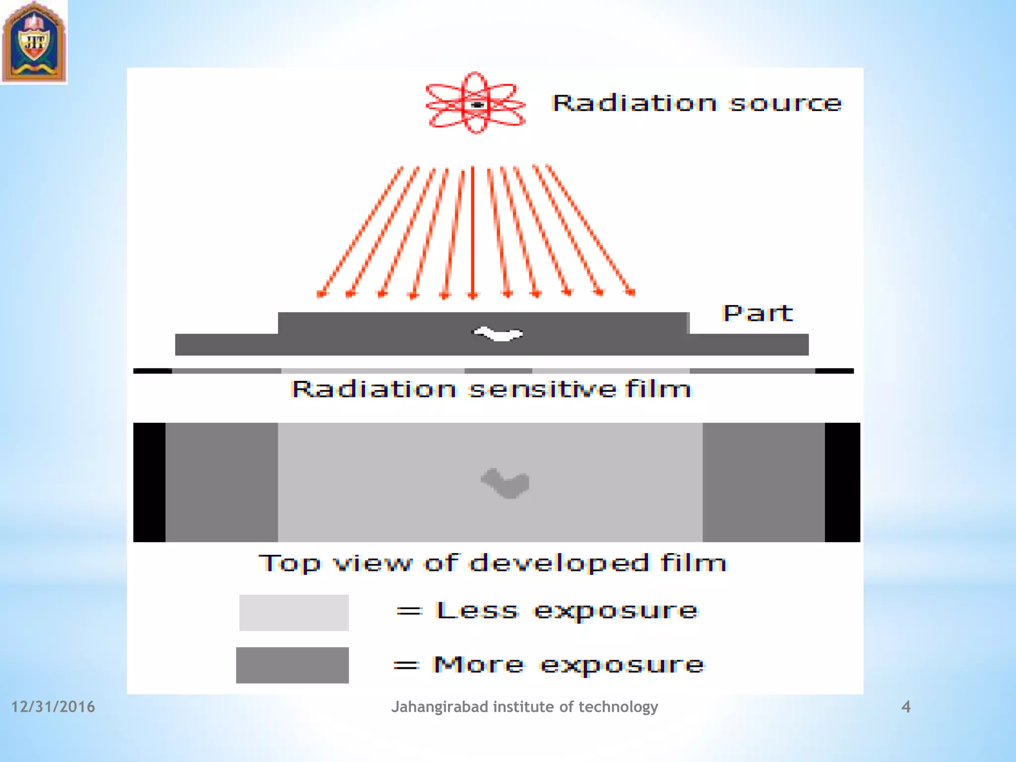
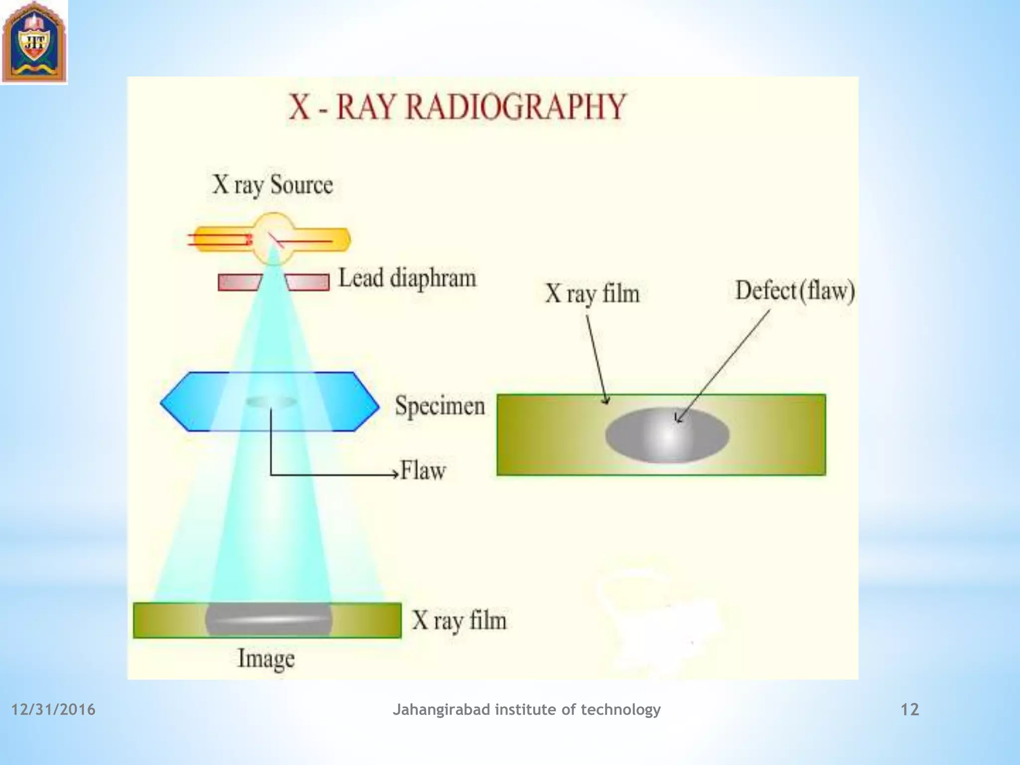
12/31/2016 Jahangirabad institute of technology 1
*
*RADIOGRAPHIC TESTING
*By:
Zoha Nasir
Assistant professor
JIT, Barabanki
*
Radiography is used in a very wide range of applications
including medicine, engineering, forensics, security, etc.
In NDT, radiography is one of the most important and widely
used methods.
Radiographic testing (RT) offers a number of advantages over
other NDT methods, however, one of its major disadvantages is
the health risk associated with the radiation.
In general, RT is method of inspecting materials for hidden
flaws by using the ability of short wavelength electromagnetic
radiation (high energy photons) to penetrate various materials.
12/31/2016 Jahangirabad institute of technology 2
*
In radiographic testing, the part to be inspected is placed between the
radiation source and a piece of radiation sensitive film.
The radiation source can either be an X ray machine or a radioactive source
(Ir-192, Co-60, or in rare cases Cs-137). The part will stop some of the
radiation where thicker and more dense areas will stop more of the radiation.
The radiation that passes through the part will expose the film and forms a
shadowgraph of the part.
The film darkness (density) will vary with the amount of radiation reaching
the film through the test object where darker areas indicate more exposure
(higher radiation intensity) and liter areas indicate less exposure (higher
radiation intensity).
This variation in the image darkness can be used to determine thickness or
composition of material and would also reveal the presence of any flaws or
discontinuities inside the material.
12/31/2016 Jahangirabad institute of technology 3
12/31/2016 Jahangirabad institute of technology 4
*
 Both surface and internal discontinuities can be detected.
 Significant variations in composition can be detected.
 It has a very few material limitations.
 Can be used for inspecting hidden areas (direct access to
surface is not required)
 Very minimal or no part preparation is required.
 Permanent test record is obtained.
 Good portability especially for gamma-ray sources.
12/31/2016 Jahangirabad institute of technology 5
*
 Hazardous to operators and other nearby personnel.
 High degree of skill and experience is required for exposure
and interpretation.
 The equipment is relatively expensive (especially for x-ray
sources).
 The process is generally slow.
 Highly directional (sensitive to flaw orientation).
 Depth of discontinuity is not indicated.
 It requires a two-sided access to the component.
12/31/2016 Jahangirabad institute of technology 6
*
Film Or Paper Radiography
Computed Radiography
Real Time Radiography
Neutron Radiography
Stereo Radiography
12/31/2016 Jahangirabad institute of technology 7
*
X-ray Radiation
Gamma Rays Radiation
Neutron Radiation
12/31/2016 Jahangirabad institute of technology 8
*
They are not detected by human senses (cannot be seen, heard,
felt, etc.).
They travel in straight lines at the speed of light.
Their paths cannot be changed by electrical or magnetic fields.
They can be diffracted, refracted to a small degree at interfaces
between two different materials, and in some cases be reflected.
They pass through matter until they have a chance to
encounter with an atomic particle.
Their degree of penetration depends on their energy and the
matter they are traveling through.
They have enough energy to ionize matter and can damage or
destroy living cells.
12/31/2016 Jahangirabad institute of technology 9
*
X-rays are just like any other kind of electromagnetic
radiation.
They can be produced in packets of energy called photons,
just like light.
There are two different atomic processes that can produce X-
ray photons. One is called Bremsstrahlung (a German term
meaning “braking radiation”) and the other is called K-shell
emission.
They can both occur in the heavy atoms of tungsten which is
often the material chosen for the target or anode of the X-ray
tube.
12/31/2016 Jahangirabad institute of technology 10
*
X-rays are passed through the specimen under inspection and
it is differentially absorbed by the specimen.
The transmitted x-rays are received by the photographic film
and the film is developed.
The dark and light shadows reveal the defects present in the
specimen and hence the defects are defected.
12/31/2016 Jahangirabad institute of technology 11
12/31/2016 Jahangirabad institute of technology 12
*
Gamma radiation is one of the three types of natural radioactivity. Gamma
rays are electromagnetic radiation just like X-rays. The other two types of
natural radioactivity are alpha and beta radiation, which are in the form of
particles. Gamma rays are the most energetic form of electromagnetic
radiation.
Gamma radiation is the product of radioactive atoms. Depending upon the
ratio of neutrons to protons within its nucleus, an isotope of a particular
element may be stable or unstable.
When the binding energy is not strong enough to hold the nucleus of an atom
together, the atom is said to be unstable. Atoms with unstable nuclei are
constantly changing as a result of the imbalance of energy within the nucleus.
Over time, the nuclei of unstable isotopes spontaneously disintegrate, or
transform, in a process known as “radioactive decay” and such material is
called “radioactive material”.
12/31/2016 Jahangirabad institute of technology 13
*
According to this law, the intensity of
reflected waves at a certain angle will be
maximum when the path difference
between two reflected waves from 2
different planes is an integral multiple of
wavelength.
12/31/2016 Jahangirabad institute of technology 14
*
Sand inclusions
Blow holes
Shrinkage
Cracks
Inclusions
12/31/2016 Jahangirabad institute of technology 15
*
No need of washing and developing films.
Low cost.
Image viewed immediately on screen.
Time consumption is less.
12/31/2016 Jahangirabad institute of technology 16
*
Poor resolution
Low image contrast
Electronic image intensifier required for increasing the contrast
12/31/2016 Jahangirabad institute of technology 17

Radiographic testing

  • 1.
    12/31/2016 Jahangirabad instituteof technology 1 * *RADIOGRAPHIC TESTING *By: Zoha Nasir Assistant professor JIT, Barabanki
  • 2.
    * Radiography is usedin a very wide range of applications including medicine, engineering, forensics, security, etc. In NDT, radiography is one of the most important and widely used methods. Radiographic testing (RT) offers a number of advantages over other NDT methods, however, one of its major disadvantages is the health risk associated with the radiation. In general, RT is method of inspecting materials for hidden flaws by using the ability of short wavelength electromagnetic radiation (high energy photons) to penetrate various materials. 12/31/2016 Jahangirabad institute of technology 2
  • 3.
    * In radiographic testing,the part to be inspected is placed between the radiation source and a piece of radiation sensitive film. The radiation source can either be an X ray machine or a radioactive source (Ir-192, Co-60, or in rare cases Cs-137). The part will stop some of the radiation where thicker and more dense areas will stop more of the radiation. The radiation that passes through the part will expose the film and forms a shadowgraph of the part. The film darkness (density) will vary with the amount of radiation reaching the film through the test object where darker areas indicate more exposure (higher radiation intensity) and liter areas indicate less exposure (higher radiation intensity). This variation in the image darkness can be used to determine thickness or composition of material and would also reveal the presence of any flaws or discontinuities inside the material. 12/31/2016 Jahangirabad institute of technology 3
  • 4.
  • 5.
    *  Both surfaceand internal discontinuities can be detected.  Significant variations in composition can be detected.  It has a very few material limitations.  Can be used for inspecting hidden areas (direct access to surface is not required)  Very minimal or no part preparation is required.  Permanent test record is obtained.  Good portability especially for gamma-ray sources. 12/31/2016 Jahangirabad institute of technology 5
  • 6.
    *  Hazardous tooperators and other nearby personnel.  High degree of skill and experience is required for exposure and interpretation.  The equipment is relatively expensive (especially for x-ray sources).  The process is generally slow.  Highly directional (sensitive to flaw orientation).  Depth of discontinuity is not indicated.  It requires a two-sided access to the component. 12/31/2016 Jahangirabad institute of technology 6
  • 7.
    * Film Or PaperRadiography Computed Radiography Real Time Radiography Neutron Radiography Stereo Radiography 12/31/2016 Jahangirabad institute of technology 7
  • 8.
    * X-ray Radiation Gamma RaysRadiation Neutron Radiation 12/31/2016 Jahangirabad institute of technology 8
  • 9.
    * They are notdetected by human senses (cannot be seen, heard, felt, etc.). They travel in straight lines at the speed of light. Their paths cannot be changed by electrical or magnetic fields. They can be diffracted, refracted to a small degree at interfaces between two different materials, and in some cases be reflected. They pass through matter until they have a chance to encounter with an atomic particle. Their degree of penetration depends on their energy and the matter they are traveling through. They have enough energy to ionize matter and can damage or destroy living cells. 12/31/2016 Jahangirabad institute of technology 9
  • 10.
    * X-rays are justlike any other kind of electromagnetic radiation. They can be produced in packets of energy called photons, just like light. There are two different atomic processes that can produce X- ray photons. One is called Bremsstrahlung (a German term meaning “braking radiation”) and the other is called K-shell emission. They can both occur in the heavy atoms of tungsten which is often the material chosen for the target or anode of the X-ray tube. 12/31/2016 Jahangirabad institute of technology 10
  • 11.
    * X-rays are passedthrough the specimen under inspection and it is differentially absorbed by the specimen. The transmitted x-rays are received by the photographic film and the film is developed. The dark and light shadows reveal the defects present in the specimen and hence the defects are defected. 12/31/2016 Jahangirabad institute of technology 11
  • 12.
  • 13.
    * Gamma radiation isone of the three types of natural radioactivity. Gamma rays are electromagnetic radiation just like X-rays. The other two types of natural radioactivity are alpha and beta radiation, which are in the form of particles. Gamma rays are the most energetic form of electromagnetic radiation. Gamma radiation is the product of radioactive atoms. Depending upon the ratio of neutrons to protons within its nucleus, an isotope of a particular element may be stable or unstable. When the binding energy is not strong enough to hold the nucleus of an atom together, the atom is said to be unstable. Atoms with unstable nuclei are constantly changing as a result of the imbalance of energy within the nucleus. Over time, the nuclei of unstable isotopes spontaneously disintegrate, or transform, in a process known as “radioactive decay” and such material is called “radioactive material”. 12/31/2016 Jahangirabad institute of technology 13
  • 14.
    * According to thislaw, the intensity of reflected waves at a certain angle will be maximum when the path difference between two reflected waves from 2 different planes is an integral multiple of wavelength. 12/31/2016 Jahangirabad institute of technology 14
  • 15.
  • 16.
    * No need ofwashing and developing films. Low cost. Image viewed immediately on screen. Time consumption is less. 12/31/2016 Jahangirabad institute of technology 16
  • 17.
    * Poor resolution Low imagecontrast Electronic image intensifier required for increasing the contrast 12/31/2016 Jahangirabad institute of technology 17