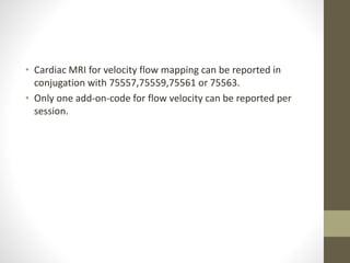
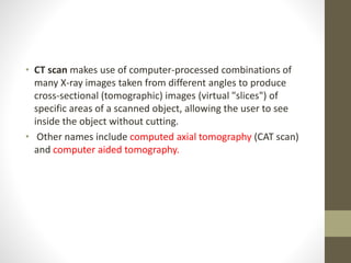
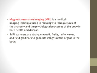
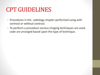
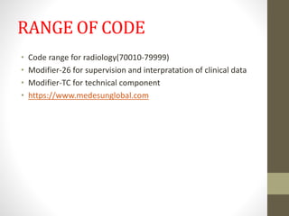
RADIOLOGY
INTRODUCTION
• Radiology is a specialty that uses medical imaging to diagnose
and treat diseases seen within the body.
• A variety of imaging techniques such as X-ray radiography,
ultrasound, computed tomography (CT), nuclear medicine
including positron emission tomography (PET), and magnetic
resonance imaging (MRI) are used to diagnose and/or treat
diseases.
• Radiography is an imaging technique using X-rays to view the
internal structure of an object. To create the image, a beam of
X-rays, a form of electromagnetic radiation, is produced by
an X-ray generator and is projected toward the object.
• Ultrasound is sound waves with frequencies higher than the
upper audible limit of human hearing.
• Ultrasound is used in many different fields. Ultrasonic devices
are used to detect objects and measure distances. Ultrasound
imaging or sonography is often used in medicine.
• CT scan makes use of computer-processed combinations of
many X-ray images taken from different angles to produce
cross-sectional (tomographic) images (virtual "slices") of
specific areas of a scanned object, allowing the user to see
inside the object without cutting.
• Other names include computed axial tomography (CAT scan)
and computer aided tomography.
• Magnetic resonance imaging (MRI) is a medical
imaging technique used in radiology to form pictures of
the anatomy and the physiological processes of the body in
both health and disease.
• MRI scanners use strong magnetic fields, radio waves,
and field gradients to generate images of the organs in the
body.
CPT GUIDELINES
• Procedures in the radiology chapter performed using with
contrast or without contrast.
• To perform a procedure various imaging techniques are used,
code are arranged based upon the type of technique.
• Neurofunctional tests are administered by a technologist or
other non-physician or non-psychologist,use 70554
• Neurofunctional tests are administered by a physician or
psychologist,use 70555
• Cardiac MRI for velocity flow mapping can be reported in
conjugation with 75557,75559,75561 or 75563.
• Only one add-on-code for flow velocity can be reported per
session.
• Two types of cardiac MRI tests will be performed
a. During rest
b. During pharmacological stress,therefore appropriate stress
testing code from the 93015-93018 series should be
reported in addition to 75559-75563.
VASCULAR PROCEDURES
• Selective vascular catheterizations should be coded to include
all lesser order selective catheterizations used in the
approach.
• Catheterization codes are not additionally reported for
diagnostic lower extremity angiography when performed
through the same access site as therapy performed in same
session
• If a different arterial puncture site is necessary report
separately.
• Diagnostic angiography codes should not be used with
interventional procedures
• Diagnostic angiography is performed along with interventional
procedures append modifier-59
• Diagnostic angiography is not reported separately when it
included in the interventional code.
• Same like above diagnostic venography is not reported
separately when it included in the interventional code.
TRANSCATHETER
PROCEDURES
• Unless specifically included in the coder descriptor,diagnostic
angiography is performed at the time of transcatheter
procedures,is separately reportable.
DIAGNOSTIC ULTRASOUND
• Use of ultrasound,without thorough evaluation of organs or
anatomic region,image documentation and final written
report is not separately reportable.
• A-mode implies a one-dimensional ultrasonic measurement
procedure.
• M-mode implies a one-dimentional ultrasonic measurement
procedure with movement of the trace for recording.
• B-scan implies a 2-dimentional ultrasonic scanning procedure
with a two dimentional display.
• Real-time scan implies a two dimensional ultrasonic scanning
procedure with display of both structure and motion with
time.
• For spectral and color doppler evaluation of the
extremities,use 93925,93926,93930,93931,93970 or 93971 as
appropriate.
• Do not report guidance codes for services in which
fluoroscopic guidance is included in the descriptor.
RADIATION ONCOLOGY
• Radiation oncology provide for teletherapy and brachytherapy
to include initial consultation,clinical treatment
planning,simulation,medical radiation
physics,dosimetry,treatment devices,special services and
clinical treatment management procedures.
• They include normal follow-up care during course of
treatment and for 3 months following its completion.
CLINICAL TREATMENT
PLANNING
• It is a complex process services including interpretation of
special testing tumor localization,treatment volume
determination time,choice of treatment
modality,determination of number of size and ports selection
of appropriate treatment devices,and other procedures.
Continued……….
• Simple planning requires a single treatment area of interest
encompassed in a single port or simple parallel opposed ports
with simple or no blocking.
• Intermediate planning requires 3 or more converging ports,2
separate treatment areas,multiple blocks or special time dose
constraints.
• Complex planning requires highly complex blocking,custom
shielding blocks,tangenital ports,special wedges or
compensators,3 or more separate treatment areas,rotational
or special beam considerations combination of therapeutic
modalities.
SIMULATION
• Simulation is the process of defining relevant normal and
abnormal target anatomy and acquiring the images and data
necessary to develop the optimal radiation treatment process
for the patient.
• Simple simulation of a single treatment area
• Intermediate simulation two separate treatment areas
• Complex three or more treatment areas.
RADIATION TREATMENT
DELIVERY
• Number of alternative methods to deliver external radiation
treatments which are described below
• X-ray
• Electron beam
• Neutron beams
• Proton beams
Radiation treatment and
management
• Simple:single treatment area,one or two ports,and two or
fewer simple blocks.
• Intermediate:2 separate treatment areas,3 or more ports on a
single treatment area,or 3 or more simple blocks
• Complex:3 or more separate treatment areas,custom
blocking,tangenital ports,wedges field on field that does not
meet IMRT guidelines
RADIATION TREATMENT
MANAGEMENT
• It is reported in units of five fractions or treatment
sessions,regardless of the actual time period in which the
services are furnished.
• Services need not be furnished on consecutive days.
• Multiple fractions representing 2 or more treatment sessions
furnished on the same day reported separately
• 77427 is also reported for 3 or 4 fractions beyond a multiple of
5 at the end of a course of treatment
• 1 or 2 fractions beyond a multiple of 5 at the end of a course
of treatment are not reported separately.
• 77469 is includes only intraoperative sessioj management and
does not include outside of that session.
• Professional services includes like
• Review of port images
• Review of dosimetry,dose delivery,and treatment parameters
• Review of patient treatment set-up
PROTON BEAM TREATMENT
DELIVERY
• Simple proton treatment delivery to a single treatment area
utilizing a single port.
• Intermediate proton treatment delivery to one or more
treatment areas using two or more ports or one or more
tangenital ports
• complex proton treatment delivery to one or more treatment
areas using two or more ports per treatment area with
matching or patching multiple isocenters,with custom blocks
and compensators
• Hyperthermia treatment given concurrently with radiation
therapy is listed separately.
• Hyperthermia is used only as an adjunct to radiation therapy
or chemotherapy
• Preliminary consultation is not included(E/M CODES)
• 0394T,0395T to report high dose rate electronic
brachytherapy.
• Modifiers of radiology chapter consist professional component
modifier and technical component as appropriate.
• Without modifier of radiology code considered as global code
• With modifier is considered as component code
RANGE OF CODE
• Code range for radiology(70010-79999)
• Modifier-26 for supervision and interpratation of clinical data
• Modifier-TC for technical component
• https://www.medesunglobal.com
THANK YOU

Radilogy

  • 1.
  • 2.
    INTRODUCTION • Radiology isa specialty that uses medical imaging to diagnose and treat diseases seen within the body. • A variety of imaging techniques such as X-ray radiography, ultrasound, computed tomography (CT), nuclear medicine including positron emission tomography (PET), and magnetic resonance imaging (MRI) are used to diagnose and/or treat diseases.
  • 3.
    • Radiography isan imaging technique using X-rays to view the internal structure of an object. To create the image, a beam of X-rays, a form of electromagnetic radiation, is produced by an X-ray generator and is projected toward the object.
  • 4.
    • Ultrasound issound waves with frequencies higher than the upper audible limit of human hearing. • Ultrasound is used in many different fields. Ultrasonic devices are used to detect objects and measure distances. Ultrasound imaging or sonography is often used in medicine.
  • 5.
    • CT scanmakes use of computer-processed combinations of many X-ray images taken from different angles to produce cross-sectional (tomographic) images (virtual "slices") of specific areas of a scanned object, allowing the user to see inside the object without cutting. • Other names include computed axial tomography (CAT scan) and computer aided tomography.
  • 6.
    • Magnetic resonanceimaging (MRI) is a medical imaging technique used in radiology to form pictures of the anatomy and the physiological processes of the body in both health and disease. • MRI scanners use strong magnetic fields, radio waves, and field gradients to generate images of the organs in the body.
  • 7.
    CPT GUIDELINES • Proceduresin the radiology chapter performed using with contrast or without contrast. • To perform a procedure various imaging techniques are used, code are arranged based upon the type of technique.
  • 8.
    • Neurofunctional testsare administered by a technologist or other non-physician or non-psychologist,use 70554 • Neurofunctional tests are administered by a physician or psychologist,use 70555
  • 9.
    • Cardiac MRIfor velocity flow mapping can be reported in conjugation with 75557,75559,75561 or 75563. • Only one add-on-code for flow velocity can be reported per session.
  • 10.
    • Two typesof cardiac MRI tests will be performed a. During rest b. During pharmacological stress,therefore appropriate stress testing code from the 93015-93018 series should be reported in addition to 75559-75563.
  • 11.
    VASCULAR PROCEDURES • Selectivevascular catheterizations should be coded to include all lesser order selective catheterizations used in the approach. • Catheterization codes are not additionally reported for diagnostic lower extremity angiography when performed through the same access site as therapy performed in same session
  • 12.
    • If adifferent arterial puncture site is necessary report separately. • Diagnostic angiography codes should not be used with interventional procedures • Diagnostic angiography is performed along with interventional procedures append modifier-59
  • 13.
    • Diagnostic angiographyis not reported separately when it included in the interventional code. • Same like above diagnostic venography is not reported separately when it included in the interventional code.
  • 14.
    TRANSCATHETER PROCEDURES • Unless specificallyincluded in the coder descriptor,diagnostic angiography is performed at the time of transcatheter procedures,is separately reportable.
  • 15.
    DIAGNOSTIC ULTRASOUND • Useof ultrasound,without thorough evaluation of organs or anatomic region,image documentation and final written report is not separately reportable. • A-mode implies a one-dimensional ultrasonic measurement procedure.
  • 16.
    • M-mode impliesa one-dimentional ultrasonic measurement procedure with movement of the trace for recording. • B-scan implies a 2-dimentional ultrasonic scanning procedure with a two dimentional display.
  • 17.
    • Real-time scanimplies a two dimensional ultrasonic scanning procedure with display of both structure and motion with time. • For spectral and color doppler evaluation of the extremities,use 93925,93926,93930,93931,93970 or 93971 as appropriate.
  • 18.
    • Do notreport guidance codes for services in which fluoroscopic guidance is included in the descriptor.
  • 19.
    RADIATION ONCOLOGY • Radiationoncology provide for teletherapy and brachytherapy to include initial consultation,clinical treatment planning,simulation,medical radiation physics,dosimetry,treatment devices,special services and clinical treatment management procedures. • They include normal follow-up care during course of treatment and for 3 months following its completion.
  • 20.
    CLINICAL TREATMENT PLANNING • Itis a complex process services including interpretation of special testing tumor localization,treatment volume determination time,choice of treatment modality,determination of number of size and ports selection of appropriate treatment devices,and other procedures.
  • 21.
    Continued………. • Simple planningrequires a single treatment area of interest encompassed in a single port or simple parallel opposed ports with simple or no blocking. • Intermediate planning requires 3 or more converging ports,2 separate treatment areas,multiple blocks or special time dose constraints.
  • 22.
    • Complex planningrequires highly complex blocking,custom shielding blocks,tangenital ports,special wedges or compensators,3 or more separate treatment areas,rotational or special beam considerations combination of therapeutic modalities.
  • 23.
    SIMULATION • Simulation isthe process of defining relevant normal and abnormal target anatomy and acquiring the images and data necessary to develop the optimal radiation treatment process for the patient.
  • 24.
    • Simple simulationof a single treatment area • Intermediate simulation two separate treatment areas • Complex three or more treatment areas.
  • 25.
    RADIATION TREATMENT DELIVERY • Numberof alternative methods to deliver external radiation treatments which are described below • X-ray • Electron beam • Neutron beams • Proton beams
  • 26.
    Radiation treatment and management •Simple:single treatment area,one or two ports,and two or fewer simple blocks. • Intermediate:2 separate treatment areas,3 or more ports on a single treatment area,or 3 or more simple blocks • Complex:3 or more separate treatment areas,custom blocking,tangenital ports,wedges field on field that does not meet IMRT guidelines
  • 27.
    RADIATION TREATMENT MANAGEMENT • Itis reported in units of five fractions or treatment sessions,regardless of the actual time period in which the services are furnished. • Services need not be furnished on consecutive days. • Multiple fractions representing 2 or more treatment sessions furnished on the same day reported separately
  • 28.
    • 77427 isalso reported for 3 or 4 fractions beyond a multiple of 5 at the end of a course of treatment • 1 or 2 fractions beyond a multiple of 5 at the end of a course of treatment are not reported separately.
  • 29.
    • 77469 isincludes only intraoperative sessioj management and does not include outside of that session. • Professional services includes like • Review of port images • Review of dosimetry,dose delivery,and treatment parameters • Review of patient treatment set-up
  • 30.
    PROTON BEAM TREATMENT DELIVERY •Simple proton treatment delivery to a single treatment area utilizing a single port. • Intermediate proton treatment delivery to one or more treatment areas using two or more ports or one or more tangenital ports • complex proton treatment delivery to one or more treatment areas using two or more ports per treatment area with matching or patching multiple isocenters,with custom blocks and compensators
  • 31.
    • Hyperthermia treatmentgiven concurrently with radiation therapy is listed separately. • Hyperthermia is used only as an adjunct to radiation therapy or chemotherapy • Preliminary consultation is not included(E/M CODES)
  • 32.
    • 0394T,0395T toreport high dose rate electronic brachytherapy. • Modifiers of radiology chapter consist professional component modifier and technical component as appropriate. • Without modifier of radiology code considered as global code • With modifier is considered as component code
  • 33.
    RANGE OF CODE •Code range for radiology(70010-79999) • Modifier-26 for supervision and interpratation of clinical data • Modifier-TC for technical component • https://www.medesunglobal.com
  • 34.