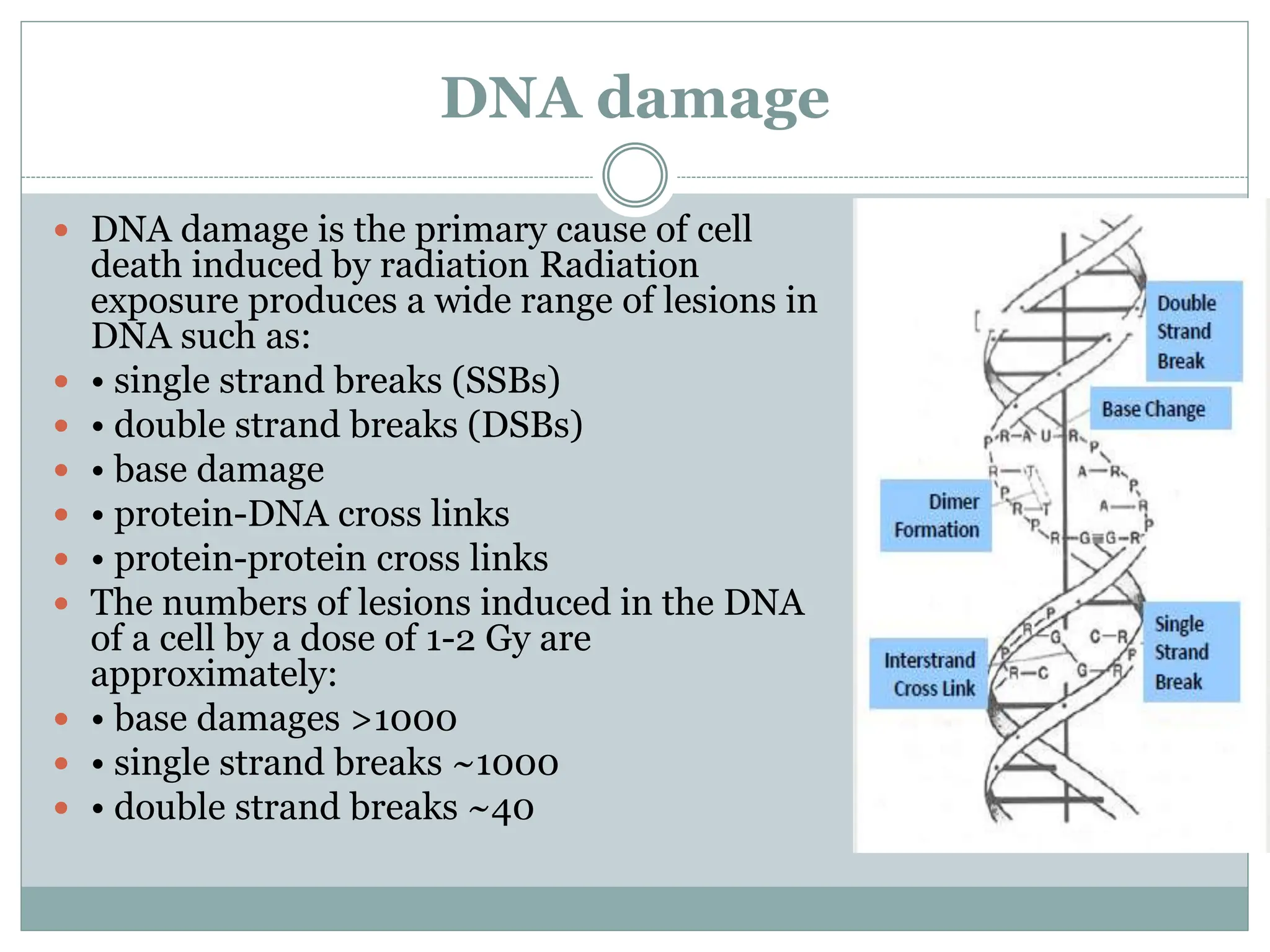
D R . S A R A H A B D U L S A L A M
Radiation effect on water, DNA
damage
1st STAGE / LEC8/ Biology theory
Radiology and Ulrasonography Techniques dep.
Radiation effect on water
 A complex series of chemical
changes occurs in water after
exposure to ionizing radiation; this
process is called water radiolysis.
 Water is the most predominant
molecule in living organisms (about
80 % of the mass of a living cell)
 • Therefore, a major proportion of
the radiation energy deposited will
be absorbed in cellular water
 • About two thirds of the biological
damage caused by low LET
radiations (sparsely ionizing
radiation) such as X rays or electrons
is due to indirect action
RADIOLYSIS OF WATER
 First step in radiolysis of water is the absorption of
radiant energy that can cause ionisation or excitation.
 IONISATION OF WATER MOLECULE
 H2O H2O+o + e-
 This reaction needs 13 eV of energy. Radical ions
formed are short lived, with life of about 1-10sec and
will soon decay to form uncharged free radical.
 H2O+o H+ + Oho
EXCITATION OF WATER MOLECULE
 H2O H2O*
 The excited water molecules are not stable and soon undergo
 radiolysis giving rise to Ho and OHo radicals.
 H2O* Ho + OH0
 The OHo free radical have a life time of 10-5 sec. They are very
powerful oxidising agent.
 The ejected energetic electrons loose their energy by collision and
are finally captured by water molecules, forming aqueous
electrons(hydrated).
 e- + H2O e- aq
 These aqueous electrons are strongly reducing agents and can
cause dissociation of water molecules forming free radicals.
 H2O + e- aq Ho + OH0
DNA damage
 DNA damage is the primary cause of cell
death induced by radiation Radiation
exposure produces a wide range of lesions in
DNA such as:
 • single strand breaks (SSBs)
 • double strand breaks (DSBs)
 • base damage
 • protein-DNA cross links
 • protein-protein cross links
 The numbers of lesions induced in the DNA
of a cell by a dose of 1-2 Gy are
approximately:
 • base damages >1000
 • single strand breaks ~1000
 • double strand breaks ~40
DNA DAMAGE
 SSB:A single Ionisation cause the break in one of the
strands of DNA molecules. The break can occur either at
the bond between sugar and phosphate or between sugar
and the base. Since most of the SSB’S are because of free
radicals, hence low LET radiations induce more SSB
because energy deposited per unit path length is less and
hence causing excitation . About 3- 4 times more SSB are
produced in a well oxygenated system, as compared to
hypoxic conditions
 DSB: If the breaks in the two strands are opposite to one
another or separated by only a few base pairs <5 DSB
may occur. DSB can formed either by single ionizing
event or by two independent events occurring in
complementary strands of DNA.
 Double strand breaks (DSBs) play a critical role in
cell killing, carcinogenesis and hereditary effects.
 There are experimental data showing that:
 • initially-produced DSBs correlate with
radiosensitivity and survival at lower dose
 • unrepaired or mis-repaired DSBs also correlate
with survival after higher doses
 • there is a causal link between the generation of
DSBs and the induction of chromosomal
translocations with carcinogenic potential
DNA DAMAGE
 When normal cell DNA is damaged by radiation provided
in the kinds of doses normally used in radiotherapy, the
cell cycle is stopped by the protein p53.
 The DNA is repaired; the cell then re-enters the cell cycle
and continues to proliferate.
 If the DNA cannot be repaired, the cell enters apoptosis –
the programmed cell death pathway.
 At high radiation doses , the molecules utilized by the
DNA repair mechanisms are damaged, so repair is not
possible, the cell loses its ability to divide, and it
subsequently dies.
non-homologous end
joining (NHEJ)
homologous recombination
(HR)
 NHEJ repair operates on blunt
ended DNA fragments
 This process involves the repair
proteins recognizing lesion
termini, cleaning up the broken
ends of the DNA molecule, and
the final ligation of the broken
ends.
 Repair by NHEJ operates
throughout the cell cycle but
dominates in G1/Sphases
 The process is error-prone
because it does not rely on
sequence homology
 HR repair utilizes sequence
homology with an
undamaged copy of the
broken region and hence
can only operate in late S-
or G2- phases.
 Undamaged DNA from
both strands is used as
templates to repair the
damage.
 The repair process of HR is
error-free.
DNA REPAIR: For double strand breaks
there are two primary repair pathways
blunt end
sticky end
 DNA repair mechanisms are important for the
recovery of cells from radiation and other damaging
agents.
 Unrepaired or mis-repaired damage to DNA will
lead in the exposed cell to:
Mutations and/or chromosome damage
might lead to: when severe often leads to:
cancer or cell death
hereditary effects
CHROMOSOMAL ABERRATIONS
 When the repair of DNA-double strand breaks is
incomplete there may be serious implications for a cell,
namely it may lead to chromosomal damage
(aberrations).
 Breaks induced by DNA may remain unrepaired or re-
join incorrectly to form abnormal configurations known
as chromosomal aberrations
 Aberrant (damaged) chromosomes:
 • rings generated when broken ends rejoin with other
broken ends
 • dicentrics (chromosomes having two centromeres)
 • translocations
 • other chromosome aberrations
 They are of two types :
 Structural Aberrations : These occur due to a loss or
genetic material, or a rearrangement in the location of
the genetic material. They include: deletions,
duplications, inversions, ring formations, and
translocations
 Numerical Aberrations : The number of chromosomes
show a change from normal diploid state .It results from
non disjunction of chromosomes during mitosis causing
unequal distribution in daughter nuclei .eg trisomy
monosomy
 DOWN’S SYNDROME : Trisomy of chromosome 21
 KLINEFELTER SYNDROME
 Radiation doses in the order of several sieverts may lead to
cell loss
 • Cells are generally regarded as having been “killed” by
radiation if they have lost reproductive integrity, even if they
physically survived
 Loss of reproductive integrity can occur by:
 • apoptosis
 • necrosis
 • mitotic catastrophe
 • induced senescence
 although all but the last of these
mechanisms ultimately results in
physical loss of the cell, this may
take a significant time to occur
CONCEPTS OF CELL DEATH
 Apoptosis or programmed cell death:
 • can occur naturally or result from insult to the cell
environment
 • occurs after low doses of irradiation in particular
cell types:
 lymphocytes
 serous salivary gland cells
 certain cells in the stem cell zone in testis and
intestinal crypts
 Necrosis
 • is a form of cell death associated with loss of cellular
membrane activity
 • cellular necrosis generally occurs after high radiation doses.
 Reproductive cell death
 • is a result of mitotic catastrophe (cells attempt to divide
without proper repair of DNA damage) which can occur in the
first few cell divisions after irradiation
 • it occurs with increasing frequency after increasing doses
 Senescent cells
 • are metabolically active but have lost the ability to divide
radiation effect on water, DNA damage lec 9.pptx

radiation effect on water, DNA damage lec 9.pptx

  • 1.
    D R .S A R A H A B D U L S A L A M Radiation effect on water, DNA damage 1st STAGE / LEC8/ Biology theory Radiology and Ulrasonography Techniques dep.
  • 2.
    Radiation effect onwater  A complex series of chemical changes occurs in water after exposure to ionizing radiation; this process is called water radiolysis.  Water is the most predominant molecule in living organisms (about 80 % of the mass of a living cell)  • Therefore, a major proportion of the radiation energy deposited will be absorbed in cellular water  • About two thirds of the biological damage caused by low LET radiations (sparsely ionizing radiation) such as X rays or electrons is due to indirect action
  • 3.
    RADIOLYSIS OF WATER First step in radiolysis of water is the absorption of radiant energy that can cause ionisation or excitation.  IONISATION OF WATER MOLECULE  H2O H2O+o + e-  This reaction needs 13 eV of energy. Radical ions formed are short lived, with life of about 1-10sec and will soon decay to form uncharged free radical.  H2O+o H+ + Oho
  • 4.
    EXCITATION OF WATERMOLECULE  H2O H2O*  The excited water molecules are not stable and soon undergo  radiolysis giving rise to Ho and OHo radicals.  H2O* Ho + OH0  The OHo free radical have a life time of 10-5 sec. They are very powerful oxidising agent.  The ejected energetic electrons loose their energy by collision and are finally captured by water molecules, forming aqueous electrons(hydrated).  e- + H2O e- aq  These aqueous electrons are strongly reducing agents and can cause dissociation of water molecules forming free radicals.  H2O + e- aq Ho + OH0
  • 7.
    DNA damage  DNAdamage is the primary cause of cell death induced by radiation Radiation exposure produces a wide range of lesions in DNA such as:  • single strand breaks (SSBs)  • double strand breaks (DSBs)  • base damage  • protein-DNA cross links  • protein-protein cross links  The numbers of lesions induced in the DNA of a cell by a dose of 1-2 Gy are approximately:  • base damages >1000  • single strand breaks ~1000  • double strand breaks ~40
  • 8.
    DNA DAMAGE  SSB:Asingle Ionisation cause the break in one of the strands of DNA molecules. The break can occur either at the bond between sugar and phosphate or between sugar and the base. Since most of the SSB’S are because of free radicals, hence low LET radiations induce more SSB because energy deposited per unit path length is less and hence causing excitation . About 3- 4 times more SSB are produced in a well oxygenated system, as compared to hypoxic conditions  DSB: If the breaks in the two strands are opposite to one another or separated by only a few base pairs <5 DSB may occur. DSB can formed either by single ionizing event or by two independent events occurring in complementary strands of DNA.
  • 10.
     Double strandbreaks (DSBs) play a critical role in cell killing, carcinogenesis and hereditary effects.  There are experimental data showing that:  • initially-produced DSBs correlate with radiosensitivity and survival at lower dose  • unrepaired or mis-repaired DSBs also correlate with survival after higher doses  • there is a causal link between the generation of DSBs and the induction of chromosomal translocations with carcinogenic potential
  • 11.
    DNA DAMAGE  Whennormal cell DNA is damaged by radiation provided in the kinds of doses normally used in radiotherapy, the cell cycle is stopped by the protein p53.  The DNA is repaired; the cell then re-enters the cell cycle and continues to proliferate.  If the DNA cannot be repaired, the cell enters apoptosis – the programmed cell death pathway.  At high radiation doses , the molecules utilized by the DNA repair mechanisms are damaged, so repair is not possible, the cell loses its ability to divide, and it subsequently dies.
  • 12.
    non-homologous end joining (NHEJ) homologousrecombination (HR)  NHEJ repair operates on blunt ended DNA fragments  This process involves the repair proteins recognizing lesion termini, cleaning up the broken ends of the DNA molecule, and the final ligation of the broken ends.  Repair by NHEJ operates throughout the cell cycle but dominates in G1/Sphases  The process is error-prone because it does not rely on sequence homology  HR repair utilizes sequence homology with an undamaged copy of the broken region and hence can only operate in late S- or G2- phases.  Undamaged DNA from both strands is used as templates to repair the damage.  The repair process of HR is error-free. DNA REPAIR: For double strand breaks there are two primary repair pathways
  • 13.
  • 14.
     DNA repairmechanisms are important for the recovery of cells from radiation and other damaging agents.  Unrepaired or mis-repaired damage to DNA will lead in the exposed cell to: Mutations and/or chromosome damage might lead to: when severe often leads to: cancer or cell death hereditary effects
  • 15.
    CHROMOSOMAL ABERRATIONS  Whenthe repair of DNA-double strand breaks is incomplete there may be serious implications for a cell, namely it may lead to chromosomal damage (aberrations).  Breaks induced by DNA may remain unrepaired or re- join incorrectly to form abnormal configurations known as chromosomal aberrations  Aberrant (damaged) chromosomes:  • rings generated when broken ends rejoin with other broken ends  • dicentrics (chromosomes having two centromeres)  • translocations  • other chromosome aberrations
  • 16.
     They areof two types :  Structural Aberrations : These occur due to a loss or genetic material, or a rearrangement in the location of the genetic material. They include: deletions, duplications, inversions, ring formations, and translocations  Numerical Aberrations : The number of chromosomes show a change from normal diploid state .It results from non disjunction of chromosomes during mitosis causing unequal distribution in daughter nuclei .eg trisomy monosomy  DOWN’S SYNDROME : Trisomy of chromosome 21  KLINEFELTER SYNDROME
  • 17.
     Radiation dosesin the order of several sieverts may lead to cell loss  • Cells are generally regarded as having been “killed” by radiation if they have lost reproductive integrity, even if they physically survived  Loss of reproductive integrity can occur by:  • apoptosis  • necrosis  • mitotic catastrophe  • induced senescence  although all but the last of these mechanisms ultimately results in physical loss of the cell, this may take a significant time to occur
  • 18.
    CONCEPTS OF CELLDEATH  Apoptosis or programmed cell death:  • can occur naturally or result from insult to the cell environment  • occurs after low doses of irradiation in particular cell types:  lymphocytes  serous salivary gland cells  certain cells in the stem cell zone in testis and intestinal crypts
  • 20.
     Necrosis  •is a form of cell death associated with loss of cellular membrane activity  • cellular necrosis generally occurs after high radiation doses.  Reproductive cell death  • is a result of mitotic catastrophe (cells attempt to divide without proper repair of DNA damage) which can occur in the first few cell divisions after irradiation  • it occurs with increasing frequency after increasing doses  Senescent cells  • are metabolically active but have lost the ability to divide

Editor's Notes

  • #3  linear energy transfer = LET
  • #8 The gray (symbol: Gy) is a derived unit of ionizing radiation dose in the International System of Units (SI).