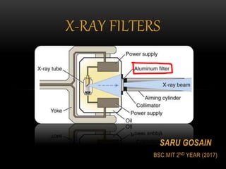
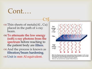
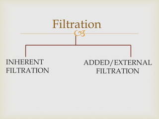
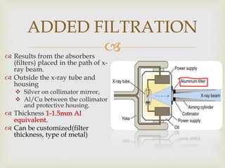
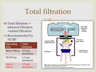
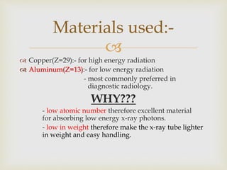
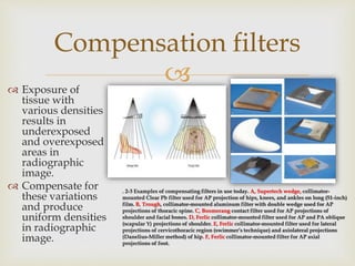
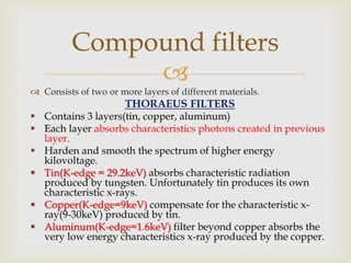
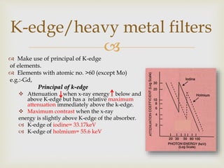
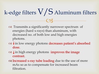
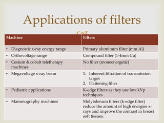
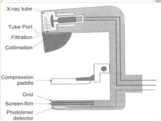
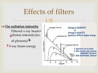
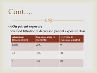
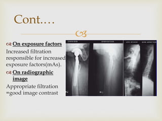
SARU GOSAIN
BSC.MIT 2ND YEAR (2017)
X-RAY FILTERS

INTRODUCTION
 Diagnostic x-ray beams are polychromatic( with
spectrum of many different energies).
 High energy photons transmitted to form the
radiographic image.
 Low energy photons get absorbed/ scattered.
 Contributes to the increase in patient radiation dose.
 Or if scattered then degrade the image quality.

Cont.…
 Thin sheets of metals(Al , Cu)
placed in the path of x-ray
beam.
 To attenuate the low energy
(soft) x-ray photons from the
spectrum before reaching to
the patient body are filters.
 And the process is known as
filtration/beam hardening.
 Unit is mm Al equivalent.

ADDED/EXTERNAL
FILTRATION
Filtration
INHERENT
FILTRATION

 Absorption of x-rays as they pass
through
 Glass/metal envelope
 The insulating oil
 The window
 Thickness 0.5-1mm Al equivalent
INHERENT FILTRATION
 Results from the absorbers
(filters) placed in the path of x-
ray beam.
 Outside the x-ray tube and
housing
 Silver on collimator mirror,
 Al/Cu between the collimator
and protective housing.
 Thickness 1-1.5mm Al
equivalent.
 Can be customized(filter
thickness, type of metal)
ADDED FILTRATION
 Total filtration =
inherent filtration
+added filtration
 Recommended by
NCRP
Total filtration
Operating
kVp
Total
filtration
Below 50kvp 0.5 mm
aluminum
50-70 kVp 1.5 mm
aluminum
Above
70kVp
2.5 mm
aluminum

 Copper(Z=29):- for high energy radiation
 Aluminum(Z=13):- for low energy radiation
- most commonly preferred in
diagnostic radiology.
WHY???
- low atomic number therefore excellent material
for absorbing low energy x-ray photons.
- low in weight therefore make the x-ray tube lighter
in weight and easy handling.
Materials used:-

 Compensation filters
 Boomerang filters
 Trough filters
 Wedge filters
 Ferlic filters
 Flattening filters
 Compound filters
 Thoraeus filters
 K-edge filers(heavy metal filters)
Other types
 Exposure of
tissue with
various densities
results in
underexposed
and overexposed
areas in
radiographic
image.
 Compensate for
these variations
and produce
uniform densities
in radiographic
image.
Compensation filters
. 2-3 Examples of compensating filters in use today. A, Supertech wedge, collimator-
mounted Clear Pb filter used for AP projection of hips, knees, and ankles on long (51-inch)
film. B, Trough, collimator-mounted aluminum filter with double wedge used for AP
projections of thoracic spine. C, Boomerang contact filter used for AP projections of
shoulder and facial bones. D, Ferlic collimator-mounted filter used for AP and PA oblique
(scapular Y) projections of shoulder. E, Ferlic collimator-mounted filter used for lateral
projections of cervicothoracic region (swimmer’s technique) and axiolateral projections
(Danelius-Miller method) of hip. F, Ferlic collimator-mounted filter for AP axial
projections of foot.

 Consists of two or more layers of different materials.
THORAEUS FILTERS
 Contains 3 layers(tin, copper, aluminum)
 Each layer absorbs characteristics photons created in previous
layer.
 Harden and smooth the spectrum of higher energy
kilovoltage.
 Tin(K-edge = 29.2keV) absorbs characteristic radiation
produced by tungsten. Unfortunately tin produces its own
characteristic x-rays.
 Copper(K-edge=9keV) compensate for the characteristic x-
ray(9-30keV) produced by tin.
 Aluminum(K-edge=1.6keV) filter beyond copper absorbs the
very low energy characteristics x-ray produced by the copper.
Compound filters

 Make use of principal of K-edge
of elements.
 Elements with atomic no. >60 (except Mo)
e.g.:-Gd,
Principal of k-edge
 Attenuation when x-ray energy below and
above K-edge but has a relative maximum
attenuation immediately above the k-edge.
 Maximum contrast when the x-ray
energy is slightly above K-edge of the absorber.
 K-edge of iodine= 33.17keV
 K-edge of holmium= 55.6 keV
K-edge/heavy metal filters

 Transmits a significantly narrower spectrum of
energies (hard x-rays) than aluminum, with
decreased no. of both low and high energies
photons.
 in low energy photons decreases patient’s absorbed
dose.
 in high energy photons improves the image
contrast.
 Increased x-ray tube loading due to the use of more
mAs so as to compensate for increased beam
filtration.
k-edge filters V/S Aluminum filters

Applications of filters
Machine Filters
• Diagnostic x-ray energy range Primary aluminum filter (mm Al)
• Orthovoltage range Compound filter (1-4mm Cu)
• Cesium & cobalt teletherapy
machines
No filter (monoenergetic)
• Megavoltage x-ray beam 1. Inherent filtration of transmission
target
2. Flattening filter
• Pediatric applications K-edge filters as they use low kVp
techniques
• Mammography machines Molybdenum filters (k-edge filter)
reduce the amount of high energies x-
rays and improve the contrast in breast
soft tissues.


 On radiation intensity
Filtered x-ray beam=
photon intensity(no.
of photons)+
X-ray beam energy
Effects of filters

 On patient exposure
Increased filtration = decreased patient exposure dose
Cont.…
Aluminum
filtration(mm)
Exposure dose to
skin(mR)
Decrease in
exposure dose(%)
None 2380 0
0.5 1850 22
3 465 80

 On exposure factors
Increased filtration
responsible for increased
exposure factors(mAs).
 On radiographic
image
Appropriate filtration
=good image contrast
Cont.…

 Thickness of absorber that
attenuate the intensity of the x-
ray beam to half its original
value.
 Indirect measure of quality of
photon energy/beam
hardness.
 The greater the HVL of the x-
ray beams, the better is the
quality of the x-ray photons.
 HVL=
0.693
𝜇
 Unit is mm of Al.
Half Value Layer (HVL)

 Reduction in the intensity of x-ray photons,
 Lengthen the time required to make an exposure,
 May absorb primary x-ray beam during excessive
filtration,
 Increase tube loading (due to more mAs).
Disadvantages

 Christensen’s PHYSICS OF DIAGNOSTIC
RADIOLOGY(4TH EDITION)
 Stewart Carlyle Bushong’s RADIOLOGIC SCIENCE FOR
TECHNOLOGISTS (10TH EDITION)
 En.m.Wikipedia.org
 www.cram.com
 Radiopaedia.org
 Qcinradiography.weebly.com
 Radiologykey.com
 www.slideshare.com
References
Any
questions???


X ray filters

  • 1.
    SARU GOSAIN BSC.MIT 2NDYEAR (2017) X-RAY FILTERS
  • 2.
     INTRODUCTION  Diagnostic x-raybeams are polychromatic( with spectrum of many different energies).  High energy photons transmitted to form the radiographic image.  Low energy photons get absorbed/ scattered.  Contributes to the increase in patient radiation dose.  Or if scattered then degrade the image quality.
  • 3.
     Cont.…  Thin sheetsof metals(Al , Cu) placed in the path of x-ray beam.  To attenuate the low energy (soft) x-ray photons from the spectrum before reaching to the patient body are filters.  And the process is known as filtration/beam hardening.  Unit is mm Al equivalent.
  • 4.
  • 5.
      Absorption ofx-rays as they pass through  Glass/metal envelope  The insulating oil  The window  Thickness 0.5-1mm Al equivalent INHERENT FILTRATION
  • 6.
     Results fromthe absorbers (filters) placed in the path of x- ray beam.  Outside the x-ray tube and housing  Silver on collimator mirror,  Al/Cu between the collimator and protective housing.  Thickness 1-1.5mm Al equivalent.  Can be customized(filter thickness, type of metal) ADDED FILTRATION
  • 7.
     Total filtration= inherent filtration +added filtration  Recommended by NCRP Total filtration Operating kVp Total filtration Below 50kvp 0.5 mm aluminum 50-70 kVp 1.5 mm aluminum Above 70kVp 2.5 mm aluminum
  • 8.
      Copper(Z=29):- forhigh energy radiation  Aluminum(Z=13):- for low energy radiation - most commonly preferred in diagnostic radiology. WHY??? - low atomic number therefore excellent material for absorbing low energy x-ray photons. - low in weight therefore make the x-ray tube lighter in weight and easy handling. Materials used:-
  • 9.
      Compensation filters Boomerang filters  Trough filters  Wedge filters  Ferlic filters  Flattening filters  Compound filters  Thoraeus filters  K-edge filers(heavy metal filters) Other types
  • 10.
     Exposure of tissuewith various densities results in underexposed and overexposed areas in radiographic image.  Compensate for these variations and produce uniform densities in radiographic image. Compensation filters . 2-3 Examples of compensating filters in use today. A, Supertech wedge, collimator- mounted Clear Pb filter used for AP projection of hips, knees, and ankles on long (51-inch) film. B, Trough, collimator-mounted aluminum filter with double wedge used for AP projections of thoracic spine. C, Boomerang contact filter used for AP projections of shoulder and facial bones. D, Ferlic collimator-mounted filter used for AP and PA oblique (scapular Y) projections of shoulder. E, Ferlic collimator-mounted filter used for lateral projections of cervicothoracic region (swimmer’s technique) and axiolateral projections (Danelius-Miller method) of hip. F, Ferlic collimator-mounted filter for AP axial projections of foot.
  • 11.
      Consists oftwo or more layers of different materials. THORAEUS FILTERS  Contains 3 layers(tin, copper, aluminum)  Each layer absorbs characteristics photons created in previous layer.  Harden and smooth the spectrum of higher energy kilovoltage.  Tin(K-edge = 29.2keV) absorbs characteristic radiation produced by tungsten. Unfortunately tin produces its own characteristic x-rays.  Copper(K-edge=9keV) compensate for the characteristic x- ray(9-30keV) produced by tin.  Aluminum(K-edge=1.6keV) filter beyond copper absorbs the very low energy characteristics x-ray produced by the copper. Compound filters
  • 12.
      Make useof principal of K-edge of elements.  Elements with atomic no. >60 (except Mo) e.g.:-Gd, Principal of k-edge  Attenuation when x-ray energy below and above K-edge but has a relative maximum attenuation immediately above the k-edge.  Maximum contrast when the x-ray energy is slightly above K-edge of the absorber.  K-edge of iodine= 33.17keV  K-edge of holmium= 55.6 keV K-edge/heavy metal filters
  • 13.
      Transmits asignificantly narrower spectrum of energies (hard x-rays) than aluminum, with decreased no. of both low and high energies photons.  in low energy photons decreases patient’s absorbed dose.  in high energy photons improves the image contrast.  Increased x-ray tube loading due to the use of more mAs so as to compensate for increased beam filtration. k-edge filters V/S Aluminum filters
  • 14.
     Applications of filters MachineFilters • Diagnostic x-ray energy range Primary aluminum filter (mm Al) • Orthovoltage range Compound filter (1-4mm Cu) • Cesium & cobalt teletherapy machines No filter (monoenergetic) • Megavoltage x-ray beam 1. Inherent filtration of transmission target 2. Flattening filter • Pediatric applications K-edge filters as they use low kVp techniques • Mammography machines Molybdenum filters (k-edge filter) reduce the amount of high energies x- rays and improve the contrast in breast soft tissues.
  • 15.
  • 16.
      On radiationintensity Filtered x-ray beam= photon intensity(no. of photons)+ X-ray beam energy Effects of filters
  • 17.
      On patientexposure Increased filtration = decreased patient exposure dose Cont.… Aluminum filtration(mm) Exposure dose to skin(mR) Decrease in exposure dose(%) None 2380 0 0.5 1850 22 3 465 80
  • 18.
      On exposurefactors Increased filtration responsible for increased exposure factors(mAs).  On radiographic image Appropriate filtration =good image contrast Cont.…
  • 19.
      Thickness ofabsorber that attenuate the intensity of the x- ray beam to half its original value.  Indirect measure of quality of photon energy/beam hardness.  The greater the HVL of the x- ray beams, the better is the quality of the x-ray photons.  HVL= 0.693 𝜇  Unit is mm of Al. Half Value Layer (HVL)
  • 20.
      Reduction inthe intensity of x-ray photons,  Lengthen the time required to make an exposure,  May absorb primary x-ray beam during excessive filtration,  Increase tube loading (due to more mAs). Disadvantages
  • 21.
      Christensen’s PHYSICSOF DIAGNOSTIC RADIOLOGY(4TH EDITION)  Stewart Carlyle Bushong’s RADIOLOGIC SCIENCE FOR TECHNOLOGISTS (10TH EDITION)  En.m.Wikipedia.org  www.cram.com  Radiopaedia.org  Qcinradiography.weebly.com  Radiologykey.com  www.slideshare.com References
  • 22.
  • 24.