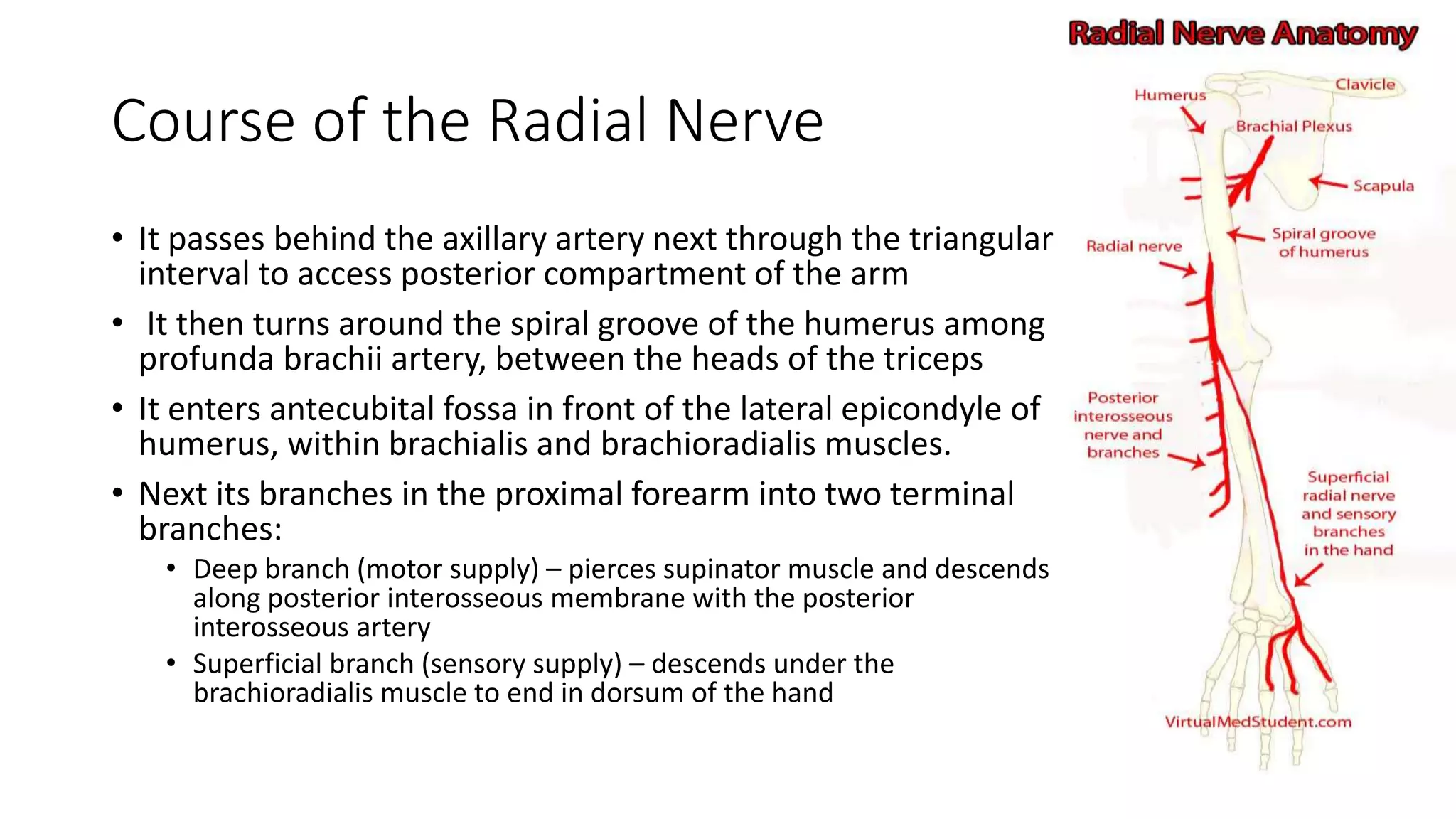
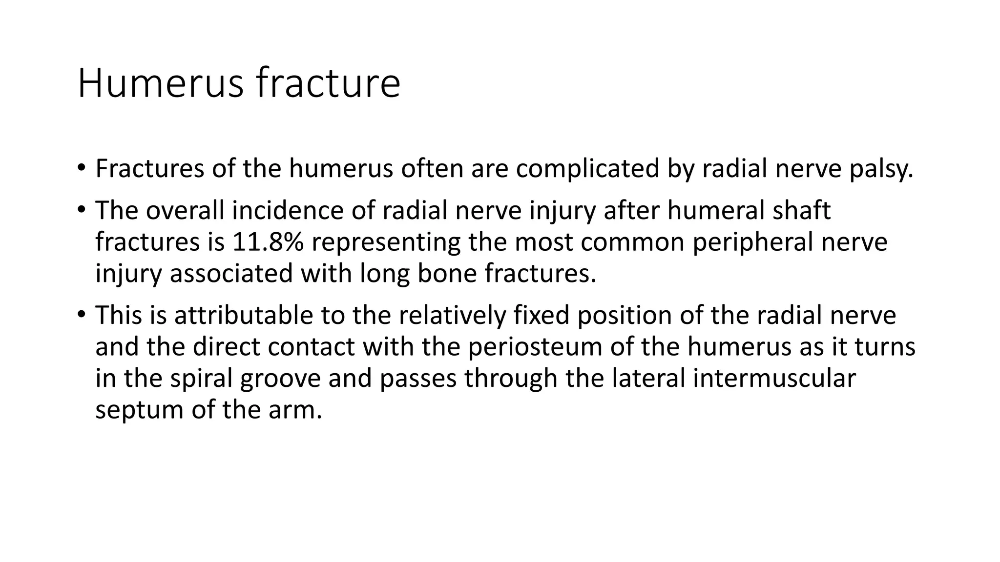
FRACTURE HUMERUS WITH
RADIAL NERVE INJURY:
PRINCIPLE OF MANAGEMENT
DR.KHADIJAH
Anatomy of shaft humerus
• Shaft of humerus: It is cylindrical in upper half and
triangular in lower half. It has three borders and three
surfaces.
• Borders
• Anterior border : continuation of lateral lip of bicipital
groove.
• Medial border: Medial lip of bicipital groove continues
downwards as medial border which then continues as medial
supracondylar ridge.
• Lateral border: Is well defined only in lower half, continues
downwards as lateral supracondylar ridge.
• Surfaces
• Anteromedial surface: between anterior and medial border.
• Anterolateral surface: between anterior and lateral border.
Just above the middle shows ‘V’ shaped tuberosity called
‘Deltoid tuberosity‘.
• Posterior surface: between medial and lateral border. In its
middle 1/3rd has a shallow groove called ‘Radial groove’.
Radial nerve
• The radial nerve is a major peripheral nerve of the upper limb.
• It originates from the brachial plexus, carrying fibers from the ventral
roots of spinal nerves C5, C6, C7, C8 & T1.
Course of the Radial Nerve
• It passes behind the axillary artery next through the triangular
interval to access posterior compartment of the arm
• It then turns around the spiral groove of the humerus among
profunda brachii artery, between the heads of the triceps
• It enters antecubital fossa in front of the lateral epicondyle of
humerus, within brachialis and brachioradialis muscles.
• Next its branches in the proximal forearm into two terminal
branches:
• Deep branch (motor supply) – pierces supinator muscle and descends
along posterior interosseous membrane with the posterior
interosseous artery
• Superficial branch (sensory supply) – descends under the
brachioradialis muscle to end in dorsum of the hand
Sensory Supply of the Radial Nerve
• Posterior aspect of arm and forearm
• Lateral ⅔ of the dorsum of hand
• Proximal dorsal aspect of lateral 3½ fingers (thumb,
index, middle and half of the ring finger)
Motor Supply of the
Radial Nerve
• Posterior compartment of the arm muscles
• Triceps muscle –adducts and extends shoulder,
extends the elbow
• Posterior compartment of the forearm muscles
• Brachioradialis muscle- elbow flexes
• Supinator muscle – supination of the forearm
• Extensor carpi radialis longus and brevis muscles–
abduct and extend the wrist
• Extensor carpi ulnaris muscle – extends and adducts
the wrist
• Extensor digitorum, extensor indicis, extensor pollicis
brevis and longus and extensor digiti minimi muscles
– extend the thumb and fingers at MCPJs and IPJs
• Abductor pollicis longus muscle – abducts the thumb
Radial nerve palsy
Posterior
interosseous nerve
syndrome
Humerus fracture
• Fractures of the humerus often are complicated by radial nerve palsy.
• The overall incidence of radial nerve injury after humeral shaft
fractures is 11.8% representing the most common peripheral nerve
injury associated with long bone fractures.
• This is attributable to the relatively fixed position of the radial nerve
and the direct contact with the periosteum of the humerus as it turns
in the spiral groove and passes through the lateral intermuscular
septum of the arm.
• At this level the bone ends can easily entrap, contuse, or even
lacerate the radial nerve. As such, radial nerve injury varies from
neurapraxias to complete neurotmesis.
• Spiral or oblique fractures of the junction between the middle and
distal third of the humeral shaft are at greater risk for radial nerve
injury.
• Iatrogenic injury to the radial nerve may also occur during
manipulations of closed reduction or at the time of surgical
intervention, during internal fixation with a compression plate or
intramedullary nail.
Management of midshaft humerus fracture
• Non operative
• CMR and splint
• Neurovascular status pre and
post documentation
• Acceptable criteria post cmr:
• Angulation up to 20 degree
• Varus up to 30 degree
• Shorthening up to 2cm
• Surgical intervention:
• Plating
• Intermedulary nail
Indication for acute surgical intervention
• Multiple other traumatic injuries
• Floating elbow (concurrent fractures in the forearm and humerus)
• Segmental fractures
• Displaced intra-articular shoulder or elbow involvement
• Bilateral humeral fractures
• New nerve or vascular injury after reduction
• Pathologic fractures
• Unacceptable alignment following closed reduction
Indication for delayed surgical intervention
• Poor pain tolerance
• Inability to perform activities of daily living
• Delayed nerve palsy
• Non-union
• Delayed union
• Loss of reduction
Management of humerus fracture with radial
nerve injury
• Classified the of nerve injury
• Complete vs partial
• Primary or secondary
• Observe clinically
• During recovery, early range of motion exercises and dynamic splinting of the
wrist, thumb, and fingers are important to help prevent joint contractures.
• Limb edema is common and must be minimized with a compressive garment,
elevate on, and range of motion exercises.
• Early exploration
• Late exploration
Early exploration
• recommended in several cases such as :
• open fracture that requires debridement and stabilization,
• irreducible fracture or unacceptable reduction, associated vascular or severe soft
tissue injury,
• radial nerve deficit after manipulation (secondary nerve palsy),
• intractable neurogenic pain suggesting nerve entrapment or compression,
• high-velocity gunshot wounds, sharp or penetrating injury,
• high suspicion of nerve laceration with spiral oblique fractures, and severe soft tissue
injury.
• During surgery, complex injury patterns should be managed in the
following order:
• fracture stabilization,
• definitive or temporary vascular repair,
• nerve repair
• Surgical intervention for radial nerve injury can be classified into:
• Neurography:
• adequate resection of surrounding scar tissue and neuromas should be performed until
healthy nerve fibers are seen under the microscope in order to facilitate recovery
• Nerve grafting:
• preferred in cases of tension at the neurography site or if there is a large nerve gap that
has to be bridged .
• Results after nerve grafting are in line with primary nerve repair.
• Factors that influence the outcome after grafting include the length of the defect that
should be bridged and the denervation time
• Nerve transfer
• tendon transfers:
• Indications for a tendon transfer include no sign of nerve recovery at 1 year, incomplete
recovery, and/or failed nerve reconstruction.
• The patient’s level of function, anatomy, and level of radial nerve injury are three factors
routinely used to decide on the appropriate tendons to use for transfer
• Wrist extension is achieved with transfer of pronator teres into extensor carpi radialis
brevis, whereas for finger extension, the flexor carpi radialis is usually used
• The factors that determine the approach to nerve repair are :
• the location
• the duration of nerve injury.
Take
home
message
Thank you
References:
1. Shao YC, Harwood P, Grotz MRW, Limb D, Giannoudis PV (2005)
Radial nerve palsy associated with fractures of the shaft of the
humerus. A systematic review. J Bone Joint Surg Br Vol 87:1647–
1652
2. M Rocchi, L Tarallo. R Mugnai, R. Adani Humerus shaft fracture
complicated by radial nerve palsy:Is surgical exploration necessary?
Musculoskelet Surg. 2016 Dec; 100(Suppl 1):53-60
3. Niver GE, Ilyas AM Management of radial nerve palsy following
fractures of the humerus. Orthop Clin North Am. 2013
Jul;44(3):419-24

Fracture humerus with radial nerve injury

  • 1.
    FRACTURE HUMERUS WITH RADIALNERVE INJURY: PRINCIPLE OF MANAGEMENT DR.KHADIJAH
  • 2.
    Anatomy of shafthumerus • Shaft of humerus: It is cylindrical in upper half and triangular in lower half. It has three borders and three surfaces. • Borders • Anterior border : continuation of lateral lip of bicipital groove. • Medial border: Medial lip of bicipital groove continues downwards as medial border which then continues as medial supracondylar ridge. • Lateral border: Is well defined only in lower half, continues downwards as lateral supracondylar ridge. • Surfaces • Anteromedial surface: between anterior and medial border. • Anterolateral surface: between anterior and lateral border. Just above the middle shows ‘V’ shaped tuberosity called ‘Deltoid tuberosity‘. • Posterior surface: between medial and lateral border. In its middle 1/3rd has a shallow groove called ‘Radial groove’.
  • 4.
    Radial nerve • Theradial nerve is a major peripheral nerve of the upper limb. • It originates from the brachial plexus, carrying fibers from the ventral roots of spinal nerves C5, C6, C7, C8 & T1.
  • 5.
    Course of theRadial Nerve • It passes behind the axillary artery next through the triangular interval to access posterior compartment of the arm • It then turns around the spiral groove of the humerus among profunda brachii artery, between the heads of the triceps • It enters antecubital fossa in front of the lateral epicondyle of humerus, within brachialis and brachioradialis muscles. • Next its branches in the proximal forearm into two terminal branches: • Deep branch (motor supply) – pierces supinator muscle and descends along posterior interosseous membrane with the posterior interosseous artery • Superficial branch (sensory supply) – descends under the brachioradialis muscle to end in dorsum of the hand
  • 6.
    Sensory Supply ofthe Radial Nerve • Posterior aspect of arm and forearm • Lateral ⅔ of the dorsum of hand • Proximal dorsal aspect of lateral 3½ fingers (thumb, index, middle and half of the ring finger)
  • 7.
    Motor Supply ofthe Radial Nerve • Posterior compartment of the arm muscles • Triceps muscle –adducts and extends shoulder, extends the elbow • Posterior compartment of the forearm muscles • Brachioradialis muscle- elbow flexes • Supinator muscle – supination of the forearm • Extensor carpi radialis longus and brevis muscles– abduct and extend the wrist • Extensor carpi ulnaris muscle – extends and adducts the wrist • Extensor digitorum, extensor indicis, extensor pollicis brevis and longus and extensor digiti minimi muscles – extend the thumb and fingers at MCPJs and IPJs • Abductor pollicis longus muscle – abducts the thumb
  • 8.
  • 9.
  • 10.
    Humerus fracture • Fracturesof the humerus often are complicated by radial nerve palsy. • The overall incidence of radial nerve injury after humeral shaft fractures is 11.8% representing the most common peripheral nerve injury associated with long bone fractures. • This is attributable to the relatively fixed position of the radial nerve and the direct contact with the periosteum of the humerus as it turns in the spiral groove and passes through the lateral intermuscular septum of the arm.
  • 11.
    • At thislevel the bone ends can easily entrap, contuse, or even lacerate the radial nerve. As such, radial nerve injury varies from neurapraxias to complete neurotmesis. • Spiral or oblique fractures of the junction between the middle and distal third of the humeral shaft are at greater risk for radial nerve injury. • Iatrogenic injury to the radial nerve may also occur during manipulations of closed reduction or at the time of surgical intervention, during internal fixation with a compression plate or intramedullary nail.
  • 12.
    Management of midshafthumerus fracture • Non operative • CMR and splint • Neurovascular status pre and post documentation • Acceptable criteria post cmr: • Angulation up to 20 degree • Varus up to 30 degree • Shorthening up to 2cm • Surgical intervention: • Plating • Intermedulary nail
  • 13.
    Indication for acutesurgical intervention • Multiple other traumatic injuries • Floating elbow (concurrent fractures in the forearm and humerus) • Segmental fractures • Displaced intra-articular shoulder or elbow involvement • Bilateral humeral fractures • New nerve or vascular injury after reduction • Pathologic fractures • Unacceptable alignment following closed reduction
  • 14.
    Indication for delayedsurgical intervention • Poor pain tolerance • Inability to perform activities of daily living • Delayed nerve palsy • Non-union • Delayed union • Loss of reduction
  • 15.
    Management of humerusfracture with radial nerve injury • Classified the of nerve injury • Complete vs partial • Primary or secondary • Observe clinically • During recovery, early range of motion exercises and dynamic splinting of the wrist, thumb, and fingers are important to help prevent joint contractures. • Limb edema is common and must be minimized with a compressive garment, elevate on, and range of motion exercises. • Early exploration • Late exploration
  • 16.
    Early exploration • recommendedin several cases such as : • open fracture that requires debridement and stabilization, • irreducible fracture or unacceptable reduction, associated vascular or severe soft tissue injury, • radial nerve deficit after manipulation (secondary nerve palsy), • intractable neurogenic pain suggesting nerve entrapment or compression, • high-velocity gunshot wounds, sharp or penetrating injury, • high suspicion of nerve laceration with spiral oblique fractures, and severe soft tissue injury. • During surgery, complex injury patterns should be managed in the following order: • fracture stabilization, • definitive or temporary vascular repair, • nerve repair
  • 18.
    • Surgical interventionfor radial nerve injury can be classified into: • Neurography: • adequate resection of surrounding scar tissue and neuromas should be performed until healthy nerve fibers are seen under the microscope in order to facilitate recovery • Nerve grafting: • preferred in cases of tension at the neurography site or if there is a large nerve gap that has to be bridged . • Results after nerve grafting are in line with primary nerve repair. • Factors that influence the outcome after grafting include the length of the defect that should be bridged and the denervation time
  • 19.
    • Nerve transfer •tendon transfers: • Indications for a tendon transfer include no sign of nerve recovery at 1 year, incomplete recovery, and/or failed nerve reconstruction. • The patient’s level of function, anatomy, and level of radial nerve injury are three factors routinely used to decide on the appropriate tendons to use for transfer • Wrist extension is achieved with transfer of pronator teres into extensor carpi radialis brevis, whereas for finger extension, the flexor carpi radialis is usually used • The factors that determine the approach to nerve repair are : • the location • the duration of nerve injury.
  • 20.
  • 21.
  • 22.
    References: 1. Shao YC,Harwood P, Grotz MRW, Limb D, Giannoudis PV (2005) Radial nerve palsy associated with fractures of the shaft of the humerus. A systematic review. J Bone Joint Surg Br Vol 87:1647– 1652 2. M Rocchi, L Tarallo. R Mugnai, R. Adani Humerus shaft fracture complicated by radial nerve palsy:Is surgical exploration necessary? Musculoskelet Surg. 2016 Dec; 100(Suppl 1):53-60 3. Niver GE, Ilyas AM Management of radial nerve palsy following fractures of the humerus. Orthop Clin North Am. 2013 Jul;44(3):419-24