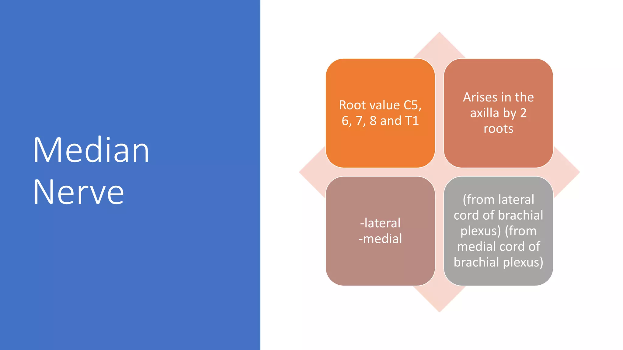
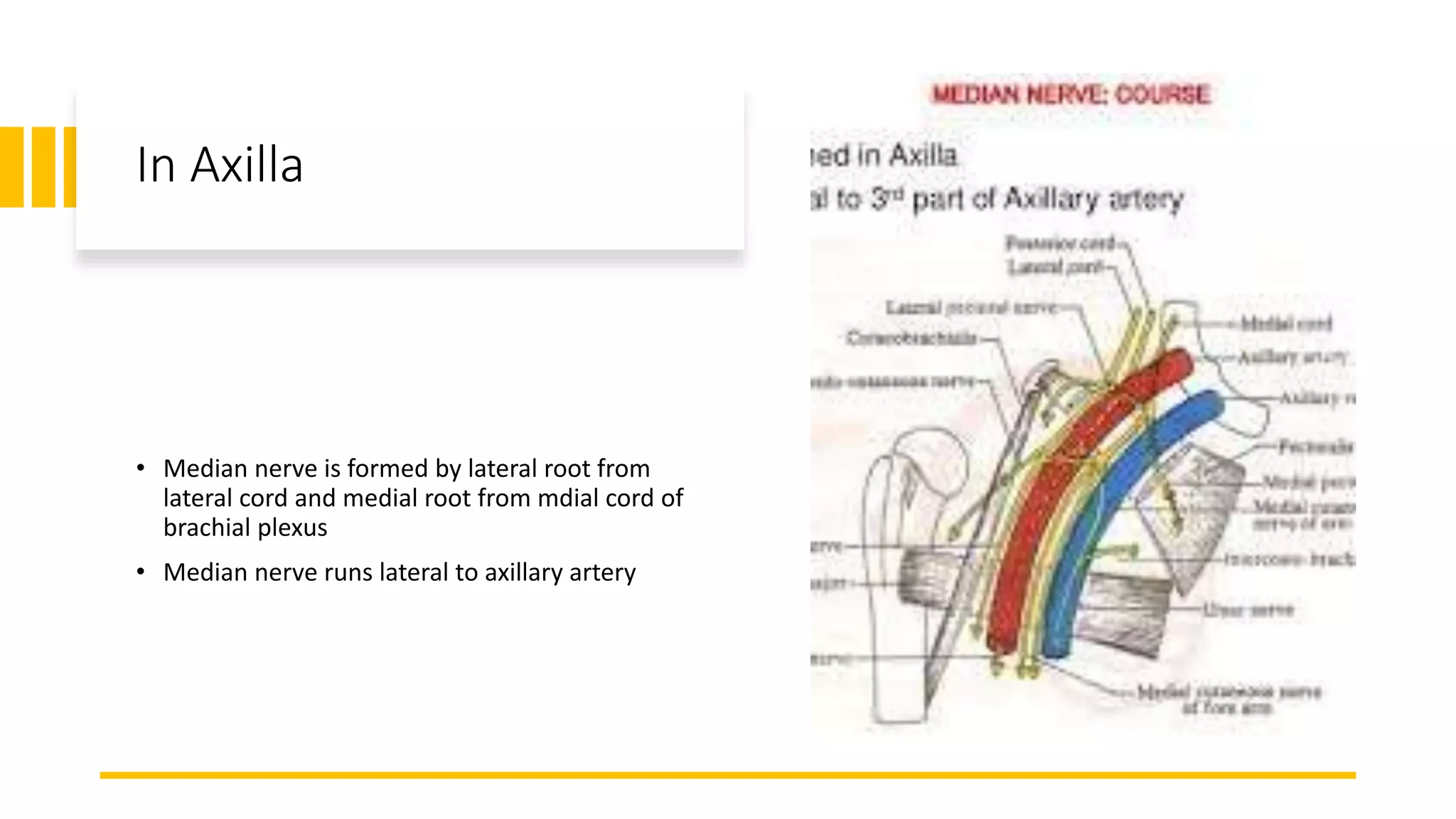
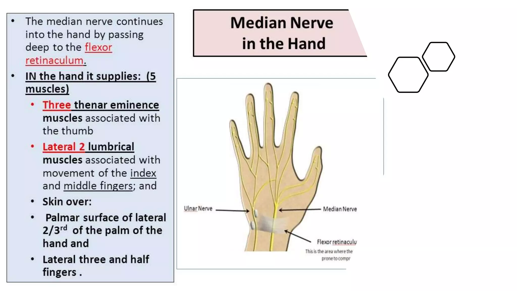
Median Nerve
-Siddhant Binyala
Median
Nerve
Root value C5,
6, 7, 8 and T1
Arises in the
axilla by 2
roots
-lateral
-medial
(from lateral
cord of brachial
plexus) (from
medial cord of
brachial plexus)
In Axilla
• Median nerve is formed by lateral root from
lateral cord and medial root from mdial cord of
brachial plexus
• Median nerve runs lateral to axillary artery
In Arm
• Median nerve continues to run on
lateral side of brachial artery till the
middle of the arm, where it crosses
infront of the artery and passes anterior
to the elbow joint into forearm
In Forearm
• Enters the forearm between the two heads of pronator
teres
• Then deep to the fibrous arch of flexor digitorum
superficialis in proximal third
• In mid forearm, descends between flexor digitorum
superficialis and flexor digitorum profundus
• About 5 cm above wrist, it coms to lie on the lateral side of
the flexor digitorum superficialis, becomes superficial just
above the wrist
In Hand
Branches
• In arm- Vascular branches to the brachial
artery
• In forearm- Muscular branches to all
superficial flexor muscles ( pronator teres,
flexor carpi radialis, palmaris longus and
flexor digitorum superficialis)
• Anterior interosseous, that comes off the
median nerve spplies lateral half of flexor
digitorum profundus, flexor pollicis longus
and pronator quadratus
• Articular branches supply the elbow joint
and prximal ulnar joint
• Palmar cutaneous branch supply skin over
thenar eminence and central part of palm
Injury to Median nerve
• Most commonly injured at the wrist or high
up in the forearm
• 1) high median nerve palsy
• 2) low median nerve palsy
Low median
nerve palsy
• Injury in the distal third of forearm
• Cuts infront of wrist or by carpal dislocation
• There will be sparing of forearm muscles, but
the muscles of the hand will be paralysed
• Thenar eminence is wasted and thumb
abduction and opposition are weak
…
High median
nerve palsy
• Injury prximal to the elbow
• Generally due to forearm fractures or elbow
dislocation
• Trauma may damage the nerve at any level
• This will cause paralysis of all muscles
supplied by the median nerve in the forearm
and hand
Flexor pollicis longus
• This muscle is tested by holding the
thumb at its base and patient is asked
to bend the terminal phalanx
Flexor digitorum
superficialis and
profundus
• Patient is asked to clasp the hands,
index finger of the affected side fails
to reflex
• Oschner.s clasping test
Flexor carpi
radialis
The hand deviates to the
ulnar side when its flexed
against resistence
Abductor pollicis
brevis
• Patient is asked to lay his hand flat
on the table, a pen is held above the
palm and the patient is asked to
touch the pen with his thumb
• Pen test
Opponens pollicis
• Bring the tip of the thumb
towards the tips of other fingers
Phelan’s maneuver
• Diagnostic test for carpal tunnel syndrome
• A person holds his forearms horizontally and
then pushes backs of the hands together (inverse
praying position to achieve maximal wrist flexion
)
Tinel’s sign
• Tinel’s sign is positive when lightly
percussing over the nerve elicits a
sensation of tingling or ‘pins and
needles’ in the distribution of nerve
Carpal compression
test
• Direct pressure over transverse
ligament for 30 seconds
• Onset of pain or paresthesia along the
course of the nerve after 30 seconds
gives a positive result for carpal tunnel
syndrome

Median nerve

  • 1.
  • 2.
    Median Nerve Root value C5, 6,7, 8 and T1 Arises in the axilla by 2 roots -lateral -medial (from lateral cord of brachial plexus) (from medial cord of brachial plexus)
  • 3.
    In Axilla • Mediannerve is formed by lateral root from lateral cord and medial root from mdial cord of brachial plexus • Median nerve runs lateral to axillary artery
  • 4.
    In Arm • Mediannerve continues to run on lateral side of brachial artery till the middle of the arm, where it crosses infront of the artery and passes anterior to the elbow joint into forearm
  • 5.
    In Forearm • Entersthe forearm between the two heads of pronator teres • Then deep to the fibrous arch of flexor digitorum superficialis in proximal third • In mid forearm, descends between flexor digitorum superficialis and flexor digitorum profundus • About 5 cm above wrist, it coms to lie on the lateral side of the flexor digitorum superficialis, becomes superficial just above the wrist
  • 6.
  • 8.
    Branches • In arm-Vascular branches to the brachial artery • In forearm- Muscular branches to all superficial flexor muscles ( pronator teres, flexor carpi radialis, palmaris longus and flexor digitorum superficialis) • Anterior interosseous, that comes off the median nerve spplies lateral half of flexor digitorum profundus, flexor pollicis longus and pronator quadratus • Articular branches supply the elbow joint and prximal ulnar joint • Palmar cutaneous branch supply skin over thenar eminence and central part of palm
  • 10.
    Injury to Mediannerve • Most commonly injured at the wrist or high up in the forearm • 1) high median nerve palsy • 2) low median nerve palsy
  • 11.
    Low median nerve palsy •Injury in the distal third of forearm • Cuts infront of wrist or by carpal dislocation • There will be sparing of forearm muscles, but the muscles of the hand will be paralysed • Thenar eminence is wasted and thumb abduction and opposition are weak …
  • 12.
    High median nerve palsy •Injury prximal to the elbow • Generally due to forearm fractures or elbow dislocation • Trauma may damage the nerve at any level • This will cause paralysis of all muscles supplied by the median nerve in the forearm and hand
  • 13.
    Flexor pollicis longus •This muscle is tested by holding the thumb at its base and patient is asked to bend the terminal phalanx
  • 14.
    Flexor digitorum superficialis and profundus •Patient is asked to clasp the hands, index finger of the affected side fails to reflex • Oschner.s clasping test
  • 15.
    Flexor carpi radialis The handdeviates to the ulnar side when its flexed against resistence
  • 16.
    Abductor pollicis brevis • Patientis asked to lay his hand flat on the table, a pen is held above the palm and the patient is asked to touch the pen with his thumb • Pen test
  • 17.
    Opponens pollicis • Bringthe tip of the thumb towards the tips of other fingers
  • 18.
    Phelan’s maneuver • Diagnostictest for carpal tunnel syndrome • A person holds his forearms horizontally and then pushes backs of the hands together (inverse praying position to achieve maximal wrist flexion )
  • 19.
    Tinel’s sign • Tinel’ssign is positive when lightly percussing over the nerve elicits a sensation of tingling or ‘pins and needles’ in the distribution of nerve
  • 20.
    Carpal compression test • Directpressure over transverse ligament for 30 seconds • Onset of pain or paresthesia along the course of the nerve after 30 seconds gives a positive result for carpal tunnel syndrome