Downloaded 166 times


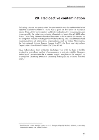
![Determination of tannins
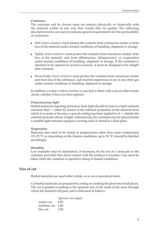
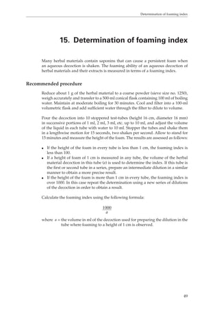
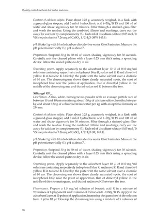
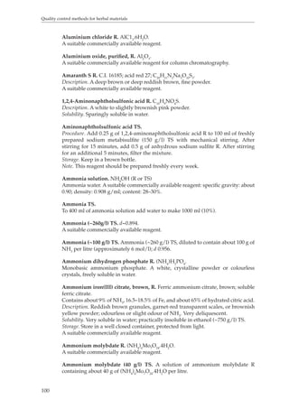
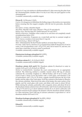
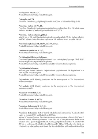
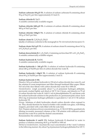
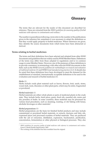
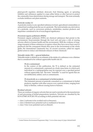
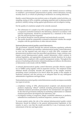
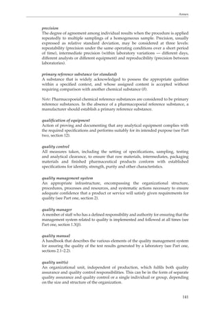
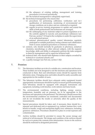
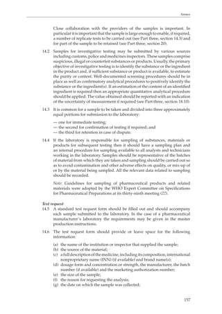
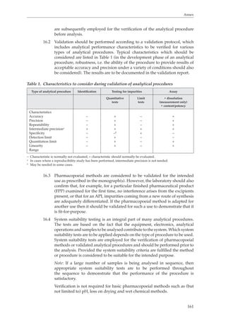
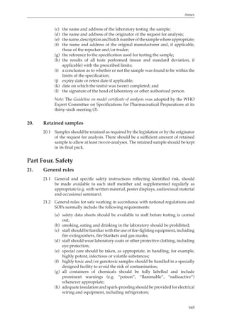
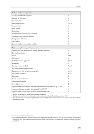
13. Determination of tannins
Tannins (or tanning substances) are substances capable of turning animal hides into
leather by binding proteins to form water-insoluble substances that are resistant
to proteolytic enzymes. This process, when applied to living tissue, is known as
an “astringent” action and is the reason for the therapeutic application of tannins.
Chemically, tannins are complex substances; they usually occur as mixtures of
polyphenols that are difficult to separate and crystallize. They are easily oxidized
and polymerized in solution; if this happens they lose much of their astringent
effect and are therefore of little therapeutic value.
45
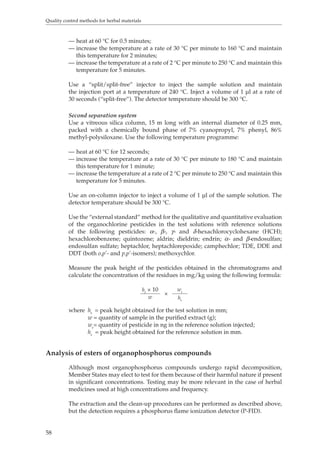
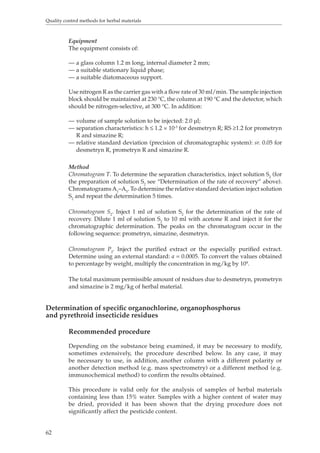
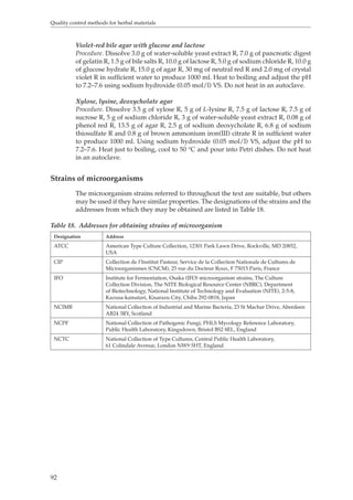
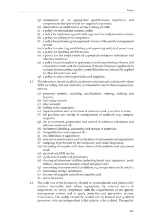
Recommended procedure
To prepare the herbal material extract, introduce the quantity specified in the test
procedure for the herbal material concerned, previously powdered to a known
fineness and weighed accurately, into a conical flask. Add 150 ml of water and
heat over a boiling water-bath for 30 minutes. Cool, transfer the mixture to a 250-
ml volumetric flask and dilute to volume with water. Allow the solid material to
settle and filter the liquid through a filter-paper, diameter 12 cm, discarding the
first 50 ml of the filtrate.
To determine the total amount of material that is extractable into water, evaporate
50.0 ml of the plant material extract to dryness, dry the residue in an oven at 105 °C
for 4 hours and weigh (T1).
To determine the amount of herbal material not bound to hide powder that is
extractable into water, take 80.0 ml of the herbal material extract, add 6.0 g of hide
powder R and shake well for 60 minutes. Filter and evaporate 50.0 ml of the clear
filtrate to dryness. Dry the residue in an oven at 105 °C and weigh (T2).
To determine the solubility of hide powder, take 6.0 g of hide powder R, add
80.0 ml of water and shake well for 60 minutes. Filter and evaporate 50.0 ml of the
clear filtrate to dryness. Dry the residue in an oven at 105 °C and weigh (T0).
Calculate the quantity of tannins as a percentage using the following formula:
[T1–(T2 – T0)] × 500
w
where w = the weight of the herbal material in grams.](https://image.slidesharecdn.com/qualitycontrolmethodsofherbalmaterials-140917115302-phpapp01/85/Quality-control-methods-of-herbal-materials-58-320.jpg)


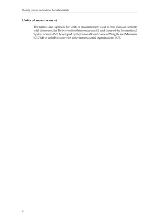
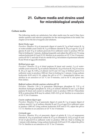
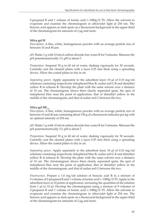
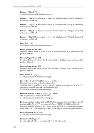
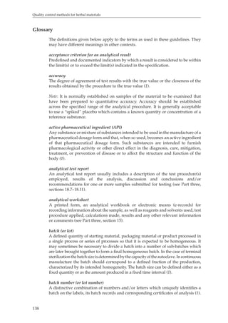
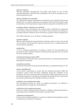
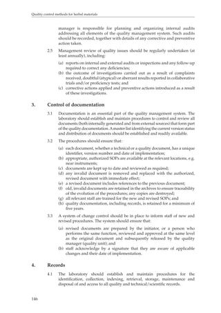
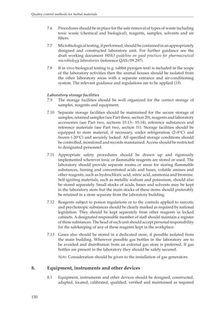
![Quality control methods for herbal materials
86
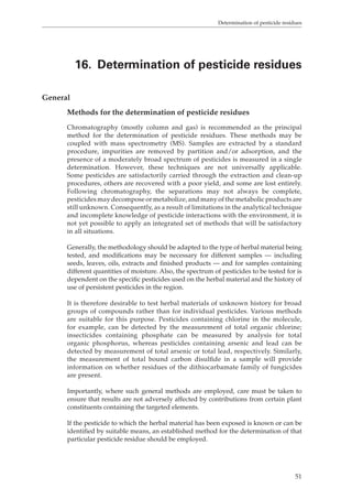
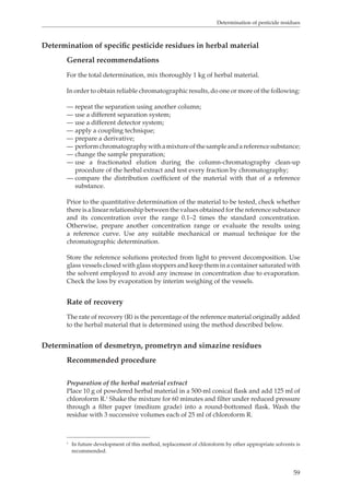
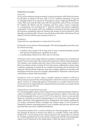
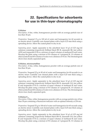
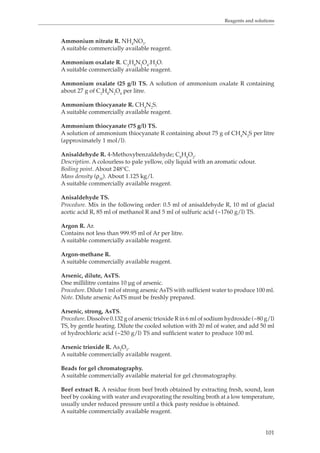
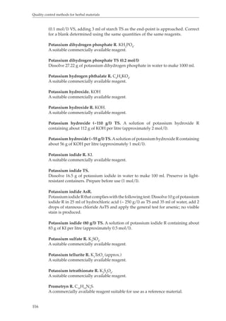
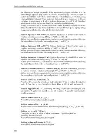
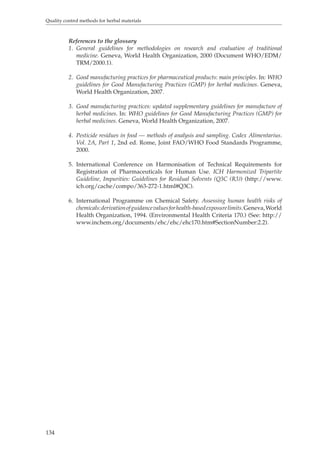
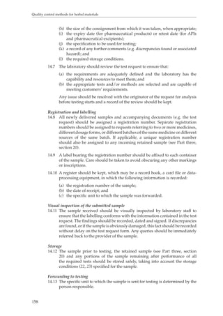
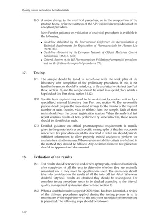
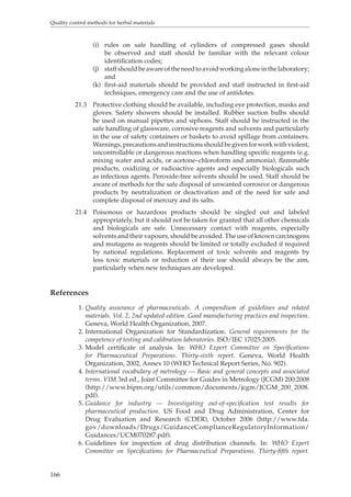
stand at room temperature for 15 min in the dark. Add 0.4 ml of acetonitrile:water
(1:9) solution to the tube. A 20-μl portion of the sample solution in the tube is
subjected to liquid chromatography analysis.
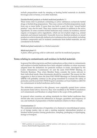
Preparation of sample
Grind the herbal material for testing to a uniform consistency using a coffee mill,
and extract a 50-g test sample with 400 ml of acetonitrile-water (9:1) by shaking
vigorously in a glass flask fitted with a stopper for 30 minutes or by using a
mechanical blender for 5 minutes. Filter the solution through a filter paper
or centrifuge. Transfer a 5-ml portion of the filtrate, or the top clean layer, to a
multifunctional column (such as a MultiSep #228 cartridge column (Romer Labs)
or an Autoprep MF-A [Showa-denko]) and pass through at a flow rate of 1 ml/
minute. The aflatoxins present in a sample are passed through the column as the
first eluate. Obtain the first 1-ml of the eluate as the test solution.
Evaporate 0.5 ml of the test solution in a glass centrifuge tube to dryness at 40 °C
or by using a nitrogen air stream to remove solvent.
To derivatize aflatoxins B1 and G1 (precolumn derivatization), add 0.1 ml of
trifluoroacetic acid (TFA) solution to the residue in the tube, tightly seal the tube
and shake vigorously. Allow the tube to stand at room temperature for 15 minutes
in the dark. Add 0.4 ml of acetonitrile-water (1:9) solution to the tube. Subject a
20-μl portion of the sample solution in the tube to liquid chromatography analysis.
Method
Liquid chromatography conditions
The mobile phase is acetonitrile-methanol-water (1:3:6).1 De-gas the mobile phase by
sonication. Connect an octadecyl-silica gel (ODS) column (4.6 mm inner diameter (ID)
× 250 mm, 3–5 μm), such as Inertsil ODS-3 (4.6 mm ID × 250 mm, 3 μm) as the liquid
chromatography column. Maintain the column at 40 °C with a flow rate of 1 ml/
minute. The aflatoxin and its derivatives are detected at the excitation and emission
wavelengths of 365 nm and 450 nm, respectively. The injection volume is 20 μl.
If an impurity peak overlaps the peaks corresponding to aflatoxins, the alternative
liquid chromatography conditions, described below, are recommended.
Alternative liquid chromatography conditions
The mobile phase is methanol-water (3:7). De-gas the mobile phase by sonication.
Connect a fluorocarbonated column, such as Wako-pack Fluofix 120E (4.6 mm ID ×
250 mm, 5 μm) as the liquid chromatography column. Maintain the column at 40 °C
with a flow rate of 1 ml/minute. The aflatoxin and its derivatives are detected at
the excitation and emission wavelengths of 365 nm and 450 nm, respectively. The
injection volume is 20 μl.
Interpretation of the results
Compare the retention time of peak area or peak heights of the aflatoxin under
study in the chromatograms. If they are bigger or higher than those obtained in a
standard solution of the aflatoxin under investigation, it should be regarded as a
positive result for the presence of aflatoxin in the sample solution.
1 If the sample solution contains a lot of impurity, the column should be washed by acetonitrile for
5–10 min and reconditioned with the mobile phase for 10 min before the next analysis.](https://image.slidesharecdn.com/qualitycontrolmethodsofherbalmaterials-140917115302-phpapp01/85/Quality-control-methods-of-herbal-materials-99-320.jpg)

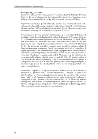
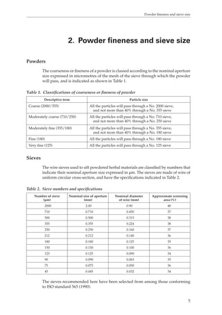
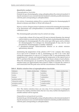
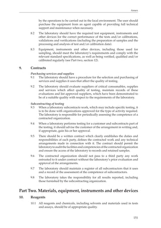
![Reagents and solutions
103
Calcium carbonate R1. CaCO3.
A suitable commercially available reagent.
Calcium carbonate R2. Calcium carbonate R1 of suitable quality to serve as a
primary standard for the standardization of disodium edetate solutions.
A suitable commercially available reagent.
Calcon R. Monosodium salt of 2-hydroxy-1-[(2-hydroxy-1-naphthyl)azo]
naphthalene-4-sulfonic acid; C.I. Mordant Black 17, C.I. 15705, Eriochrome Blue
Black R, Solochrome Dark Blue; C20H13NaO5S.
A suitable commercially available reagent.
Calcon carboxylic acid R. 2-Hydroxy-1-(2-hydroxy-4-sulfo-1-naphthylazo)-3-
naphthoic acid; C21H14N2O7S,3H2O.
Description. A dark-brown powder with a violet tint.
Solubility. Practically insoluble in water; slightly soluble in methanol R and in
ethanol (~750 g/l) TS; freely soluble in solutions of alkali hydroxides.
A suitable commercially available reagent.
Calcon carboxylic acid indicator mixture R.
Procedure. Mix 0.1 g of calcon carboxylic acid R with 10 g of anhydrous sodium
sulfate R.
Calcon indicator mixture R.
Procedure. Mix 0.1 g of calcon R with 10g of anhydrous sodium sulfate R.
Carbophenothion. CHClOPS. O,O-Diethyl S-[[(4-chlorophenyl)thio] methyl]-
111623phosphorodithionate.
Yellowish liquid, practically insoluble in water, miscible with organic solvents. d25:
4
about 1.27.
A suitable commercially available reagent.
Cetrimide R. Contains not less than 96.0% and not more than 101.0% of
alkyltrimethylammonium bromide, calculated as C17H38BrN with reference to
the dried substance.
Description. A white or almost white, voluminous, free-flowing powder; slight
characteristic odour.
Solubility. Soluble in two parts of water; freely soluble in ethanol (~750 g/l) TS.
A suitable commercially available reagent.
Chinese ink TS. Indian ink.
A suitable commercially available reagent.
Note. Before use, dilute 1 ml of black Chinese ink TS with 2 ml of water; if necessary,
further dilute up to 1:10. It must be freshly prepared.
Chloral hydrate R. C2H3Cl3O2.
Description. Colourless, hygroscopic crystals with a sharp odour.
Melting temperature. About 55°C.
A suitable commercially available reagent.
Chloral hydrate TS.
Procedure. Dissolve 50 g of chloral hydrate R in 20 ml of water.](https://image.slidesharecdn.com/qualitycontrolmethodsofherbalmaterials-140917115302-phpapp01/85/Quality-control-methods-of-herbal-materials-116-320.jpg)
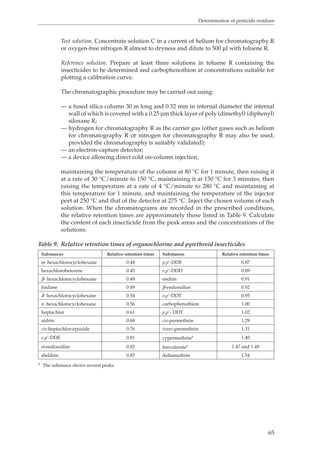
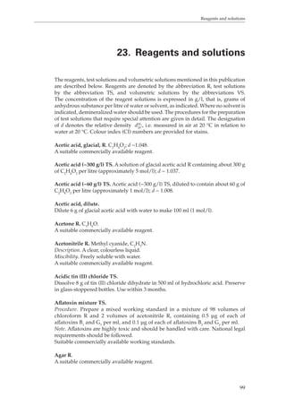
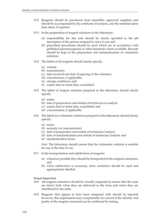
![Reagents and solutions
107
Ferrous sulfate. FeSO4.7 H2O
A suitable commercially available reagent.
Florisil R.
A suitable commercially available material for column chromatography.
Formic acid (~1080 g/l) TS. CH2O2; d ~ 1.2.
A suitable commercially available reagent.
D-Fructose R. C6H12O6.
Description. A white, crystalline powder.
Melting point. About 103°C with decomposition.
Specific optical rotation. Use a 0.10 g/ml solution in water containing 0.05 ml of
ammonia (~100 g/l) TS; [α]D20 °C = about -92°.
A suitable commercially available reagent.
D-Galactose R. C6H12O6.
Description. A white, crystalline powder.
Melting point. About 164°C.
Specific optical rotation. Use a 0.10 g/ml solution in water; [α]D20 °C = about +80°.
A suitable commercially available reagent.
Glucose R. Dextrose; C6H12O6. Quality conforms to the monograph in The
international pharmacopoeia (5).
Glucose hydrate R. Monohydrate of α-D’glucopyranose, CHO.HO. Contains
61262not less than 99.0% and not more than 101.5% of CHO, calculated with reference
6126to the dried substance.
Description. Colourless crystals or a white crystalline or granular powder; odourless.
Solubility. Soluble in about 1 part of water and in about 60 parts of ethanol (~750 g/l)
TS; more soluble in boiling water and in boiling ethanol (~750 g/l) TS.
Acidity. Dissolve 5 g in 50 ml of carbon-dioxide-free water R. Neutralization
requires not more than 0.5 ml of carbonate-free sodium hydroxide (0.02 mol/l) VS,
phenolphthalein/ethanol TS being used as indicator.
Specific optical rotation. Dissolve 100 mg, previously dried to constant weight, in 1
ml of water, and add a few drops of ammonia (~100 g/l) TS; [α]20 °C
= + 52 to + 53°.
DSoluble starch or sulfites. Dissolve 1 g in 10 ml of water and add 1 drop of iodine TS;
the liquid is coloured yellow.
Loss on drying. Dry to constant weight at 105 °C; loses not less than 80 mg/g and
not more than 100 mg/g.
Sulfated ash. Not more than 1.0 mg/g.
Assay. Dissolve about 0.1 g, accurately weighed, in 50 ml of water, add 30 ml of
iodine (0.1 mol/l) VS and 10 ml of sodium carbonate (50 g/l) TS, and allow to stand
for 20 minutes. Add 15 ml of hydrochloric acid (~70 g/l) TS and titrate the excess
of iodine with sodium thiosulfate (0.1 mol/l) VS, using starch TS as indicator.
Perform a blank determination and make any necessary corrections. Each ml of
iodine (0.1 mol/l) VS is equivalent to 9.008 mg of C6H12O6.
Glycerol R. Propane- 1,2,3-triol with small amounts of water, C3H8O3. Contains not
less than 970 g/kg of C3H8O3.
Description. A clear, almost colourless, syrupy and hygroscopic liquid; odourless.](https://image.slidesharecdn.com/qualitycontrolmethodsofherbalmaterials-140917115302-phpapp01/85/Quality-control-methods-of-herbal-materials-120-320.jpg)
![Reagents and solutions
Solubility. Soluble in water; very slightly soluble in ethanol (~750 g/l) TS; insoluble
in ether R.
Melting point. About 213 °C with decomposition.
Specific optical rotation. Dissolve 0.2 g in 10 ml of hydrochloric acid (~250 g/l) TS;
[α]D20 °C
111
= about +21.5 °.
A suitable commercially available reagent.
Magnesium chloride R. MgCl2.6H2O.
A suitable commercially available reagent.
Magnesium nitrate R. Mg(NO3)2
See Magnesium nitrate hexahydrate.
Magnesium nitrate hexahydrate R. Mg(NO3)2.6H2O
A suitable commercially available reagent.
Mercuric bromide R. HgBr2.
A suitable commercially available reagent.
Mercuric bromide AsTS.
Procedure. Dissolve 5 g of mercuric bromide R in sufficient ethanol (~ 750 g/l) TS
to produce 100 ml.
Mercuric bromide paper AsR.
Procedure. Use smooth, white filter-paper weighing 65–120 g/m2. The thickness
of the paper in mm should be approximately equal numerically to the weight
expressed as above, divided by 400. Soak pieces of filter-paper, not less than
25 mm in width, in mercuric bromide AsTS, decant the superfluous liquid,
suspend the paper over a non-metallic thread and allow it to dry, protected from
light.
Storage. Store the mercuric bromide paper AsR in stoppered bottles in the dark.
Note. Paper that has been exposed to sunlight or to vapours of ammonia must not
be used as it produces only a pale stain or no stain at all.
Mercuric nitrate TS. Millon’s reagent; nitric acid solution of mercury.
Procedure. Dissolve 1 ml of mercury R in 9 ml of fuming nitric acid R, keeping the
mixture well cooled during the reaction. When the reaction is complete, dilute the
solution with an equal volume of water. It should be protected from light and used
within two months of preparation.
Mercuric thiocyanate R. C2HgN2S2.
A suitable commercially available reagent.
Mercuric thiocyanate TS. A saturated solution of mercuric thiocyanate R in ethanol
(~ 750 g/l) TS.
Mercury R. Hg.
A suitable commercially available reagent.
Methane R. CH4.
A suitable commercially available reagent.](https://image.slidesharecdn.com/qualitycontrolmethodsofherbalmaterials-140917115302-phpapp01/85/Quality-control-methods-of-herbal-materials-124-320.jpg)
![Reagents and solutions
123
Thionine R. C.I. 52000; C12H10ClN3S.
Description. Blackish green glistening crystals.
Solubility. Freely soluble in hot water.
A suitable commercially available reagent.
Thionine TS.
Procedure. Dissolve 0.2 g of thionine R in 100 ml of ethanol (~188 g/l) TS.
Toluene R. C7H8. Methylbenzene.
A clear, colourless, flammable liquid, very slightly soluble in water, miscible with
alcohol. d20
20: 0.865 to 0.870.
bp: about 110 °C.
A suitable commercially available reagent.
Trifluoroacetic acid (TFA). CF3COOH.
A suitable commercially available reagent.
2,2,4-Trimethylpentane R. C8H18.
A suitable commercially available reagent.
Trinitrophenol R. C6H3N3O7.
A suitable commercially available reagent.
Trinitrophenol, ethanolic, TS.
Procedure. Dissolve 1 g of trinitrophenol R in 100 ml of ethanol (~750 g/l) TS.
Tropaeolin O R. C.I. 14270; E103: resorcin yellow; chrysoin S; sulpho orange; acid
orange 6; C12H9N2NaO5S.
Description. Produces a yellow colour in moderately alkaline solutions and an
orange colour in strongly alkaline solutions (pH range 11.0-12.7).
A suitable commercially available reagent.
Water, carbon-dioxide-free, R. Water that has been boiled vigorously for a few
minutes and protected from the atmosphere during cooling and storage.
Xylene R. C8H10.
A suitable commercially available reagent.
D-Xylose R. C5H10O5.
Description. A white, crystalline powder.
Specific optical rotation. Dissolve 1 g in 10 ml of water; [α]D20 °C = about +20°.
A suitable commercially available reagent.
Yeast extract, water-soluble, R.
A suitable commercially available reagent.
Zinc R. Zn; granulate, powder, or dust.
A suitable commercially available reagent.
Zinc, AsR, granulated. Granulated zinc R that complies with the following tests:](https://image.slidesharecdn.com/qualitycontrolmethodsofherbalmaterials-140917115302-phpapp01/85/Quality-control-methods-of-herbal-materials-136-320.jpg)


This document provides guidelines for quality control testing of herbal materials. It describes general considerations for methods, including use of the metric system, precision of measurements, calculation of results, and establishment of limits. Reagents and solutions are given specific designations. Temperature is generally room temperature unless otherwise specified. The document aims to support development of national standards for herbal materials.


