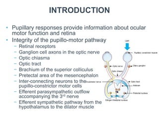
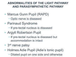
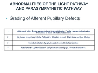
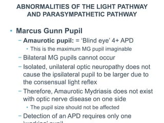
Pupils
COMMON EYE DISORDERS
NEUROLOGICAL EYE DISEASE
CHAPTER CONTENTS
 Introduction
 Normal pupillary phenomena
 Pupillary testing
 Abnormalities of the light reflex and
Parasympathetic Pathway
 The oculo-sympathetic pathway
INTRODUCTION
• Pupillary responses provide information about ocular
motor function and retina
• Integrity of the pupillo-motor pathway
− Retinal receptors
− Ganglion cell axons in the optic nerve
− Optic chiasma
− Optic tract
− Brachium of the superior colliculus
− Pretectal area of the mesencephalon
− Inter-connecting neurons to the
pupillo-constrictor motor cells
− Efferent parasympathetic outflow
accompanying the 3rd nerve
− Efferent sympathetic pathway from the
hypothalamus to the dilator muscle
INTRODUCTION
• Light reflex pathway
− Afferent neurons from retinal ganglion cells to
pretectal area
− Inter-neurons from pretectal complex to
Edinger-Westphal nuclei
− Parasympathetic outflow with 3rd nerve to
ciliary ganglion
− From ciliary ganglion to iris sphincter
RESPONSE INDUCED BY ONE EYE
INTRODUCTION
• A blurred retinal image or the conscious
visual fixation of a near object results in
accommodative effort
• A near synkinesis is evoked and includes:
− Increased accommodation of the lens
− Convergence of the visual axes of the eyes
− Pupillary constriction
• The accommodative drive comes from the
frontal lobe
INTRODUCTION
• The Edinger-Westphal nucleus is divided
into the:
− Rostral portion: involved in accommodation
− Caudal portion: elicits pupil constriction
− Middle segment: stimulation results in
accommodation and pupil constriction
• The final pathway for pupil constriction
consists of:
− Oculo-motor nerve
− Ciliary ganglion
NORMAL PUPILLARY PHENOMENA
• Pupil size changes with age
− At birth the pupils are larger and get smaller
• Pupil size is controlled by the
Parasympathetic and Sympathetic
Nervous Systems
• The pupil has three main optical functions:
− Regulate amount of light reaching the retina
− Diminish chromatic and spherical aberrations
produced by the peripheral imperfections of
the optical system of the cornea and lens
NORMAL PUPILLARY PHENOMENA
• Normal size of the pupil ranges between 2-6 mm
• Shape of the pupils is examined to assess if they
are regular or irregular and symmetrical
• Direct and indirect responses to bright light are
evaluated
• The accommodation reaction should be tested
NORMAL PUPILLARY PHENOMENA
• Physiologic anisocoria occurs in 20% of
general population
• Less than 0.5 mm anisocoria is considered as
a normal or a physiological pupillary
phenomenon
− Can be up to 1.0 mm
• Degree of anisocoria can vary from day to day
and even switch eyes
NORMAL PUPILLARY PHENOMENA
• Pupillary unrest (Hippus) occurs during
distance fixation with constant, ambient
illumination
• The pupils should be bilaterally
symmetrical
− Non-rhythmical unrest or variation in size
should usually be less than 1 mm
• Near Synkinesis is a normal pupillary
phenomenon
• Light produces miosis greater than when a
PUPILLARY TESTING
• When observing the pupils we should
assess:
− Size
− Shape
− Symmetry
− Responses to light stimuli
− Accommodative reaction of the pupils
PUPILLARY TESTING
• Perform pupillary testing in dim
illumination
• Reflexes assessed:
− Direct Light Reflex
• Shine the light in a pupil and look at the same pupil
− Consensual Reflex
• Shine the light in a pupil and look at constriction in
the other pupil
− Swinging Flashlight Test
• Bright light used in a darkened room
• Patient should fixate on a distant object
ABNORMALITIES OF THE LIGHT PATHWAY
AND PARASYMPATHETIC PATHWAY
• Marcus Gunn Pupil (RAPD)
− Optic nerve is diseased
• Parinaud Syndrome
− If pre-tectal nucleus is diseased
• Argyll Robertson Pupil
− If pre-tectal nucleus is diseased but
accommodation is intact
• 3rd nerve palsy
• Holmes Adie Pupil (Adie’s tonic pupil)
− Dilated pupil on one side and otherwise
ABNORMALITIES OF THE LIGHT PATHWAY
AND PARASYMPATHETIC PATHWAY
• Marcus Gunn Pupil
− RAPD where amount of light transmitted from one eye is
less than the other
− During the swinging flashlight test if an afferent pathway
problem exists, there will be dilatation instead of
constriction
− There will be a sluggish direct response and so the
corresponding consensual response of the other eye will
also be sluggish
− Requires marked asymmetry or unilateral disease
− Bilateral APDs can occur but only unilateral RAPDs exist
ABNORMALITIES OF THE LIGHT PATHWAY
AND PARASYMPATHETIC PATHWAY
• Grading of Afferent Pupillary Defects
1+ Initial constriction. Greater escape to larger intermediate size. Pupillary escape indicating that
the number of fibers affected is not as many
2+ No change in pupil size initially. Followed by dilatation of pupil. Slight delay and then dilation.
3+ Immediate dilation of pupil, instead of normal initial constriction
4+ Patient has No Light Perception. Completely amaurotic pupil. Immediate dilatation.
ABNORMALITIES OF THE LIGHT PATHWAY
AND PARASYMPATHETIC PATHWAY
• Marcus Gunn Pupil
− Optic neuropathy must be unilateral or
markedly asymmetric to get an RAPD
− Ocular media opacities will not cause a MG
pupil
− Maculopathy or amblyopia will not cause MG
pupil unless the VA < 20/200
• In which case only a 1+ APD will occur
− Retinal damage will cause a significant MG
pupil
• Has to be extensive
ABNORMALITIES OF THE LIGHT PATHWAY
AND PARASYMPATHETIC PATHWAY
• Marcus Gunn Pupil
− Amaurotic pupil: = ‘Blind eye’ 4+ APD
• This is the maximum MG pupil imaginable
− Bilateral MG pupils cannot occur
− Isolated, unilateral optic neuropathy does not
cause the ipsilateral pupil to be larger due to
the consensual light reflex
− Therefore, Amaurotic Mydriasis does not exist
with optic nerve disease on one side
• The pupil size should not be affected
− Detection of an APD requires only one
ABNORMALITIES OF THE LIGHT PATHWAY
AND PARASYMPATHETIC PATHWAY
• Argyll Robertson pupil
− Miotic, irregular pupils
− Light-near dissociation
• Pupils constrict to convergence but not to direct
light
− Poor dilation in darkness
− Poor response to mydriatic agents
ABNORMALITIES OF THE LIGHT PATHWAY
AND PARASYMPATHETIC PATHWAY
• Argyll Robertson pupil
− Usually bilateral, and often asymmetric
− Main cause is neuro-syphilis
• Other causes include diabetes mellitus, MS
• Occur in secondary and tertiary syphilis
− Likely site of the lesion is the region of the
Sylvian Aqueduct
− With tropicamide, the normal pupil will dilate
and but the abnormal pupil will not
− Treatment of neuro-syphilis is with penicillin
ABNORMALITIES OF THE LIGHT PATHWAY
AND PARASYMPATHETIC PATHWAY
• Adie’s tonic pupil
− Idiopathic, benign case of internal
ophthalmoplegia
− Probably due to viral infection at the ciliary
ganglion with destruction of fibers going to the
iris
• Remaining fibers compensate by an increase in
receptors
• Denervation super-sensitivity
• Compensation takes a couple of days to occur
− A large, dilated pupil on one side is
characteristic
ABNORMALITIES OF THE LIGHT PATHWAY
AND PARASYMPATHETIC PATHWAY
• Adie’s tonic pupil
− Characteristics of the condition include:
• A dilated pupil with poor to absent direct light
reaction
• Slow constriction to prolonged near effort
• Slow redilation
− Constriction in the abnormal pupil lasts much
longer
• Hence the term ‘Tonic pupil’
− Constriction on convergence
− Initial accommodative paresis which resolves
ABNORMALITIES OF THE LIGHT PATHWAY
AND PARASYMPATHETIC PATHWAY
• Adie’s tonic pupil
− Etiology is unknown but is possibly due to
viral infection or is post infectious
− Lesion occurs in the ciliary ganglion or short
posterior ciliary nerves
− There is aberrant regeneration of more
numerous fibers innervating the ciliary muscle
(97%) than the iris sphincter (3%)
• This is only found in Adie’s pupil cases
THE OCULO-SYMPATHETIC
PATHWAY
• 1st order neuron
− From the hypothalamus to the C8-T2 level
• 2nd order neuron (pre-ganglionic)
− From the spinal cord to the para-vertebral
sympathetic chain and ends at the superior
cervical ganglion (SCG)
• 3rd order neuron (post-ganglionic)
− From the SCG, along the internal carotid artery,
joins the ophthalmic nerve and enters the
orbit through the superior orbital fissure
− Terminates at the iris, Müller’s muscle
and the inferior tarsal muscle
− Sudomotor (cholinergic) and Vasomotor
(noradrenergic) fibers to the face travel with
the external carotid artery and its branches
THE OCULO-SYMPATHETIC
PATHWAY
• The dilator muscle is innervated by sympathetic fibers
• A three neuron pathway exists
− Starts in the brain, down the brainstem to the lateral part of levels C8
to T2 of the spinal cord
− To the sympathetic plexus over the top of the lung
− To the superior cervical ganglion (SCG)
• It re-enters the head along the carotid artery to the
cavernous sinus
• Enters the orbit to supply the dilator fibers of the iris
• This pathway remains ipsi-lateral throughout its course
THE OCULO-SYMPATHETIC
PATHWAY
• A lesion of the sympathetic supply to the eye results in
Horner Syndrome, which is characterized by:
− Miosis
− Partial ptosis of the upper and lower lids
− Anhidrosis
− Enophthalmos
• The causative lesion is always ipsi-lateral to the side of the
Horner’s eye
THE OCULO-SYMPATHETIC
PATHWAY
• Congenital Horner syndrome is characterized by iris
heterochromia
− Due to loss of the sympathetic drive required for iris pigmentation
• This is due to the affectation of the sympathetic system
during childbirth
• The face will appear flushed on one side, with a sharp
dividing line that runs precisely along the sagittal midline
THE OCULO-SYMPATHETIC
PATHWAY
• Depending on the company it keeps, the site of lesion causing the
Horner Syndrome can be localized
• Following synapse in the SCG, some of the fibers continue along the
external carotid artery to the face and are responsible for facial sweating
− Therefore, a Horner Syndrome plus lack of sweating (anhidrosis) implies a
more proximal lesion along the sympathetic pathway
• The sympathetic fibers innervate the dilator muscles of the iris, the
inferior tarsal muscle in the lower lid and Muller’s muscle in the upper lid
− Consequently, a partial ptosis and a lower lid that moves up causes an apparent
enophthalmos
THE OCULO-SYMPATHETIC
PATHWAY
• Causes of Horner syndrome include:
− Hemispheric lesions involving the hypothalamus (e.g. stroke)
− Brainstem lesions (e.g. stroke)
− Cervical cord lesions
− Root lesions at T1 (root compression)
− Sympathetic chain lesions
• Whiplash can result in internal carotid artery dissection
− Cavernous sinus lesions
− Orbit lesions
− Migraine and cluster headache
THE OCULO-SYMPATHETIC
PATHWAY
• Assessment of Horner syndrome
− 4% cocaine has no effect on an abnormal pupil whereas the
normal pupil dilates
− Due to denervation super-sensitivity, the abnormal pupil dilates
to dilute adrenaline and the lid elevates
− No response to dilute adrenaline occurs in the normal pupil
− Unlike cocaine, that has a negative effect on the abnormal pupil,
adrenaline has a positive effect on the abnormal pupil to confirm
the presence of Horner Syndrome
− Apraclonidine (Iopadine) is a weak alpha-1 agonist and has a
similar response to adrenaline
THE OCULO-SYMPATHETIC
PATHWAY
• Assessment of Horner syndrome
− With 1% hydroxyamphetamine:
• A 3rd order neuron lesion does not result in pupil dilatation
whereas a 1st and 2nd order neuron lesion does
− Hydroxyamphetamine, like cocaine, is not readily
available and its routine use in localizing Horner
syndrome has become less popular
− The Parker Pen Test
• A pen is run over the skin on both sides
• There is less smooth movement on the affected (rougher) side
THE OCULO-SYMPATHETIC
PATHWAY
• Assessment of Horner syndrome
− Aniscoria greater in dim light suggests a sympathetic problem and
that Horner pupils constrict to light
− In darkness, a Horner pupil may demonstrate dilation lag
• On switching off the lights, at 5 seconds there is marked anisocoria but by 20
seconds, no anisocoria may be present which is especially so in mild disorders
− There is no response to cocaine in the abnormal pupils
• Aniscoria which is greater in bright light is a
parasympathetic problem and can occur in an oculomotor
nerve palsy
− The abnormal pupil does not constrict to bright light
PUPIL EXAMINATION IN AN
UNCONSCIOUS PATIENT
• The pupil reflex is an objective clinical sign
• A normal pupil reflex in a comatose patient
suggests a metabolic cause
− Such as hypoglycemia, liver failure or kidney
failure
• Abnormal pupil reflexes in a comatose
patient suggest a structural cause
• Normal pupils
− Metabolic encephalopathy (70%) DM
− Hypoglycemia
PUPIL EXAMINATION IN AN
UNCONSCIOUS PATIENT
• Unequal pupils
− May indicate brain pathology
− A dilated pupil can indicate a herniated
temporal lobe
− Hutchinson’s pupil occurs due to raised intra-
cranial pressure which causes a 3rd nerve
compression at the tentorium cerebellum
• Bilateral dilated pupils (unreactive)
− Progressive tentorial herniation
− Implies irreversible brain damage

Pupils (1).pptx

  • 1.
  • 2.
    CHAPTER CONTENTS  Introduction Normal pupillary phenomena  Pupillary testing  Abnormalities of the light reflex and Parasympathetic Pathway  The oculo-sympathetic pathway
  • 3.
    INTRODUCTION • Pupillary responsesprovide information about ocular motor function and retina • Integrity of the pupillo-motor pathway − Retinal receptors − Ganglion cell axons in the optic nerve − Optic chiasma − Optic tract − Brachium of the superior colliculus − Pretectal area of the mesencephalon − Inter-connecting neurons to the pupillo-constrictor motor cells − Efferent parasympathetic outflow accompanying the 3rd nerve − Efferent sympathetic pathway from the hypothalamus to the dilator muscle
  • 4.
    INTRODUCTION • Light reflexpathway − Afferent neurons from retinal ganglion cells to pretectal area − Inter-neurons from pretectal complex to Edinger-Westphal nuclei − Parasympathetic outflow with 3rd nerve to ciliary ganglion − From ciliary ganglion to iris sphincter
  • 8.
  • 9.
    INTRODUCTION • A blurredretinal image or the conscious visual fixation of a near object results in accommodative effort • A near synkinesis is evoked and includes: − Increased accommodation of the lens − Convergence of the visual axes of the eyes − Pupillary constriction • The accommodative drive comes from the frontal lobe
  • 10.
    INTRODUCTION • The Edinger-Westphalnucleus is divided into the: − Rostral portion: involved in accommodation − Caudal portion: elicits pupil constriction − Middle segment: stimulation results in accommodation and pupil constriction • The final pathway for pupil constriction consists of: − Oculo-motor nerve − Ciliary ganglion
  • 11.
    NORMAL PUPILLARY PHENOMENA •Pupil size changes with age − At birth the pupils are larger and get smaller • Pupil size is controlled by the Parasympathetic and Sympathetic Nervous Systems • The pupil has three main optical functions: − Regulate amount of light reaching the retina − Diminish chromatic and spherical aberrations produced by the peripheral imperfections of the optical system of the cornea and lens
  • 12.
    NORMAL PUPILLARY PHENOMENA •Normal size of the pupil ranges between 2-6 mm • Shape of the pupils is examined to assess if they are regular or irregular and symmetrical • Direct and indirect responses to bright light are evaluated • The accommodation reaction should be tested
  • 13.
    NORMAL PUPILLARY PHENOMENA •Physiologic anisocoria occurs in 20% of general population • Less than 0.5 mm anisocoria is considered as a normal or a physiological pupillary phenomenon − Can be up to 1.0 mm • Degree of anisocoria can vary from day to day and even switch eyes
  • 14.
    NORMAL PUPILLARY PHENOMENA •Pupillary unrest (Hippus) occurs during distance fixation with constant, ambient illumination • The pupils should be bilaterally symmetrical − Non-rhythmical unrest or variation in size should usually be less than 1 mm • Near Synkinesis is a normal pupillary phenomenon • Light produces miosis greater than when a
  • 15.
    PUPILLARY TESTING • Whenobserving the pupils we should assess: − Size − Shape − Symmetry − Responses to light stimuli − Accommodative reaction of the pupils
  • 16.
    PUPILLARY TESTING • Performpupillary testing in dim illumination • Reflexes assessed: − Direct Light Reflex • Shine the light in a pupil and look at the same pupil − Consensual Reflex • Shine the light in a pupil and look at constriction in the other pupil − Swinging Flashlight Test • Bright light used in a darkened room • Patient should fixate on a distant object
  • 17.
    ABNORMALITIES OF THELIGHT PATHWAY AND PARASYMPATHETIC PATHWAY • Marcus Gunn Pupil (RAPD) − Optic nerve is diseased • Parinaud Syndrome − If pre-tectal nucleus is diseased • Argyll Robertson Pupil − If pre-tectal nucleus is diseased but accommodation is intact • 3rd nerve palsy • Holmes Adie Pupil (Adie’s tonic pupil) − Dilated pupil on one side and otherwise
  • 18.
    ABNORMALITIES OF THELIGHT PATHWAY AND PARASYMPATHETIC PATHWAY • Marcus Gunn Pupil − RAPD where amount of light transmitted from one eye is less than the other − During the swinging flashlight test if an afferent pathway problem exists, there will be dilatation instead of constriction − There will be a sluggish direct response and so the corresponding consensual response of the other eye will also be sluggish − Requires marked asymmetry or unilateral disease − Bilateral APDs can occur but only unilateral RAPDs exist
  • 19.
    ABNORMALITIES OF THELIGHT PATHWAY AND PARASYMPATHETIC PATHWAY • Grading of Afferent Pupillary Defects 1+ Initial constriction. Greater escape to larger intermediate size. Pupillary escape indicating that the number of fibers affected is not as many 2+ No change in pupil size initially. Followed by dilatation of pupil. Slight delay and then dilation. 3+ Immediate dilation of pupil, instead of normal initial constriction 4+ Patient has No Light Perception. Completely amaurotic pupil. Immediate dilatation.
  • 20.
    ABNORMALITIES OF THELIGHT PATHWAY AND PARASYMPATHETIC PATHWAY • Marcus Gunn Pupil − Optic neuropathy must be unilateral or markedly asymmetric to get an RAPD − Ocular media opacities will not cause a MG pupil − Maculopathy or amblyopia will not cause MG pupil unless the VA < 20/200 • In which case only a 1+ APD will occur − Retinal damage will cause a significant MG pupil • Has to be extensive
  • 21.
    ABNORMALITIES OF THELIGHT PATHWAY AND PARASYMPATHETIC PATHWAY • Marcus Gunn Pupil − Amaurotic pupil: = ‘Blind eye’ 4+ APD • This is the maximum MG pupil imaginable − Bilateral MG pupils cannot occur − Isolated, unilateral optic neuropathy does not cause the ipsilateral pupil to be larger due to the consensual light reflex − Therefore, Amaurotic Mydriasis does not exist with optic nerve disease on one side • The pupil size should not be affected − Detection of an APD requires only one
  • 22.
    ABNORMALITIES OF THELIGHT PATHWAY AND PARASYMPATHETIC PATHWAY • Argyll Robertson pupil − Miotic, irregular pupils − Light-near dissociation • Pupils constrict to convergence but not to direct light − Poor dilation in darkness − Poor response to mydriatic agents
  • 23.
    ABNORMALITIES OF THELIGHT PATHWAY AND PARASYMPATHETIC PATHWAY • Argyll Robertson pupil − Usually bilateral, and often asymmetric − Main cause is neuro-syphilis • Other causes include diabetes mellitus, MS • Occur in secondary and tertiary syphilis − Likely site of the lesion is the region of the Sylvian Aqueduct − With tropicamide, the normal pupil will dilate and but the abnormal pupil will not − Treatment of neuro-syphilis is with penicillin
  • 24.
    ABNORMALITIES OF THELIGHT PATHWAY AND PARASYMPATHETIC PATHWAY • Adie’s tonic pupil − Idiopathic, benign case of internal ophthalmoplegia − Probably due to viral infection at the ciliary ganglion with destruction of fibers going to the iris • Remaining fibers compensate by an increase in receptors • Denervation super-sensitivity • Compensation takes a couple of days to occur − A large, dilated pupil on one side is characteristic
  • 25.
    ABNORMALITIES OF THELIGHT PATHWAY AND PARASYMPATHETIC PATHWAY • Adie’s tonic pupil − Characteristics of the condition include: • A dilated pupil with poor to absent direct light reaction • Slow constriction to prolonged near effort • Slow redilation − Constriction in the abnormal pupil lasts much longer • Hence the term ‘Tonic pupil’ − Constriction on convergence − Initial accommodative paresis which resolves
  • 26.
    ABNORMALITIES OF THELIGHT PATHWAY AND PARASYMPATHETIC PATHWAY • Adie’s tonic pupil − Etiology is unknown but is possibly due to viral infection or is post infectious − Lesion occurs in the ciliary ganglion or short posterior ciliary nerves − There is aberrant regeneration of more numerous fibers innervating the ciliary muscle (97%) than the iris sphincter (3%) • This is only found in Adie’s pupil cases
  • 27.
    THE OCULO-SYMPATHETIC PATHWAY • 1storder neuron − From the hypothalamus to the C8-T2 level • 2nd order neuron (pre-ganglionic) − From the spinal cord to the para-vertebral sympathetic chain and ends at the superior cervical ganglion (SCG) • 3rd order neuron (post-ganglionic) − From the SCG, along the internal carotid artery, joins the ophthalmic nerve and enters the orbit through the superior orbital fissure − Terminates at the iris, Müller’s muscle and the inferior tarsal muscle − Sudomotor (cholinergic) and Vasomotor (noradrenergic) fibers to the face travel with the external carotid artery and its branches
  • 28.
    THE OCULO-SYMPATHETIC PATHWAY • Thedilator muscle is innervated by sympathetic fibers • A three neuron pathway exists − Starts in the brain, down the brainstem to the lateral part of levels C8 to T2 of the spinal cord − To the sympathetic plexus over the top of the lung − To the superior cervical ganglion (SCG) • It re-enters the head along the carotid artery to the cavernous sinus • Enters the orbit to supply the dilator fibers of the iris • This pathway remains ipsi-lateral throughout its course
  • 29.
    THE OCULO-SYMPATHETIC PATHWAY • Alesion of the sympathetic supply to the eye results in Horner Syndrome, which is characterized by: − Miosis − Partial ptosis of the upper and lower lids − Anhidrosis − Enophthalmos • The causative lesion is always ipsi-lateral to the side of the Horner’s eye
  • 30.
    THE OCULO-SYMPATHETIC PATHWAY • CongenitalHorner syndrome is characterized by iris heterochromia − Due to loss of the sympathetic drive required for iris pigmentation • This is due to the affectation of the sympathetic system during childbirth • The face will appear flushed on one side, with a sharp dividing line that runs precisely along the sagittal midline
  • 31.
    THE OCULO-SYMPATHETIC PATHWAY • Dependingon the company it keeps, the site of lesion causing the Horner Syndrome can be localized • Following synapse in the SCG, some of the fibers continue along the external carotid artery to the face and are responsible for facial sweating − Therefore, a Horner Syndrome plus lack of sweating (anhidrosis) implies a more proximal lesion along the sympathetic pathway • The sympathetic fibers innervate the dilator muscles of the iris, the inferior tarsal muscle in the lower lid and Muller’s muscle in the upper lid − Consequently, a partial ptosis and a lower lid that moves up causes an apparent enophthalmos
  • 32.
    THE OCULO-SYMPATHETIC PATHWAY • Causesof Horner syndrome include: − Hemispheric lesions involving the hypothalamus (e.g. stroke) − Brainstem lesions (e.g. stroke) − Cervical cord lesions − Root lesions at T1 (root compression) − Sympathetic chain lesions • Whiplash can result in internal carotid artery dissection − Cavernous sinus lesions − Orbit lesions − Migraine and cluster headache
  • 33.
    THE OCULO-SYMPATHETIC PATHWAY • Assessmentof Horner syndrome − 4% cocaine has no effect on an abnormal pupil whereas the normal pupil dilates − Due to denervation super-sensitivity, the abnormal pupil dilates to dilute adrenaline and the lid elevates − No response to dilute adrenaline occurs in the normal pupil − Unlike cocaine, that has a negative effect on the abnormal pupil, adrenaline has a positive effect on the abnormal pupil to confirm the presence of Horner Syndrome − Apraclonidine (Iopadine) is a weak alpha-1 agonist and has a similar response to adrenaline
  • 34.
    THE OCULO-SYMPATHETIC PATHWAY • Assessmentof Horner syndrome − With 1% hydroxyamphetamine: • A 3rd order neuron lesion does not result in pupil dilatation whereas a 1st and 2nd order neuron lesion does − Hydroxyamphetamine, like cocaine, is not readily available and its routine use in localizing Horner syndrome has become less popular − The Parker Pen Test • A pen is run over the skin on both sides • There is less smooth movement on the affected (rougher) side
  • 35.
    THE OCULO-SYMPATHETIC PATHWAY • Assessmentof Horner syndrome − Aniscoria greater in dim light suggests a sympathetic problem and that Horner pupils constrict to light − In darkness, a Horner pupil may demonstrate dilation lag • On switching off the lights, at 5 seconds there is marked anisocoria but by 20 seconds, no anisocoria may be present which is especially so in mild disorders − There is no response to cocaine in the abnormal pupils • Aniscoria which is greater in bright light is a parasympathetic problem and can occur in an oculomotor nerve palsy − The abnormal pupil does not constrict to bright light
  • 36.
    PUPIL EXAMINATION INAN UNCONSCIOUS PATIENT • The pupil reflex is an objective clinical sign • A normal pupil reflex in a comatose patient suggests a metabolic cause − Such as hypoglycemia, liver failure or kidney failure • Abnormal pupil reflexes in a comatose patient suggest a structural cause • Normal pupils − Metabolic encephalopathy (70%) DM − Hypoglycemia
  • 37.
    PUPIL EXAMINATION INAN UNCONSCIOUS PATIENT • Unequal pupils − May indicate brain pathology − A dilated pupil can indicate a herniated temporal lobe − Hutchinson’s pupil occurs due to raised intra- cranial pressure which causes a 3rd nerve compression at the tentorium cerebellum • Bilateral dilated pupils (unreactive) − Progressive tentorial herniation − Implies irreversible brain damage

Editor's Notes