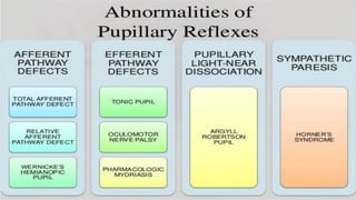
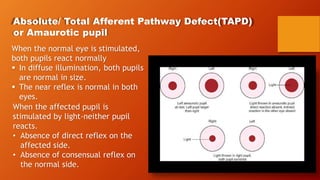
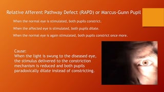
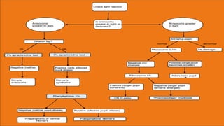
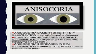
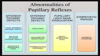
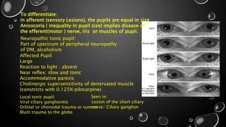
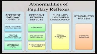
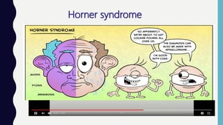
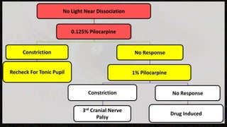
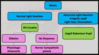
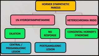
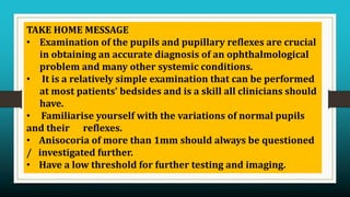
 Anatomy
Pupillary reflexes
 Systemic examination of pupil
 Abnormality Of Pupillary Reflexes
Pharmacology Of Pupil
Why?
To find disorders of the pupillary function.
To detect disorders of the afferent visual system and
autonomic innervation of eye.
 A systemic approach for interpretation of the findings.
The examination should be done in a logical order
since the pupillary system responds in a logically
predictable way
Pupil: Aperture in the centre of Iris
Number :
Normal : One pupil In each eye
Abnormal : More than one pupil in
each eye (polycoria)
True Polycoria:
Each Pupil will have its own, intact
sphincter muscle. Each pupil will
individually constrict and dilate.
False or pseudopolycoria:
Don’t have separate sphincter
muscles.
Holes in Iris look like
additional pupils
Location:
Normal: Almost in the centre (slightly nasal) of iris.
Abnormal: Placed Eccentrically (Eccentric Pupil)
(correctopia)
Sector Iris HypoplasiaColobomatous lesions
Correctopia+ Lens subluxation:
Ectopia lentis et pupillae
Shape:
Normal: Circular
Abnormal: Tadpole shaped Pupil
Hypersympathetic activity affects only a portion of the pupillary dilators, the pupil loses its normal, circular shape.
Irregular oval shape or a circle with a narrow segment that extends in the direction of the affected dilators.
Pear shaped in
adherent leukoma
Physiological changes in size:
Pupils tend to dilate during
emotional stress and constrict
during sleep.
Pupillary unrest:
Refers to the constant fluctuation
in pupillary diameter under
normal environmental condition.
Hippus(jumping Pupil);
Exaggeration of the pupillary
unrest i.e. fluctuation in
pupillary diameter which can be
easily detected on visual
inspection without
magnification.
Abnormal:
Congenital Miosis:
tapetoretinal degenrations can be isolated finding.
Persistent Mydriasis:
Develops during the second or third decade.
Anisocoria:
Difference in the size of two pupils (cor: pupil, aniso: difference)
COLOUR
Functions:
Limits the amount of light reaching the retina.
Controls the amount of chromaticand spheric aberration in the
retinal image.
 Allows the passage of aqueous humor from the anterior to the
posterior chamber.
Pupillary reflexes
Light reflex
Darkness
reflex
Psychosensor
y reflex
Near reflex Lid closure reflex
Pupillary light reaction
When light is shown in one eye, both pupils constrict.
Constriction of the pupil to which light is shown Direct Light Reflex
Constriction of the other pupil Consensual (indirect) light reflex
Why?
 Pupillary constriction in response to light protects against excessive bleaching of the
visual pigment by reducing the amount of light entering the eye.
 Helps in light and dark adaptation, thus maximises visual acuity at different light
levels.
Darkness reflex
Psychosensory reflex
Dilatation of pupil in response to sensory and psychic stimuli
Fully developed by six months of age
Pathways are unknown
Components:
Sympathetic discharge to dilator pupillae muscle
Inhibition of parasympathetic discharge to sphincter pupillae muscle
Performing the exam
Tip 1: Seating Position
Ask the patient to sit comfortably on a chair while you sit on one side of the patient & never directly in front
of the patient. Sit to the side to avoid obstructing the patient’s view when you ask the patient to fixate at a
distant target.
Tip 2: Fixation
Ask the patient to maintain fixation on a distant target. The pupils constrict when
the eyes focus at near. The patient must look at a distant target in order to prevent
accommodation associated miosis that can confound your pupil exam.
Tip 3: Room Lighting
Examination of pupil should be done in a room with dim illumination in order to avoid
constriction caused by a brightly lit room.
Tip 4: Illumination
Use an evenly bright source of illumination for examining the light reflexes. Lights
with uneven illumination patterns may cause variable constriction of the pupils.
Tip 5: Dark Irises
It is often difficult to view a consensual pupillary response in patients with dark
irises in dark rooms. In these cases, you can use a dim secondary light held below
the pupils in addition to your bright primary light that you use to elicit the pupillary
response. If your secondary light is too bright it will cause undesirable constriction
of the pupil.
Systemic Examination Of the Pupils
Step 1: Confirm that the pupils respond to light
Dim Light
Ask the patient to look into distance to limit intrusion by miosis of the near reflex.
Using a strong light source, stimulate both the eyes simultaneously.
Both pupils will constrict symmetrically.
Step 2: Compare the Pupillary diameters to one another.
Determines whether the autonomic (efferent) innervation of the eye is intact.
If there is anisocoria, repeat testing of both pupil’s responses to a strong, binocular
light stimulus.
Step 3: Swinging flashlight test
Compares the afferent pupillary responses of one eye to the other.
Step 4: Examination of the pathologic findings
Pathologic states are:
 A relative afferent pupillary defect (RAPD)
 An anisocoria with normal responses to light in both eyes.
 A monocular or bilateral deficit in light responses.
Absolute/ Total Afferent Pathway Defect(TAPD)
or Amaurotic pupil
When the normal eye is stimulated,
both pupils react normally
 In diffuse illumination, both pupils
are normal in size.
 The near reflex is normal in both
eyes.
When the affected pupil is
stimulated by light-neither pupil
reacts.
• Absence of direct reflex on the
affected side.
• Absence of consensual reflex on
the normal side.
Cause:
Due to lesions of the optic nerve , the affected eye is completed
blind ( no perception of light)
Seen in: Complete optic nerve lesions( Optic atrophy)
• Ischaemic optic neuropathy
• Glaucomatous optic neuropathy
• Acute optic neuritis
• Traumatic optic nerve avulsion
Relative Afferent Pathway Defect (RAPD) or Marcus-Gunn Pupil
When the normal eye is stimulated, both pupils constrict.
When the affected eye is stimulated, both pupils dilate.
When the normal eye is again stimulated, both pupils constrict once more.
Cause:
When the light is swung to the diseased eye,
the stimulus delivered to the constriction
mechanism is reduced and both pupils
paradoxically dilate instead of constricting.
Seen in :
Optic Nerve disorders:
 Optic Neuritis
 Ischemic optic neuropathies- AION/NAAION
 Glaucoma: If severe and unilateral disc involvement.
 Traumatic optic neuropathy
 Compressive optic neuropathy
 Surgical damage to Optic nerve
 Heritable: LHON
 Radiation optic nerve damage
Retinal Disorders:
 RD (if macula is detached, or if atleast two quadrants of retina are detached)
 OIS
 Ischemic CRVO/Severe BRVO
 CRAO
 Unilateral Severe ARMD
 Intraocular tumors
Grades of RAPD
Grades of RAPD Observation
I Weak initial constriction and greater redilatation.
II Initial stall and greater redilatation
III Immediate pupillary dilatation
IV Immediate pupillary dilatation following prolonged
illumination of the good eye for 6 seconds.
V Immediate pupillary dilatation with no secondary
constriction
RAPD
When an RAPD is found, the cause must be identified.
If the cause cannot be found, perimetric examination of both eyes is necessary
Wernicke’s Hemianopic Pupil
Light reflex( ipsilateral direct and contralateral consensual)
 absent when light is thrown on the temporal half of the retina of the
affected side and nasal half of the retina of the opposite side.
 Present when light is thrown on the nasal half of the affected side
and temporal half of the opposite side.
Cause:
 The temporal visual field is larger than the nasal field.
 Temporal retina has more pupillomotor sensitivity than nasal retina.
Seen in:
Lesions of Optic Tract
To differentiate:
In afferent (sensory Lesions), the pupils are equal in size
Anisocoria ( inequality in pupil size) implies disease of
the efferent(motor ) nerve, iris or muscles of pupil.
Lesion of the short ciliary
nerve/ Ciliary ganglion
Affected Pupil
Large
Reaction to light : absent
Near reflex: slow and tonic
Accommodative paresis
Cholinergic supersensitivity of denervated muscle
(constricts with 0.125% pilocarpine)
Seen in:Local tonic pupil:
Viral ciliary ganglionitis
Orbital or choroidal trauma or tumors
Blunt trauma to the globe
Neuropathic tonic pupil:
Part of spectrum of peripheral neuropathy
of DM, alcoholism
Testing with pilocarpine 1% and 0.1%
Pilocarpine 0.1%
Indication: Diagnosis of a
tonic pupil is suspected
The tonic pupil has a
characteristic denervation
hypersensitivity to a
cholinergic stimulus, which
can be associated with adie’s
syndrome or a paresis of the
oculomotor nerve.
Pilocarpine 1%
Indications:
Light
Maximal accommodative effort or
Weak pilocarpine will not cause the
pupil to constrict.
Providing a drug induced
mydriasis, if an anticholinergic
drug( such as atropine or
scopolamine) has produced a
pharmacologic dilatation.
Fails to constrict pupil
Problem within the iris and pupil
itself
Pupillary miosis
Oculomotor palsy
Pupillary involvement in oculomotor paralysis
Cause: Damage to the third cranial nerve at locations between the oculomotor nucleus and the ciliary
ganglion.
Pupillary mydriasis: Internal ophthalmoplegia
Compressive mechanism, such as by a tumour or an aneurysm
No pupillary company with a progressive loss of third nerve function : primary aberrancy
Sign of a slowly growing mass lesion in the cavernous sinus.
Adie’s Tonic pupil Argyll Robertson pupil
Unilateral Bilateral
Dilated pupil Small, irregular pupils
Accommodation reflec slow and prolonged Accommodation reflex present
Absent or sluggish light reflex Absent light reflex
Cholinergic hypersensitivity No such phenomenon
Horner syndrome
 Monocular loss of sympathetic innervation to the eye.
 Loss of function in all of the ocular structures that are sympathetically controlled.
 Pupil is smaller, but the light reaction remains normal.
 A ptosis of the upper lid, caused by paresis of muller’s muscle.
 A small elevation of the lower lid.
 Apparent enophthalmos due to narrowing of the palpebral fissure.
 Impaired sweating and temperature regulation in the face: if the site of damage
to the sympathetic path lies proximal to the branching of the fibres.
1% Hydroxyamphetamine, 2.5% tyramine test
Stimulating the release of noradrenalin into the
synaptic cleft at the terminus of the end neuron of the
sympathetic chain.
Measure diameter of both pupils:
Before 45 min after instillation of the drops.
Unaffected pupil dilates well and the affected pupil
dilates by 0.5mm or less.
Site of damage
Third (terminal) neuron of the sympathetic pathway, i.e.
at or above the ganglion cervicale superius of the
sympathetic chain.
(post ganglionic)
Affected pupil dilates as well or better than its
contralateral partner.
Site of Damage
Below the level of the ganglion cervicale superius, i.e. in the first
or second order neurons of the sympathetic pathway. (pre
ganglionic)
Abnormal Pupil
Mydriasis
Normal light
reaction
Abnormal light
reaction
Physiologic
Anisocoria
Light Near
Dissociation
No Light Near
Dissociation
Miosis
Unilateral
Parinaud Syndrome
Bilateral Adie’s
Pupil
Unilateral Adie’s
Pupil
• Convergence retraction Nystagmus
• Vertical Gaze Deficit
• Convergence Deficit
• Lid Retraction
• Segmental Pupillary Constriction
• Impaired Dark Adaptation
• Impaired Near Reflex
• Tonic Redilation
Bilateral
Light Near Dissociation
Constriction
Recheck For Tonic Pupil
Constriction
3rd Cranial Nerve
Palsy
Drug Induced
No Response
1% Pilocarpine
No Response
0.125% Pilocarpine
No Light Near Dissociation
4% Cocaine
Physiologic
Anisocoria
No ResponseDilation
Horner Sympathetic
paresis
Argyll Robertson Pupil
Abnormal Light Reaction
Irregular pupil
Light Near Dissociation
Normal Light Reaction
Miosis
HORNER SYMPATHETIC
PARESIS
1% HYDROXYAMPHETAMINE HETEROCHROMIA IRIDIS
CONGENITAL HORNER’S
SYNDROME
NO
RESPONSE
DILATION
POSTGANGLIONIC
LESION
CENTRAL /
PREGANGLIONIC
LESION
TAKE HOME MESSAGE
• Examination of the pupils and pupillary reflexes are crucial
in obtaining an accurate diagnosis of an ophthalmological
problem and many other systemic conditions.
• It is a relatively simple examination that can be performed
at most patients’ bedsides and is a skill all clinicians should
have.
• Familiarise yourself with the variations of normal pupils
and their reflexes.
• Anisocoria of more than 1mm should always be questioned
/ investigated further.
• Have a low threshold for further testing and imaging.
PUPIL AND ITS DISORDERS

PUPIL AND ITS DISORDERS

  • 2.
     Anatomy Pupillary reflexes Systemic examination of pupil  Abnormality Of Pupillary Reflexes Pharmacology Of Pupil
  • 3.
    Why? To find disordersof the pupillary function. To detect disorders of the afferent visual system and autonomic innervation of eye.  A systemic approach for interpretation of the findings. The examination should be done in a logical order since the pupillary system responds in a logically predictable way
  • 4.
    Pupil: Aperture inthe centre of Iris Number : Normal : One pupil In each eye Abnormal : More than one pupil in each eye (polycoria)
  • 5.
    True Polycoria: Each Pupilwill have its own, intact sphincter muscle. Each pupil will individually constrict and dilate. False or pseudopolycoria: Don’t have separate sphincter muscles. Holes in Iris look like additional pupils
  • 6.
    Location: Normal: Almost inthe centre (slightly nasal) of iris. Abnormal: Placed Eccentrically (Eccentric Pupil) (correctopia) Sector Iris HypoplasiaColobomatous lesions Correctopia+ Lens subluxation: Ectopia lentis et pupillae
  • 7.
    Shape: Normal: Circular Abnormal: Tadpoleshaped Pupil Hypersympathetic activity affects only a portion of the pupillary dilators, the pupil loses its normal, circular shape. Irregular oval shape or a circle with a narrow segment that extends in the direction of the affected dilators.
  • 8.
  • 10.
    Physiological changes insize: Pupils tend to dilate during emotional stress and constrict during sleep. Pupillary unrest: Refers to the constant fluctuation in pupillary diameter under normal environmental condition. Hippus(jumping Pupil); Exaggeration of the pupillary unrest i.e. fluctuation in pupillary diameter which can be easily detected on visual inspection without magnification.
  • 11.
    Abnormal: Congenital Miosis: tapetoretinal degenrationscan be isolated finding. Persistent Mydriasis: Develops during the second or third decade. Anisocoria: Difference in the size of two pupils (cor: pupil, aniso: difference)
  • 12.
  • 13.
    Functions: Limits the amountof light reaching the retina. Controls the amount of chromaticand spheric aberration in the retinal image.  Allows the passage of aqueous humor from the anterior to the posterior chamber.
  • 14.
  • 15.
    Pupillary light reaction Whenlight is shown in one eye, both pupils constrict. Constriction of the pupil to which light is shown Direct Light Reflex Constriction of the other pupil Consensual (indirect) light reflex Why?  Pupillary constriction in response to light protects against excessive bleaching of the visual pigment by reducing the amount of light entering the eye.  Helps in light and dark adaptation, thus maximises visual acuity at different light levels.
  • 17.
  • 18.
    Psychosensory reflex Dilatation ofpupil in response to sensory and psychic stimuli Fully developed by six months of age Pathways are unknown Components: Sympathetic discharge to dilator pupillae muscle Inhibition of parasympathetic discharge to sphincter pupillae muscle
  • 19.
    Performing the exam Tip1: Seating Position Ask the patient to sit comfortably on a chair while you sit on one side of the patient & never directly in front of the patient. Sit to the side to avoid obstructing the patient’s view when you ask the patient to fixate at a distant target.
  • 20.
    Tip 2: Fixation Askthe patient to maintain fixation on a distant target. The pupils constrict when the eyes focus at near. The patient must look at a distant target in order to prevent accommodation associated miosis that can confound your pupil exam.
  • 21.
    Tip 3: RoomLighting Examination of pupil should be done in a room with dim illumination in order to avoid constriction caused by a brightly lit room.
  • 22.
    Tip 4: Illumination Usean evenly bright source of illumination for examining the light reflexes. Lights with uneven illumination patterns may cause variable constriction of the pupils.
  • 23.
    Tip 5: DarkIrises It is often difficult to view a consensual pupillary response in patients with dark irises in dark rooms. In these cases, you can use a dim secondary light held below the pupils in addition to your bright primary light that you use to elicit the pupillary response. If your secondary light is too bright it will cause undesirable constriction of the pupil.
  • 24.
    Systemic Examination Ofthe Pupils Step 1: Confirm that the pupils respond to light Dim Light Ask the patient to look into distance to limit intrusion by miosis of the near reflex. Using a strong light source, stimulate both the eyes simultaneously. Both pupils will constrict symmetrically. Step 2: Compare the Pupillary diameters to one another. Determines whether the autonomic (efferent) innervation of the eye is intact. If there is anisocoria, repeat testing of both pupil’s responses to a strong, binocular light stimulus.
  • 25.
    Step 3: Swingingflashlight test Compares the afferent pupillary responses of one eye to the other.
  • 26.
    Step 4: Examinationof the pathologic findings Pathologic states are:  A relative afferent pupillary defect (RAPD)  An anisocoria with normal responses to light in both eyes.  A monocular or bilateral deficit in light responses.
  • 28.
    Absolute/ Total AfferentPathway Defect(TAPD) or Amaurotic pupil When the normal eye is stimulated, both pupils react normally  In diffuse illumination, both pupils are normal in size.  The near reflex is normal in both eyes. When the affected pupil is stimulated by light-neither pupil reacts. • Absence of direct reflex on the affected side. • Absence of consensual reflex on the normal side.
  • 29.
    Cause: Due to lesionsof the optic nerve , the affected eye is completed blind ( no perception of light) Seen in: Complete optic nerve lesions( Optic atrophy) • Ischaemic optic neuropathy • Glaucomatous optic neuropathy • Acute optic neuritis • Traumatic optic nerve avulsion
  • 30.
    Relative Afferent PathwayDefect (RAPD) or Marcus-Gunn Pupil When the normal eye is stimulated, both pupils constrict. When the affected eye is stimulated, both pupils dilate. When the normal eye is again stimulated, both pupils constrict once more. Cause: When the light is swung to the diseased eye, the stimulus delivered to the constriction mechanism is reduced and both pupils paradoxically dilate instead of constricting.
  • 32.
    Seen in : OpticNerve disorders:  Optic Neuritis  Ischemic optic neuropathies- AION/NAAION  Glaucoma: If severe and unilateral disc involvement.  Traumatic optic neuropathy  Compressive optic neuropathy  Surgical damage to Optic nerve  Heritable: LHON  Radiation optic nerve damage Retinal Disorders:  RD (if macula is detached, or if atleast two quadrants of retina are detached)  OIS  Ischemic CRVO/Severe BRVO  CRAO  Unilateral Severe ARMD  Intraocular tumors
  • 33.
    Grades of RAPD Gradesof RAPD Observation I Weak initial constriction and greater redilatation. II Initial stall and greater redilatation III Immediate pupillary dilatation IV Immediate pupillary dilatation following prolonged illumination of the good eye for 6 seconds. V Immediate pupillary dilatation with no secondary constriction RAPD When an RAPD is found, the cause must be identified. If the cause cannot be found, perimetric examination of both eyes is necessary
  • 34.
    Wernicke’s Hemianopic Pupil Lightreflex( ipsilateral direct and contralateral consensual)  absent when light is thrown on the temporal half of the retina of the affected side and nasal half of the retina of the opposite side.  Present when light is thrown on the nasal half of the affected side and temporal half of the opposite side. Cause:  The temporal visual field is larger than the nasal field.  Temporal retina has more pupillomotor sensitivity than nasal retina. Seen in: Lesions of Optic Tract
  • 38.
    To differentiate: In afferent(sensory Lesions), the pupils are equal in size Anisocoria ( inequality in pupil size) implies disease of the efferent(motor ) nerve, iris or muscles of pupil. Lesion of the short ciliary nerve/ Ciliary ganglion Affected Pupil Large Reaction to light : absent Near reflex: slow and tonic Accommodative paresis Cholinergic supersensitivity of denervated muscle (constricts with 0.125% pilocarpine) Seen in:Local tonic pupil: Viral ciliary ganglionitis Orbital or choroidal trauma or tumors Blunt trauma to the globe Neuropathic tonic pupil: Part of spectrum of peripheral neuropathy of DM, alcoholism
  • 42.
    Testing with pilocarpine1% and 0.1% Pilocarpine 0.1% Indication: Diagnosis of a tonic pupil is suspected The tonic pupil has a characteristic denervation hypersensitivity to a cholinergic stimulus, which can be associated with adie’s syndrome or a paresis of the oculomotor nerve. Pilocarpine 1% Indications: Light Maximal accommodative effort or Weak pilocarpine will not cause the pupil to constrict. Providing a drug induced mydriasis, if an anticholinergic drug( such as atropine or scopolamine) has produced a pharmacologic dilatation. Fails to constrict pupil Problem within the iris and pupil itself Pupillary miosis Oculomotor palsy
  • 43.
    Pupillary involvement inoculomotor paralysis Cause: Damage to the third cranial nerve at locations between the oculomotor nucleus and the ciliary ganglion. Pupillary mydriasis: Internal ophthalmoplegia Compressive mechanism, such as by a tumour or an aneurysm No pupillary company with a progressive loss of third nerve function : primary aberrancy Sign of a slowly growing mass lesion in the cavernous sinus.
  • 47.
    Adie’s Tonic pupilArgyll Robertson pupil Unilateral Bilateral Dilated pupil Small, irregular pupils Accommodation reflec slow and prolonged Accommodation reflex present Absent or sluggish light reflex Absent light reflex Cholinergic hypersensitivity No such phenomenon
  • 49.
    Horner syndrome  Monocularloss of sympathetic innervation to the eye.  Loss of function in all of the ocular structures that are sympathetically controlled.  Pupil is smaller, but the light reaction remains normal.  A ptosis of the upper lid, caused by paresis of muller’s muscle.  A small elevation of the lower lid.  Apparent enophthalmos due to narrowing of the palpebral fissure.  Impaired sweating and temperature regulation in the face: if the site of damage to the sympathetic path lies proximal to the branching of the fibres.
  • 50.
    1% Hydroxyamphetamine, 2.5%tyramine test Stimulating the release of noradrenalin into the synaptic cleft at the terminus of the end neuron of the sympathetic chain. Measure diameter of both pupils: Before 45 min after instillation of the drops. Unaffected pupil dilates well and the affected pupil dilates by 0.5mm or less. Site of damage Third (terminal) neuron of the sympathetic pathway, i.e. at or above the ganglion cervicale superius of the sympathetic chain. (post ganglionic) Affected pupil dilates as well or better than its contralateral partner. Site of Damage Below the level of the ganglion cervicale superius, i.e. in the first or second order neurons of the sympathetic pathway. (pre ganglionic)
  • 54.
    Abnormal Pupil Mydriasis Normal light reaction Abnormallight reaction Physiologic Anisocoria Light Near Dissociation No Light Near Dissociation Miosis
  • 55.
    Unilateral Parinaud Syndrome Bilateral Adie’s Pupil UnilateralAdie’s Pupil • Convergence retraction Nystagmus • Vertical Gaze Deficit • Convergence Deficit • Lid Retraction • Segmental Pupillary Constriction • Impaired Dark Adaptation • Impaired Near Reflex • Tonic Redilation Bilateral Light Near Dissociation
  • 56.
    Constriction Recheck For TonicPupil Constriction 3rd Cranial Nerve Palsy Drug Induced No Response 1% Pilocarpine No Response 0.125% Pilocarpine No Light Near Dissociation
  • 57.
    4% Cocaine Physiologic Anisocoria No ResponseDilation HornerSympathetic paresis Argyll Robertson Pupil Abnormal Light Reaction Irregular pupil Light Near Dissociation Normal Light Reaction Miosis
  • 58.
    HORNER SYMPATHETIC PARESIS 1% HYDROXYAMPHETAMINEHETEROCHROMIA IRIDIS CONGENITAL HORNER’S SYNDROME NO RESPONSE DILATION POSTGANGLIONIC LESION CENTRAL / PREGANGLIONIC LESION
  • 59.
    TAKE HOME MESSAGE •Examination of the pupils and pupillary reflexes are crucial in obtaining an accurate diagnosis of an ophthalmological problem and many other systemic conditions. • It is a relatively simple examination that can be performed at most patients’ bedsides and is a skill all clinicians should have. • Familiarise yourself with the variations of normal pupils and their reflexes. • Anisocoria of more than 1mm should always be questioned / investigated further. • Have a low threshold for further testing and imaging.