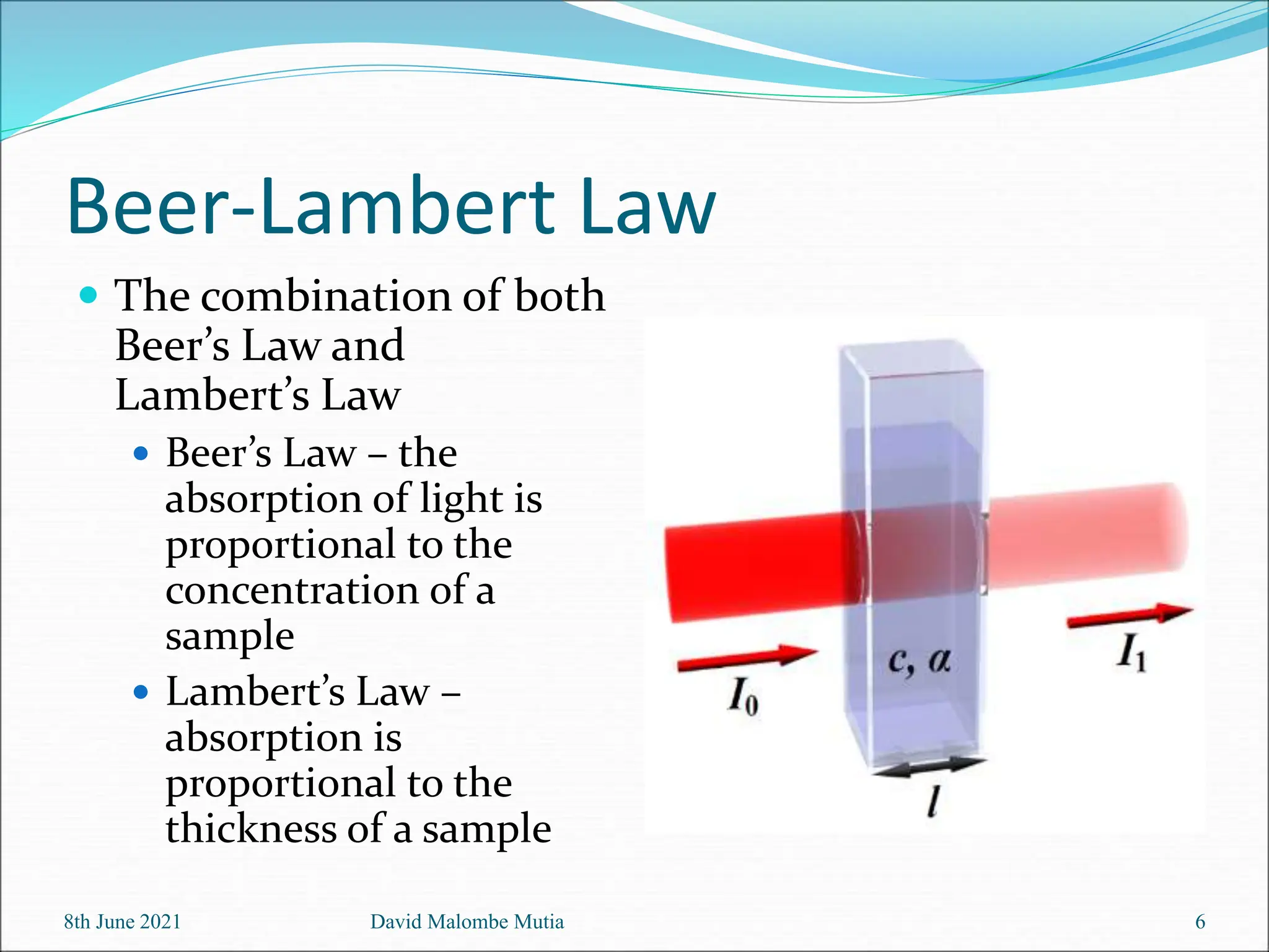
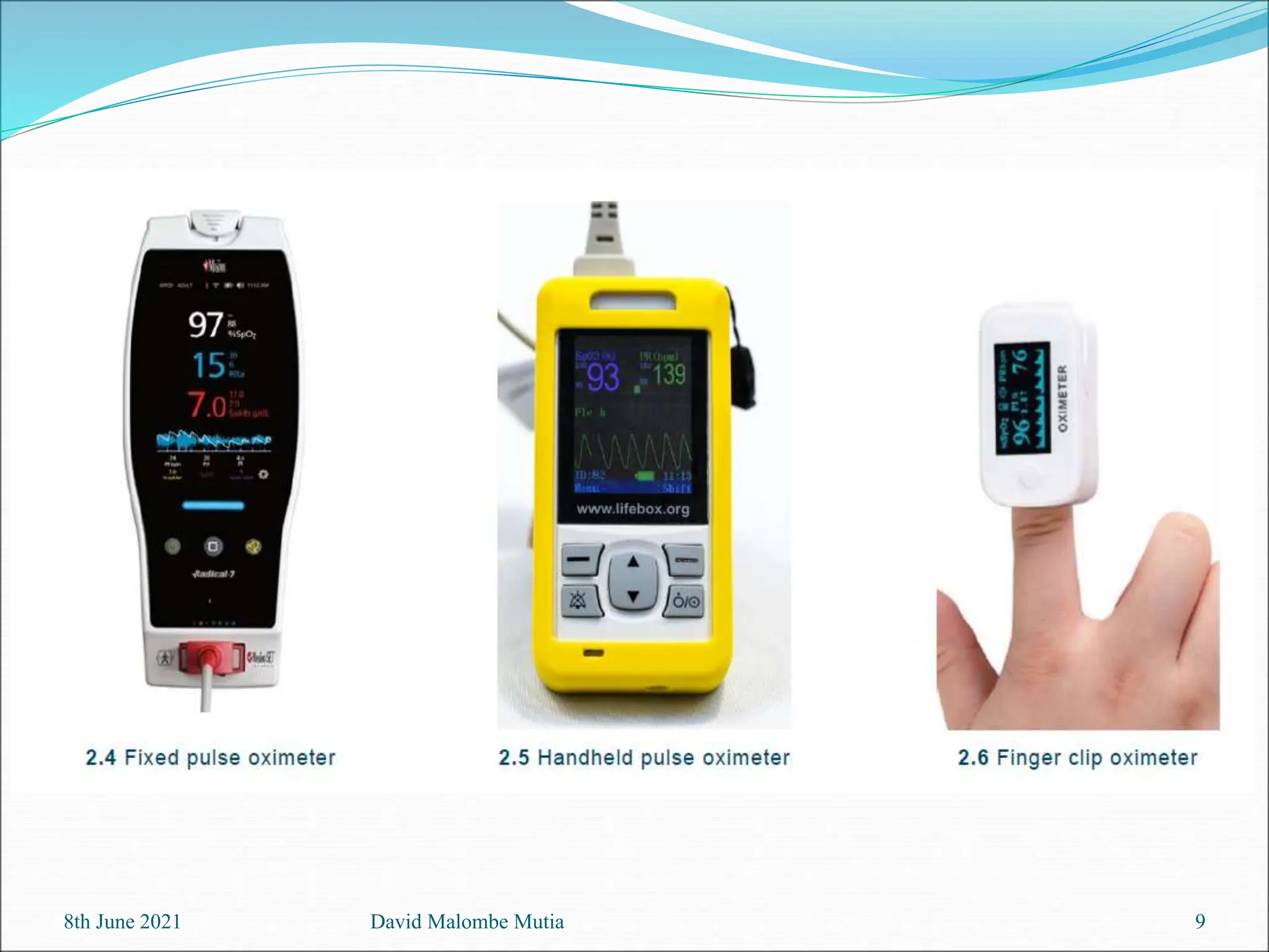
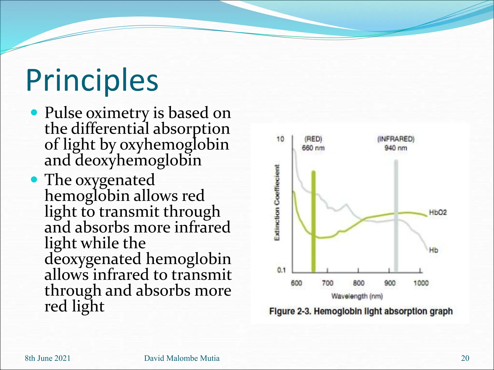
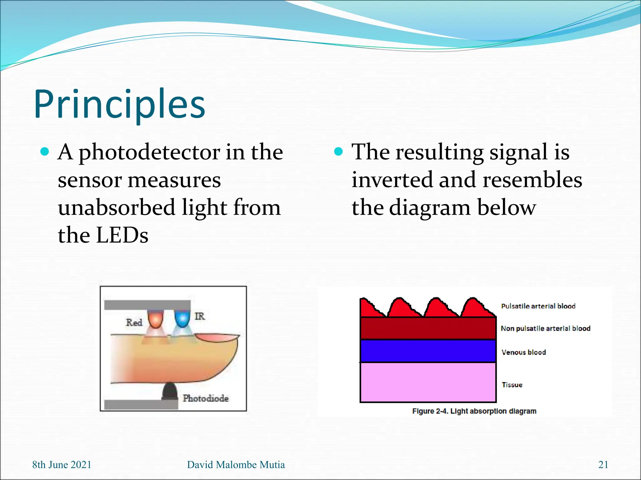
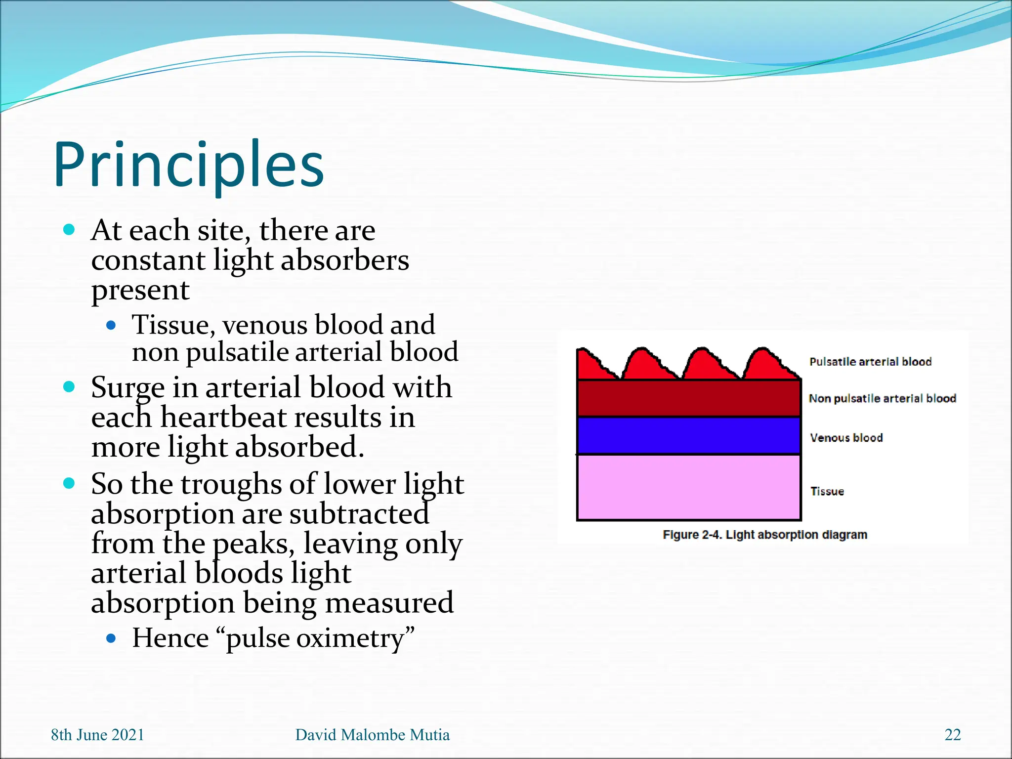
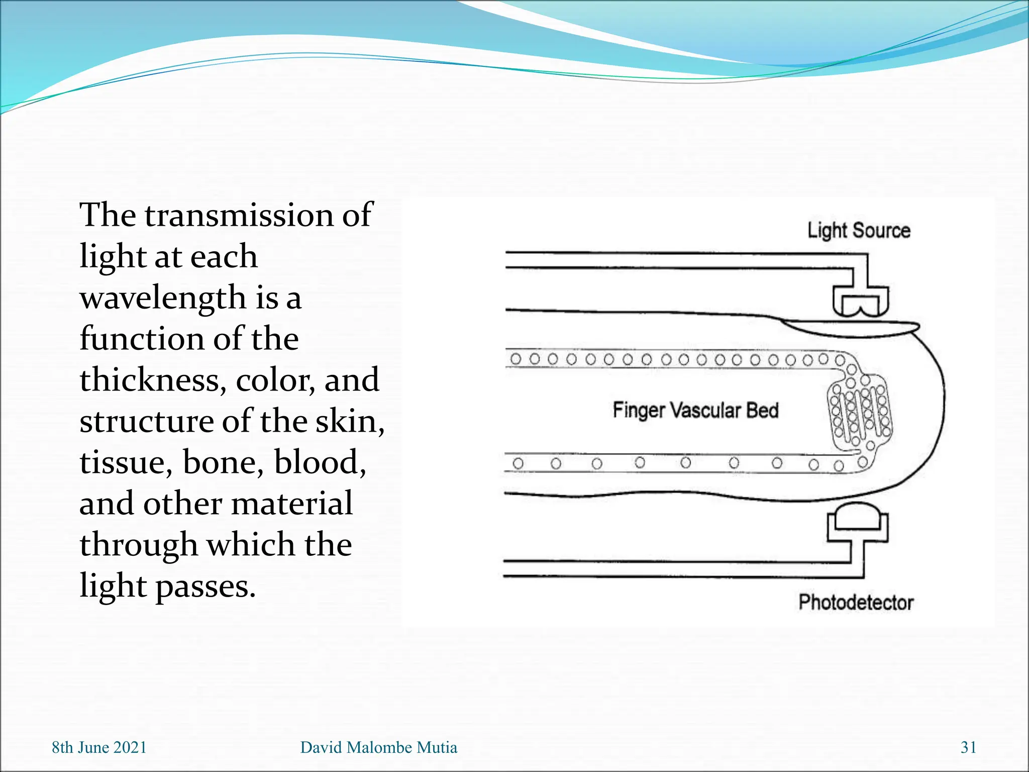
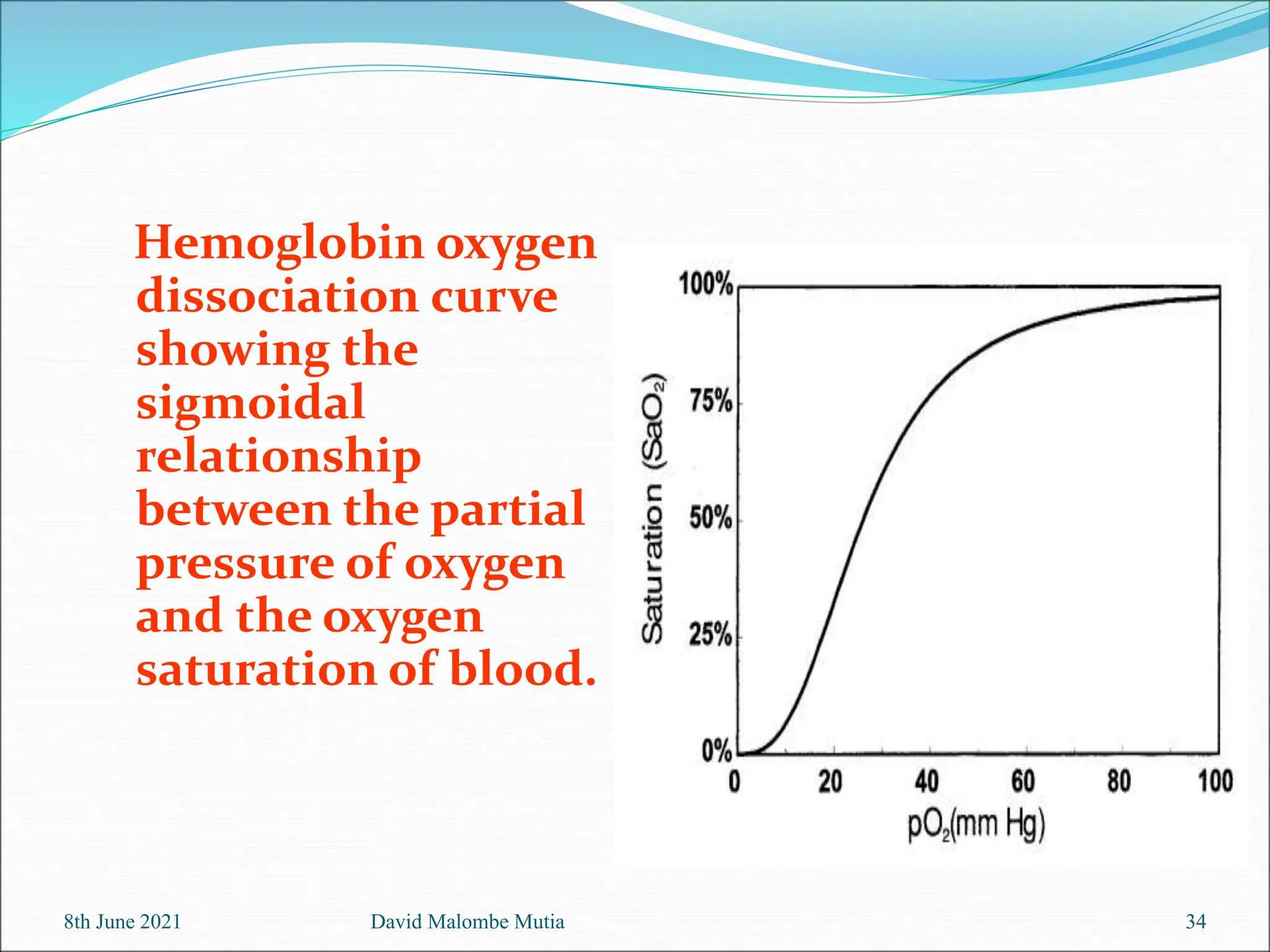
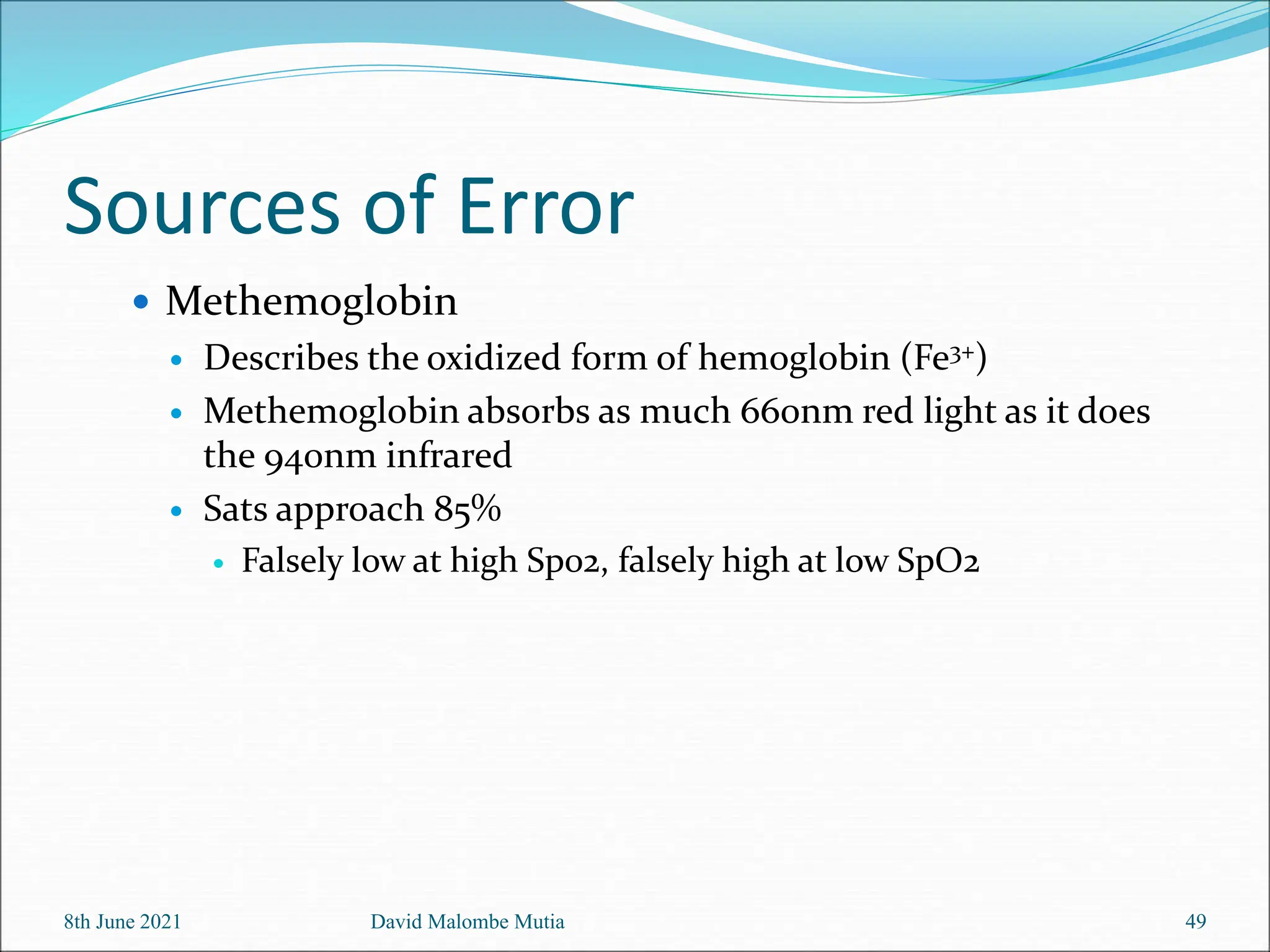
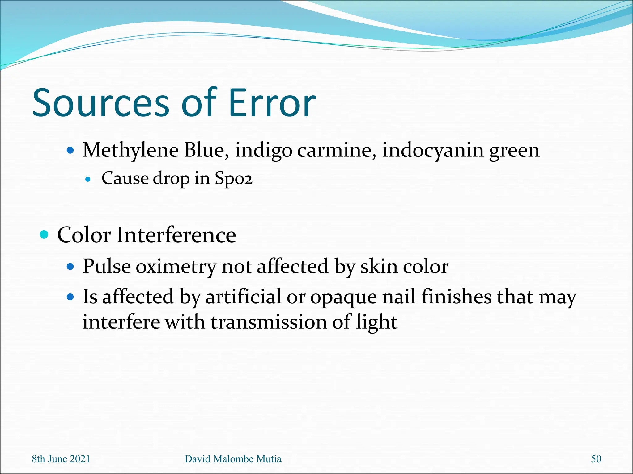
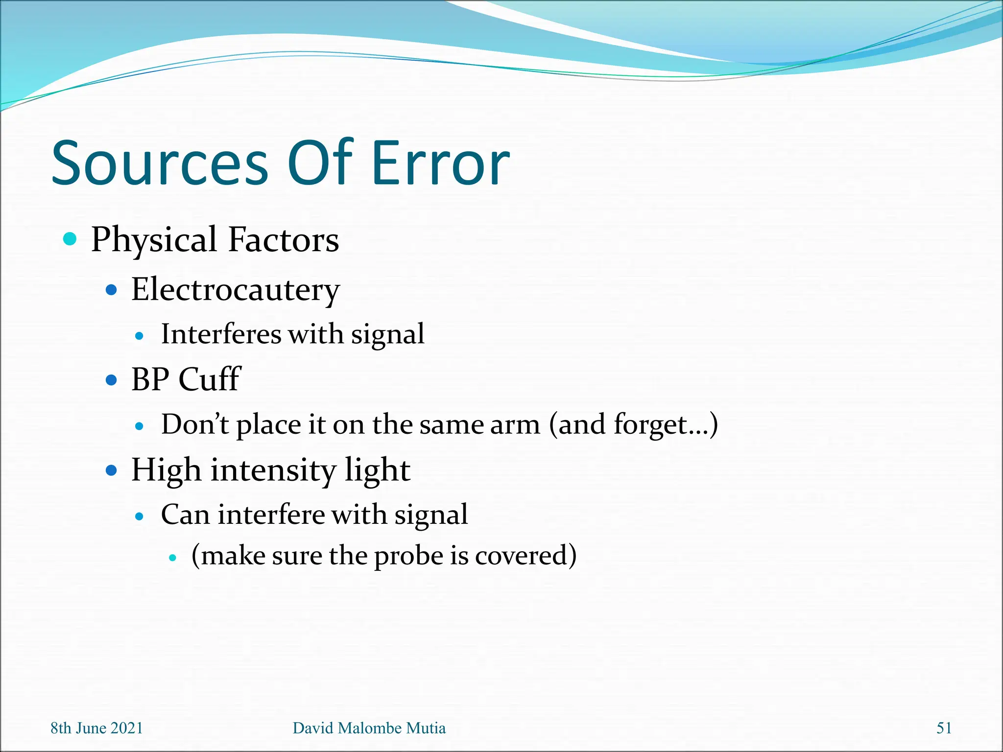
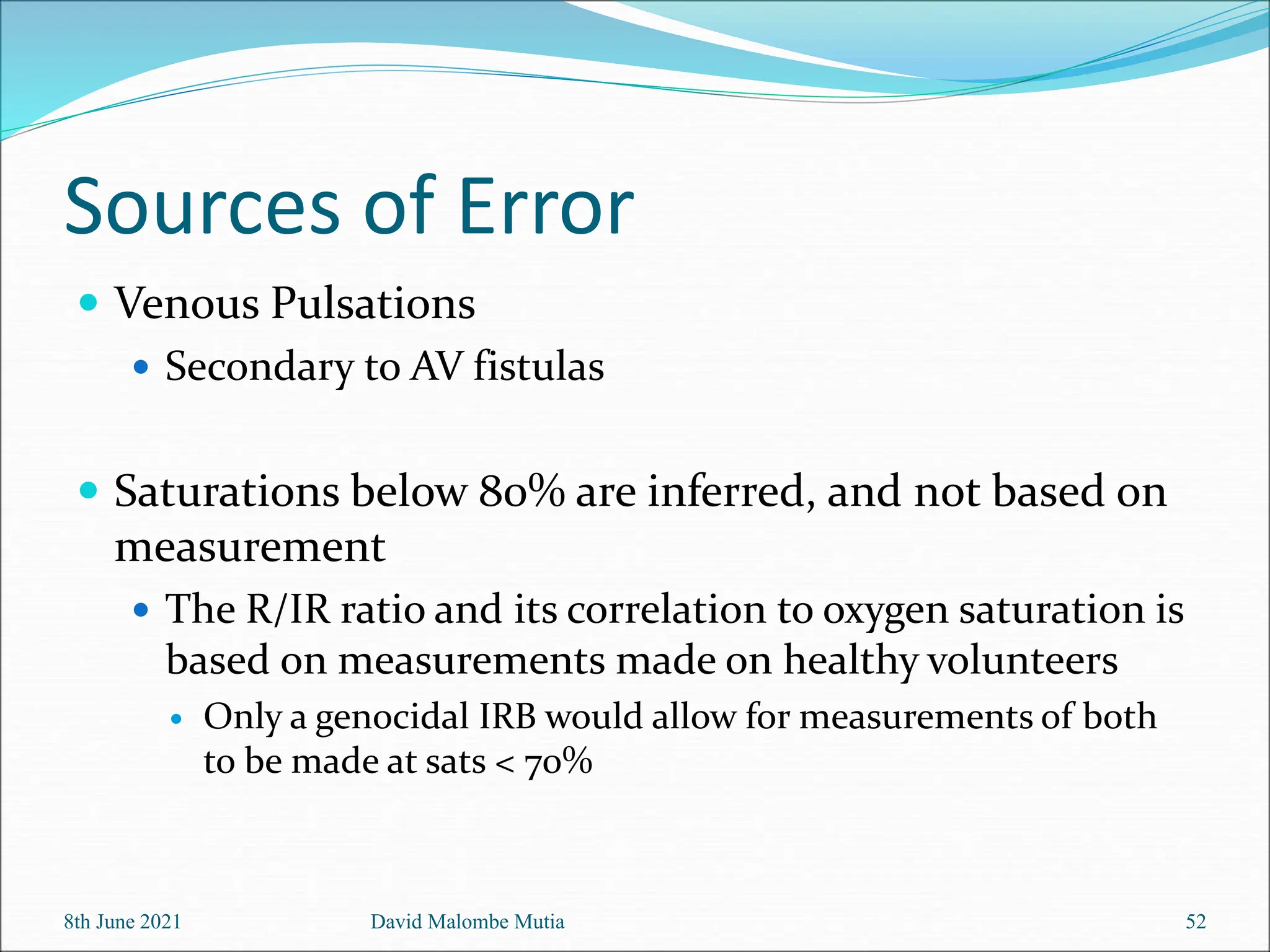
8th June 2021 David Malombe Mutia 1
DIAGNOSTIC EQUIPMENT
OXIMETRY AND PULSE
OXIMETRY
Contents
 Aims & Objectives
 Introduction
 History
 Anatomy and Physiology
 Principle
 Application
 Operation
 Maintenance & Cleaning
 Safety Precautions
 Troubleshooting
8th June 2021 David Malombe Mutia 2
Aims
 To provide basic
understanding on
the use of pulse
oximetry
 To perform and
understand the basic
user’s troubleshooting
steps
8th June 2021 David Malombe Mutia 3
Objectives
- as a result of completing this module, the user should be able to:
 describe what an pulse
oximetry is including its
applications
 Perform basic user
maintenance
 perform and identify basic
problems, errors and basic
troubleshooting solutions.
8th June 2021 David Malombe Mutia 4
Beer’s Law
8th June 2021 David Malombe Mutia 5
Beer-Lambert Law
 The combination of both
Beer’s Law and
Lambert’s Law
 Beer’s Law – the
absorption of light is
proportional to the
concentration of a
sample
 Lambert’s Law –
absorption is
proportional to the
thickness of a sample
8th June 2021 David Malombe Mutia 6
Introduction: What is Pulse Oximetry?
 Oximetry is the determination of the oxygen
content of blood of tissues, normally by optical
means.
 In the clinical laboratory the oxygen content of
whole blood can be determined by a bench-top
cooximeter or blood gas analyzer.
8th June 2021 David Malombe Mutia 7
 Pulse oximeters may be used to help determine the
severity of an infant’s illness by evaluating if blood
oxygen saturation is low and if respiratory support is
needed.
 Pulse oximeters also may be used to assess the success
of treatment and determine a need for increasing or
decreasing respiratory interventions to achieve
target SpO2.
8th June 2021 David Malombe Mutia 8
8th June 2021 David Malombe Mutia 9
8th June 2021 David Malombe Mutia 10
To be sure the reported SpO2 is accurate, it is important to ensure the
patient signal is both strong and stable. Patient movement, blood flow
and external sources of light may impact the stability of the patient’s
trace.
Examples of a “normal signal” and potential “poor traces”
are below
Functions
 Pulse oximetry is the most significant technological
advance ever made in monitoring the well-being and
safety of patients during anesthesia, recovery and
critical care
 Pulse oximetry is an extremely easy to-use,
noninvasive, and accurate measurement of real-time
arterial oxygen saturation.
8th June 2021 David Malombe Mutia 11
 Normal SpO2 for neonatal patients should be:
▪ 90%–100% if not on oxygen
▪ 90–95% on oxygen (Alert 2.1)
 If SpO2 readings are less than 90%, the patient should
be considered for supplemental oxygen therapy (see
oxygen concentrator module). Oxygen saturations,
heart rate, and clinical condition should all
correspond.
8th June 2021 David Malombe Mutia 12
Basic Construction
 Pulse oximetry is an extremely easy to-use,
noninvasive, and accurate measurement of real-
time arterial oxygen saturation.
 Pulse oximeter sensors consist of a pair of small
and inexpensive R and IR LEDs and a highly
sensitive silicon photodiode. These components
are mounted inside a reusable rigid spring-loaded
clip, a flexible probe, or a disposable adhesive
wrap.
8th June 2021 David Malombe Mutia 13
History
 Karl Matthes – 1935
 First oximeter to measure O2 saturation
 Subsequent oximeters developed by Hewlett Packard were
bulky and expensive ($10,000)
 1972 – Takuo Aoyagi
 Pulsatile changes in absorption of red and infra-red light to
measure arterial oxygen saturation
 BiOx, Nellcor (started by anesthesiologist Bill New) and
Novametrix began manufacturing in 1980’s
 1987 – ASA recommends inclusion of pulse oximetry and
capnography into operating room as standard of care
 Prior to this, morbidity and mortality related to hypoxemia
estimated at 1/2000 – 1/7000 cases
8th June 2021 David Malombe Mutia 14
History
1970’s First modern pulse oximetry was
invented.
1980 Its commercial development and
application and noninvasive oximetry became
practical.
1986 The explosive growth of this new technology
and its considerable utility led John Severinghaus
and Poul Astrup
8th June 2021 David Malombe Mutia 15
Questions!!!
8th June 2021 David Malombe Mutia 16
HOW IT WORKS
 Red blood cells contain hemoglobin, which carries
oxygen. Oxygen is carried in the bloodstream by
binding to hemoglobin in the red blood cells.
 Each hemoglobin can carry four oxygen molecules
and at that point becomes 100% saturated.
8th June 2021 David Malombe Mutia 17
 The color of blood depends on how much hemoglobin
is saturated with oxygen. Hemoglobin that is carrying
oxygen is called oxygenated hemoglobin
(oxyhemoglobin) and appears bright red while
deoxygenated hemoglobin (deoxyhemoglobin)
appears dark red.
 Pulse oximeters are able to differentiate between the
light absorbing properties of oxygenated hemoglobin
and deoxygenated hemoglobin, therefore calculating
blood oxygen saturation of pulsating arterial
blood.
8th June 2021 David Malombe Mutia 18
8th June 2021 David Malombe Mutia 19
Pulse oximetry is based on two principles:
 The difference in light absorbance by
hemoglobin at two different wavelengths in red
spectrum due to the degree of oxygenation
(deoxyhaemoglobin and oxyhaemoglobin).
 The pulsatile nature of light signals coming from
the arterial blood component of body tissue due
to changes in local blood volume resulting from
heartbeat pulses.
Principles
 Pulse oximetry is based on
the differential absorption
of light by oxyhemoglobin
and deoxyhemoglobin
 The oxygenated
hemoglobin allows red
light to transmit through
and absorbs more infrared
light while the
deoxygenated hemoglobin
allows infrared to transmit
through and absorbs more
red light
8th June 2021 David Malombe Mutia 20
Principles
 A photodetector in the
sensor measures
unabsorbed light from
the LEDs
 The resulting signal is
inverted and resembles
the diagram below
8th June 2021 David Malombe Mutia 21
Principles
 At each site, there are
constant light absorbers
present
 Tissue, venous blood and
non pulsatile arterial blood
 Surge in arterial blood with
each heartbeat results in
more light absorbed.
 So the troughs of lower light
absorption are subtracted
from the peaks, leaving only
arterial bloods light
absorption being measured
 Hence “pulse oximetry”
8th June 2021 David Malombe Mutia 22
Principles
 After the photodetector, the Red/Infrared ratio is
calculated
 This is compared to an algorithm that is specific to each
company/device and is based on measurements
obtained in healthy volunteers
 This ratio corresponds to Sp02
 0.5 is approx 100%
 1 is approx 85%
 2 is approx 0%
8th June 2021 David Malombe Mutia 23
 The absorption of visible light by a haemoglobin
solution varies with oxygenation. This is because the
two common forms of the molecule, oxidised
haemoglobin (HbO2) and reduced haemoglobin (Hb),
have significantly different optical spectra in the
wavelength range from 500nm to 1000nm,
8th June 2021 David Malombe Mutia 24
8th June 2021 David Malombe Mutia 25
Principles of operation
 Pulse oximetry is based on the fractional
change in light transmission during an arterial
pulse at two different wavelengths.
 In this method the fractional change in the
signal is due only to the arterial blood itself,
and therefore the complicated nonpulsatile
and highly variable optical characteristics of
tissue are eliminated.
8th June 2021 David Malombe Mutia 26
 In a typical configuration, light at two different
wavelengths illuminating one side of a finger will
be detected on the other side, after having
traversed the intervening vascular tissues
8th June 2021 David Malombe Mutia 27
 By measuring the light transmitted through the
fingertip (or the earlobe) at two different
wavelengths, one in the red and the other in the
near infra−red part of the spectrum, the oxygen
saturation of the arterial blood in the finger (or
ear).
8th June 2021 David Malombe Mutia 28
 If we assume initially that the transmission of light
through the arterial bed is influenced only by the
relative concentrations of HbO2 and Hb and their
absorption coefficients at the two measurement
wavelengths, then the light intensity will decrease
logarithmically with path length according to the
well−known Beer−Lambert law.
8th June 2021 David Malombe Mutia 29
 When a monochromatic light passes through a colored
solution, the amount of light transmitted decreases
exponentially with the increase in concencentration of
the solution and with the increase in the thickness of
the layer of the solution through which the light passes
8th June 2021 David Malombe Mutia 30
The transmission of
light at each
wavelength is a
function of the
thickness, color, and
structure of the skin,
tissue, bone, blood,
and other material
through which the
light passes.
8th June 2021 David Malombe Mutia 31
 The transmission of light at each wavelength is a
function of the thickness, color, and structure of the
skin, tissue, bone, blood, and other material through
which the light passes.
 The absorbance of light by a sample is defined as the
negative logarithm of the ratio of the light intensity in
the presence of the sample (I) to that without (Io): A=
–log(I/Io)
8th June 2021 David Malombe Mutia 32
 Failure to provide adequate oxygen to tissues—
hypoxia—can in a matter of minutes result in
reduced work capacity of muscles, depressed
mental activity, and ultimately cell death.
8th June 2021 David Malombe Mutia 33
Hemoglobin oxygen
dissociation curve
showing the
sigmoidal
relationship
between the partial
pressure of oxygen
and the oxygen
saturation of blood.
8th June 2021 David Malombe Mutia 34
 The higher the pO2 in blood, the higher the SaO2. But
due to the highly cooperative binding of four oxygen
molecules to each hemoglobin molecule, the oxygen
binding curve is sigmoidal, and consequently the SaO2
value is particularly sensitive to dangerously low
pO2levels.
 With a normal arterial blood pO2 above 90 mmHg,
the oxygen saturation should be at least 95%, and a
pulse oximeter can readily verify a safe oxygen level.
8th June 2021 35
David Malombe Mutia
 If oxygen content falls, say to a pO2 below 40
mmHg, metabolic needs may not be met, and the
corresponding oxygen saturation will drop below
80%.
 Pulse oximetry therefore provides a indirect
measure of oxygen sufficiency and will alert the
clinician to any danger of imminent hypoxia in a
patient.
8th June 2021 David Malombe Mutia 36
Questions!!!
8th June 2021 David Malombe Mutia 37
 Noninvasive monitoring of SaO2 by pulse oximetry
is a rapidly growing practice in many fields of
clinical medicine.
 The most important advantage of this technique is
the capability to provide continuous, safe, and
effective monitoring of blood oxygenation.
8th June 2021 David Malombe Mutia 38
Application
 Pulse oximetry is also being used in the
monitoring of pulmonary disease in adults and in
the investigation of sleep disorders.
 For patients at risk of respiratory failure, it is
important to monitor the efficiency of gas
exchange in the lungs, ie how well the arterial
blood is oxygenated (as opposed to whether or not
air is going in and out of the lungs).
8th June 2021 David Malombe Mutia 39
 Both of these requirements can be met non−invasively with
the technology of pulse oximetry
8th June 2021 David Malombe Mutia 40
 Pulse oximetry relies on the detection of time-variant
photoplethysmographic (PPG) signals, caused by changes
in arterial blood volume associated with cardiac
contraction.
 The SaO2 is derived by analyzing the time-variant changes
in absorbance caused by the pulsating arterial blood at the
same R and IR wavelength used in conventional invasive-
type oximeters.
8th June 2021 David Malombe Mutia 41
 A normalization process is commonly performed
by which the pulsatile (ac) component at each
wavelength, which results from the expansion and
relaxation of the arterial bed, is divided by the
corresponding nonpulsatile (dc) component of the
PPG, which is composed of the light absorbed by
the blood-less tissue and the nonpulsatile portion
of the blood compartment.
8th June 2021 David Malombe Mutia 42
8th June 2021 David Malombe Mutia 43
This effective scaling process results in a normalized
R/IR ratio, which is dependent on SaO2, but is largely
independent of the incident light intensity, skin
pigmentation, tissue thickness, and other
nonpulsatile variables.
Questions!!!
8th June 2021 David Malombe Mutia 44
Uses
Arterial oxygenation, and, because the variation in light
absorption is proportional to the volume of arterial blood with
8th June 2021 David Malombe Mutia 45
Sources of Error
 Strength of Arterial Pulse
 Any factor that reduces arterial pulsations will reduce
the ability of the instrument to obtain and analyze the
signal
 Hypothermia
 Hypotension
 Vasopressor use
8th June 2021 David Malombe Mutia 46
Sources of Error
 Body Movement
 Extraneous movements can cause intermittent changes
in absorbance
 Shivering
 Parkinsonian tremors
8th June 2021 David Malombe Mutia 47
Sources of Error
 Dyshemoglobinemias
 Carboxyhemoglobin
 CO binds to heme competitively with 250 times the affinity of
oxygen
 COHgb has same absorption pattern of 660nm light as
O2Hgb
 Readings are artificially high
8th June 2021 David Malombe Mutia 48
Sources of Error
 Methemoglobin
 Describes the oxidized form of hemoglobin (Fe3+)
 Methemoglobin absorbs as much 660nm red light as it does
the 940nm infrared
 Sats approach 85%
 Falsely low at high Sp02, falsely high at low SpO2
8th June 2021 David Malombe Mutia 49
Sources of Error
 Methylene Blue, indigo carmine, indocyanin green
 Cause drop in Sp02
 Color Interference
 Pulse oximetry not affected by skin color
 Is affected by artificial or opaque nail finishes that may
interfere with transmission of light
8th June 2021 David Malombe Mutia 50
Sources Of Error
 Physical Factors
 Electrocautery
 Interferes with signal
 BP Cuff
 Don’t place it on the same arm (and forget…)
 High intensity light
 Can interfere with signal
 (make sure the probe is covered)
8th June 2021 David Malombe Mutia 51
Sources of Error
 Venous Pulsations
 Secondary to AV fistulas
 Saturations below 80% are inferred, and not based on
measurement
 The R/IR ratio and its correlation to oxygen saturation is
based on measurements made on healthy volunteers
 Only a genocidal IRB would allow for measurements of both
to be made at sats < 70%
8th June 2021 David Malombe Mutia 52
Questions?
8th June 2021 David Malombe Mutia 53
THANK YOU
54
8th June 2021 David Malombe Mutia

Notes on the Pulse Oximetry in medical engineering

  • 1.
    8th June 2021David Malombe Mutia 1 DIAGNOSTIC EQUIPMENT OXIMETRY AND PULSE OXIMETRY
  • 2.
    Contents  Aims &Objectives  Introduction  History  Anatomy and Physiology  Principle  Application  Operation  Maintenance & Cleaning  Safety Precautions  Troubleshooting 8th June 2021 David Malombe Mutia 2
  • 3.
    Aims  To providebasic understanding on the use of pulse oximetry  To perform and understand the basic user’s troubleshooting steps 8th June 2021 David Malombe Mutia 3
  • 4.
    Objectives - as aresult of completing this module, the user should be able to:  describe what an pulse oximetry is including its applications  Perform basic user maintenance  perform and identify basic problems, errors and basic troubleshooting solutions. 8th June 2021 David Malombe Mutia 4
  • 5.
    Beer’s Law 8th June2021 David Malombe Mutia 5
  • 6.
    Beer-Lambert Law  Thecombination of both Beer’s Law and Lambert’s Law  Beer’s Law – the absorption of light is proportional to the concentration of a sample  Lambert’s Law – absorption is proportional to the thickness of a sample 8th June 2021 David Malombe Mutia 6
  • 7.
    Introduction: What isPulse Oximetry?  Oximetry is the determination of the oxygen content of blood of tissues, normally by optical means.  In the clinical laboratory the oxygen content of whole blood can be determined by a bench-top cooximeter or blood gas analyzer. 8th June 2021 David Malombe Mutia 7
  • 8.
     Pulse oximetersmay be used to help determine the severity of an infant’s illness by evaluating if blood oxygen saturation is low and if respiratory support is needed.  Pulse oximeters also may be used to assess the success of treatment and determine a need for increasing or decreasing respiratory interventions to achieve target SpO2. 8th June 2021 David Malombe Mutia 8
  • 9.
    8th June 2021David Malombe Mutia 9
  • 10.
    8th June 2021David Malombe Mutia 10 To be sure the reported SpO2 is accurate, it is important to ensure the patient signal is both strong and stable. Patient movement, blood flow and external sources of light may impact the stability of the patient’s trace. Examples of a “normal signal” and potential “poor traces” are below
  • 11.
    Functions  Pulse oximetryis the most significant technological advance ever made in monitoring the well-being and safety of patients during anesthesia, recovery and critical care  Pulse oximetry is an extremely easy to-use, noninvasive, and accurate measurement of real-time arterial oxygen saturation. 8th June 2021 David Malombe Mutia 11
  • 12.
     Normal SpO2for neonatal patients should be: ▪ 90%–100% if not on oxygen ▪ 90–95% on oxygen (Alert 2.1)  If SpO2 readings are less than 90%, the patient should be considered for supplemental oxygen therapy (see oxygen concentrator module). Oxygen saturations, heart rate, and clinical condition should all correspond. 8th June 2021 David Malombe Mutia 12
  • 13.
    Basic Construction  Pulseoximetry is an extremely easy to-use, noninvasive, and accurate measurement of real- time arterial oxygen saturation.  Pulse oximeter sensors consist of a pair of small and inexpensive R and IR LEDs and a highly sensitive silicon photodiode. These components are mounted inside a reusable rigid spring-loaded clip, a flexible probe, or a disposable adhesive wrap. 8th June 2021 David Malombe Mutia 13
  • 14.
    History  Karl Matthes– 1935  First oximeter to measure O2 saturation  Subsequent oximeters developed by Hewlett Packard were bulky and expensive ($10,000)  1972 – Takuo Aoyagi  Pulsatile changes in absorption of red and infra-red light to measure arterial oxygen saturation  BiOx, Nellcor (started by anesthesiologist Bill New) and Novametrix began manufacturing in 1980’s  1987 – ASA recommends inclusion of pulse oximetry and capnography into operating room as standard of care  Prior to this, morbidity and mortality related to hypoxemia estimated at 1/2000 – 1/7000 cases 8th June 2021 David Malombe Mutia 14
  • 15.
    History 1970’s First modernpulse oximetry was invented. 1980 Its commercial development and application and noninvasive oximetry became practical. 1986 The explosive growth of this new technology and its considerable utility led John Severinghaus and Poul Astrup 8th June 2021 David Malombe Mutia 15
  • 16.
    Questions!!! 8th June 2021David Malombe Mutia 16
  • 17.
    HOW IT WORKS Red blood cells contain hemoglobin, which carries oxygen. Oxygen is carried in the bloodstream by binding to hemoglobin in the red blood cells.  Each hemoglobin can carry four oxygen molecules and at that point becomes 100% saturated. 8th June 2021 David Malombe Mutia 17
  • 18.
     The colorof blood depends on how much hemoglobin is saturated with oxygen. Hemoglobin that is carrying oxygen is called oxygenated hemoglobin (oxyhemoglobin) and appears bright red while deoxygenated hemoglobin (deoxyhemoglobin) appears dark red.  Pulse oximeters are able to differentiate between the light absorbing properties of oxygenated hemoglobin and deoxygenated hemoglobin, therefore calculating blood oxygen saturation of pulsating arterial blood. 8th June 2021 David Malombe Mutia 18
  • 19.
    8th June 2021David Malombe Mutia 19 Pulse oximetry is based on two principles:  The difference in light absorbance by hemoglobin at two different wavelengths in red spectrum due to the degree of oxygenation (deoxyhaemoglobin and oxyhaemoglobin).  The pulsatile nature of light signals coming from the arterial blood component of body tissue due to changes in local blood volume resulting from heartbeat pulses.
  • 20.
    Principles  Pulse oximetryis based on the differential absorption of light by oxyhemoglobin and deoxyhemoglobin  The oxygenated hemoglobin allows red light to transmit through and absorbs more infrared light while the deoxygenated hemoglobin allows infrared to transmit through and absorbs more red light 8th June 2021 David Malombe Mutia 20
  • 21.
    Principles  A photodetectorin the sensor measures unabsorbed light from the LEDs  The resulting signal is inverted and resembles the diagram below 8th June 2021 David Malombe Mutia 21
  • 22.
    Principles  At eachsite, there are constant light absorbers present  Tissue, venous blood and non pulsatile arterial blood  Surge in arterial blood with each heartbeat results in more light absorbed.  So the troughs of lower light absorption are subtracted from the peaks, leaving only arterial bloods light absorption being measured  Hence “pulse oximetry” 8th June 2021 David Malombe Mutia 22
  • 23.
    Principles  After thephotodetector, the Red/Infrared ratio is calculated  This is compared to an algorithm that is specific to each company/device and is based on measurements obtained in healthy volunteers  This ratio corresponds to Sp02  0.5 is approx 100%  1 is approx 85%  2 is approx 0% 8th June 2021 David Malombe Mutia 23
  • 24.
     The absorptionof visible light by a haemoglobin solution varies with oxygenation. This is because the two common forms of the molecule, oxidised haemoglobin (HbO2) and reduced haemoglobin (Hb), have significantly different optical spectra in the wavelength range from 500nm to 1000nm, 8th June 2021 David Malombe Mutia 24
  • 25.
    8th June 2021David Malombe Mutia 25
  • 26.
    Principles of operation Pulse oximetry is based on the fractional change in light transmission during an arterial pulse at two different wavelengths.  In this method the fractional change in the signal is due only to the arterial blood itself, and therefore the complicated nonpulsatile and highly variable optical characteristics of tissue are eliminated. 8th June 2021 David Malombe Mutia 26
  • 27.
     In atypical configuration, light at two different wavelengths illuminating one side of a finger will be detected on the other side, after having traversed the intervening vascular tissues 8th June 2021 David Malombe Mutia 27
  • 28.
     By measuringthe light transmitted through the fingertip (or the earlobe) at two different wavelengths, one in the red and the other in the near infra−red part of the spectrum, the oxygen saturation of the arterial blood in the finger (or ear). 8th June 2021 David Malombe Mutia 28
  • 29.
     If weassume initially that the transmission of light through the arterial bed is influenced only by the relative concentrations of HbO2 and Hb and their absorption coefficients at the two measurement wavelengths, then the light intensity will decrease logarithmically with path length according to the well−known Beer−Lambert law. 8th June 2021 David Malombe Mutia 29
  • 30.
     When amonochromatic light passes through a colored solution, the amount of light transmitted decreases exponentially with the increase in concencentration of the solution and with the increase in the thickness of the layer of the solution through which the light passes 8th June 2021 David Malombe Mutia 30
  • 31.
    The transmission of lightat each wavelength is a function of the thickness, color, and structure of the skin, tissue, bone, blood, and other material through which the light passes. 8th June 2021 David Malombe Mutia 31
  • 32.
     The transmissionof light at each wavelength is a function of the thickness, color, and structure of the skin, tissue, bone, blood, and other material through which the light passes.  The absorbance of light by a sample is defined as the negative logarithm of the ratio of the light intensity in the presence of the sample (I) to that without (Io): A= –log(I/Io) 8th June 2021 David Malombe Mutia 32
  • 33.
     Failure toprovide adequate oxygen to tissues— hypoxia—can in a matter of minutes result in reduced work capacity of muscles, depressed mental activity, and ultimately cell death. 8th June 2021 David Malombe Mutia 33
  • 34.
    Hemoglobin oxygen dissociation curve showingthe sigmoidal relationship between the partial pressure of oxygen and the oxygen saturation of blood. 8th June 2021 David Malombe Mutia 34
  • 35.
     The higherthe pO2 in blood, the higher the SaO2. But due to the highly cooperative binding of four oxygen molecules to each hemoglobin molecule, the oxygen binding curve is sigmoidal, and consequently the SaO2 value is particularly sensitive to dangerously low pO2levels.  With a normal arterial blood pO2 above 90 mmHg, the oxygen saturation should be at least 95%, and a pulse oximeter can readily verify a safe oxygen level. 8th June 2021 35 David Malombe Mutia
  • 36.
     If oxygencontent falls, say to a pO2 below 40 mmHg, metabolic needs may not be met, and the corresponding oxygen saturation will drop below 80%.  Pulse oximetry therefore provides a indirect measure of oxygen sufficiency and will alert the clinician to any danger of imminent hypoxia in a patient. 8th June 2021 David Malombe Mutia 36
  • 37.
    Questions!!! 8th June 2021David Malombe Mutia 37
  • 38.
     Noninvasive monitoringof SaO2 by pulse oximetry is a rapidly growing practice in many fields of clinical medicine.  The most important advantage of this technique is the capability to provide continuous, safe, and effective monitoring of blood oxygenation. 8th June 2021 David Malombe Mutia 38 Application
  • 39.
     Pulse oximetryis also being used in the monitoring of pulmonary disease in adults and in the investigation of sleep disorders.  For patients at risk of respiratory failure, it is important to monitor the efficiency of gas exchange in the lungs, ie how well the arterial blood is oxygenated (as opposed to whether or not air is going in and out of the lungs). 8th June 2021 David Malombe Mutia 39
  • 40.
     Both ofthese requirements can be met non−invasively with the technology of pulse oximetry 8th June 2021 David Malombe Mutia 40
  • 41.
     Pulse oximetryrelies on the detection of time-variant photoplethysmographic (PPG) signals, caused by changes in arterial blood volume associated with cardiac contraction.  The SaO2 is derived by analyzing the time-variant changes in absorbance caused by the pulsating arterial blood at the same R and IR wavelength used in conventional invasive- type oximeters. 8th June 2021 David Malombe Mutia 41
  • 42.
     A normalizationprocess is commonly performed by which the pulsatile (ac) component at each wavelength, which results from the expansion and relaxation of the arterial bed, is divided by the corresponding nonpulsatile (dc) component of the PPG, which is composed of the light absorbed by the blood-less tissue and the nonpulsatile portion of the blood compartment. 8th June 2021 David Malombe Mutia 42
  • 43.
    8th June 2021David Malombe Mutia 43 This effective scaling process results in a normalized R/IR ratio, which is dependent on SaO2, but is largely independent of the incident light intensity, skin pigmentation, tissue thickness, and other nonpulsatile variables.
  • 44.
    Questions!!! 8th June 2021David Malombe Mutia 44
  • 45.
    Uses Arterial oxygenation, and,because the variation in light absorption is proportional to the volume of arterial blood with 8th June 2021 David Malombe Mutia 45
  • 46.
    Sources of Error Strength of Arterial Pulse  Any factor that reduces arterial pulsations will reduce the ability of the instrument to obtain and analyze the signal  Hypothermia  Hypotension  Vasopressor use 8th June 2021 David Malombe Mutia 46
  • 47.
    Sources of Error Body Movement  Extraneous movements can cause intermittent changes in absorbance  Shivering  Parkinsonian tremors 8th June 2021 David Malombe Mutia 47
  • 48.
    Sources of Error Dyshemoglobinemias  Carboxyhemoglobin  CO binds to heme competitively with 250 times the affinity of oxygen  COHgb has same absorption pattern of 660nm light as O2Hgb  Readings are artificially high 8th June 2021 David Malombe Mutia 48
  • 49.
    Sources of Error Methemoglobin  Describes the oxidized form of hemoglobin (Fe3+)  Methemoglobin absorbs as much 660nm red light as it does the 940nm infrared  Sats approach 85%  Falsely low at high Sp02, falsely high at low SpO2 8th June 2021 David Malombe Mutia 49
  • 50.
    Sources of Error Methylene Blue, indigo carmine, indocyanin green  Cause drop in Sp02  Color Interference  Pulse oximetry not affected by skin color  Is affected by artificial or opaque nail finishes that may interfere with transmission of light 8th June 2021 David Malombe Mutia 50
  • 51.
    Sources Of Error Physical Factors  Electrocautery  Interferes with signal  BP Cuff  Don’t place it on the same arm (and forget…)  High intensity light  Can interfere with signal  (make sure the probe is covered) 8th June 2021 David Malombe Mutia 51
  • 52.
    Sources of Error Venous Pulsations  Secondary to AV fistulas  Saturations below 80% are inferred, and not based on measurement  The R/IR ratio and its correlation to oxygen saturation is based on measurements made on healthy volunteers  Only a genocidal IRB would allow for measurements of both to be made at sats < 70% 8th June 2021 David Malombe Mutia 52
  • 53.
    Questions? 8th June 2021David Malombe Mutia 53
  • 54.
    THANK YOU 54 8th June2021 David Malombe Mutia