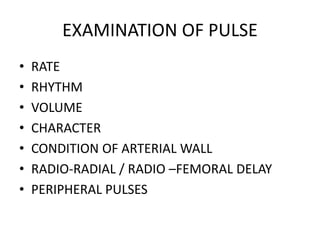
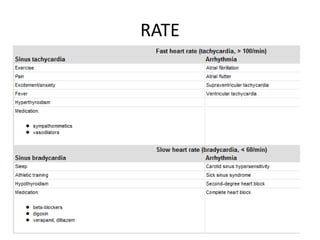
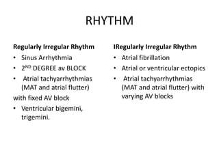
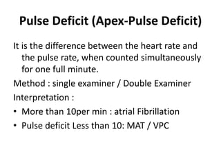
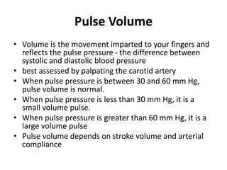
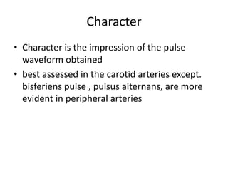
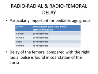
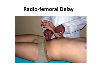
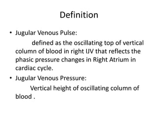
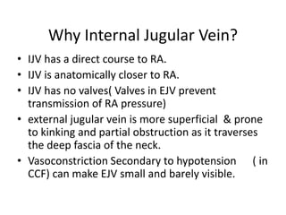
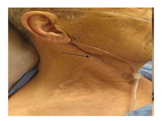
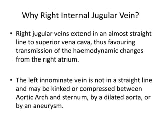
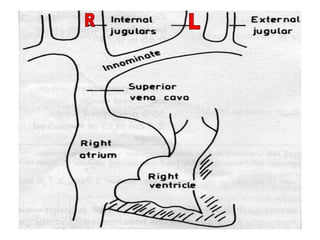
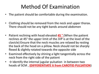
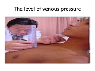
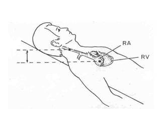
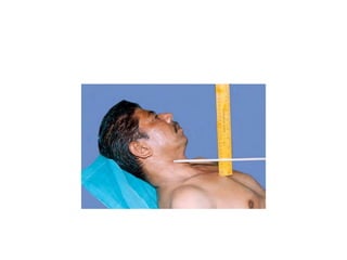
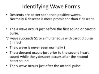
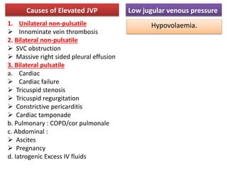
The document provides a review of examining pulse and jugular venous pressure (JVP). It defines pulse as the expansion and elongation of the arterial wall due to blood pressure, and JVP as the oscillating top of blood in the right internal jugular vein reflecting right atrial pressure changes.
When examining pulse, the rate, rhythm, volume, character, arterial wall condition, and peripheral pulses are assessed. Abnormal rhythms and characteristics like pulsus paradoxus are also described. Examining JVP involves observing the pressure level and waveform pattern. An elevated or changed waveform can indicate cardiac or pulmonary issues. The document outlines how to properly examine both pulse and JVP and interpret the findings.
The jugular venous pressure (JVP, sometimes referred to as jugular venous pulse) is the indirectly observed pressure over the venous system via visualization of the internal jugular vein. It can be useful in the differentiation of different forms of heart and lung disease.
Brief Presentation on clinical examination of Cardio Vascular System with Report of Normal case
references:
macleod's clinical examination 13th edition
hutchinson clinical methods
Brief Presentation on clinical examination of Cardio Vascular System with Report of Normal case
references:
macleod's clinical examination 13th edition
hutchinson clinical methods
The venous system contains about 70–80% of the circulating blood volume which is non-pulsatile. However, changes in flow and pressure caused by the right atrial and right ventricular filling produce pulsations in the central veins that are transmitted to the peripheral veins (e.g. jugular veins) and are opposite to the direction of the blood flow.
●
The arterial pulse and blood pressure reflects the dynamics of the left side of the heart, while the jugular veins provide the information about the hemodynamic events from the right side of the heart-right atrial pressure during systole and right ventricular filling pressure during diastole.
●
Hence, an accurate assessment of the venous pulse, the jugular venous pulse (JVP) reflects the dynamics of the right side of the heart.1
History ●
Lancis (1728) first described the cervical venous pulse of the external jugular vein in a patient with tricuspid regurgitation (see Table 16.1).
●
However, the classic graphic recordings of the JVP were done by Chauvea and Marey (1863).
●
But it was Potain (1869) who accurately described the wave pattern in the internal jugular vein.
Giant a Waves or Cannon Waves
These occur whenever the RA contracts against the closed TV during RV systole. Paul Wood described the giant a wave as ‘venous Corrigan’. Cannon waves may occur either regularly or irregularly and are most common in the presence of arrhythmias. ●
Regular cannon waves occur in – Junctional rhythm – Ventricular tachycardia (VT) 1:1 retrograde conduction – Isorhythmic AV dissociation
●
Irregular cannon waves occur in – Complete heart block (see Fig. 16.6) – Classic AV dissociation –VT – Ventricular pacing – Ventricular ectopics
This presentation is about normal wave patterns of JVP and their variations. It includes definition, mechanism, abnormalities and clinical significance of jugular venous pressure.
Useful for medical and biology students who want to study the cardiac cycle in a short time with big benefits !!
CVS physiology - Wigger Diagram - ECG of cardiac cycle - Heart sounds
Clinical examination in cardiolgy starts with the examination of arterial pulse.
In this PowerPoint presentation we have discussed all about this important topic
The dimensions of healthcare quality refer to various attributes or aspects that define the standard of healthcare services. These dimensions are used to evaluate, measure, and improve the quality of care provided to patients. A comprehensive understanding of these dimensions ensures that healthcare systems can address various aspects of patient care effectively and holistically. Dimensions of Healthcare Quality and Performance of care include the following; Appropriateness, Availability, Competence, Continuity, Effectiveness, Efficiency, Efficacy, Prevention, Respect and Care, Safety as well as Timeliness.
Telehealth Psychology Building Trust with Clients.pptxThe Harvest Clinic
Telehealth psychology is a digital approach that offers psychological services and mental health care to clients remotely, using technologies like video conferencing, phone calls, text messaging, and mobile apps for communication.
Medical Technology Tackles New Health Care Demand - Research Report - March 2...pchutichetpong
M Capital Group (“MCG”) predicts that with, against, despite, and even without the global pandemic, the medical technology (MedTech) industry shows signs of continuous healthy growth, driven by smaller, faster, and cheaper devices, growing demand for home-based applications, technological innovation, strategic acquisitions, investments, and SPAC listings. MCG predicts that this should reflects itself in annual growth of over 6%, well beyond 2028.
According to Chris Mouchabhani, Managing Partner at M Capital Group, “Despite all economic scenarios that one may consider, beyond overall economic shocks, medical technology should remain one of the most promising and robust sectors over the short to medium term and well beyond 2028.”
There is a movement towards home-based care for the elderly, next generation scanning and MRI devices, wearable technology, artificial intelligence incorporation, and online connectivity. Experts also see a focus on predictive, preventive, personalized, participatory, and precision medicine, with rising levels of integration of home care and technological innovation.
The average cost of treatment has been rising across the board, creating additional financial burdens to governments, healthcare providers and insurance companies. According to MCG, cost-per-inpatient-stay in the United States alone rose on average annually by over 13% between 2014 to 2021, leading MedTech to focus research efforts on optimized medical equipment at lower price points, whilst emphasizing portability and ease of use. Namely, 46% of the 1,008 medical technology companies in the 2021 MedTech Innovator (“MTI”) database are focusing on prevention, wellness, detection, or diagnosis, signaling a clear push for preventive care to also tackle costs.
In addition, there has also been a lasting impact on consumer and medical demand for home care, supported by the pandemic. Lockdowns, closure of care facilities, and healthcare systems subjected to capacity pressure, accelerated demand away from traditional inpatient care. Now, outpatient care solutions are driving industry production, with nearly 70% of recent diagnostics start-up companies producing products in areas such as ambulatory clinics, at-home care, and self-administered diagnostics.
Deep Leg Vein Thrombosis (DVT): Meaning, Causes, Symptoms, Treatment, and Mor...The Lifesciences Magazine
Deep Leg Vein Thrombosis occurs when a blood clot forms in one or more of the deep veins in the legs. These clots can impede blood flow, leading to severe complications.
Explore our infographic on 'Essential Metrics for Palliative Care Management' which highlights key performance indicators crucial for enhancing the quality and efficiency of palliative care services.
This visual guide breaks down important metrics across four categories: Patient-Centered Metrics, Care Efficiency Metrics, Quality of Life Metrics, and Staff Metrics. Each section is designed to help healthcare professionals monitor and improve care delivery for patients facing serious illnesses. Understand how to implement these metrics in your palliative care practices for better outcomes and higher satisfaction levels.
CHAPTER 1 SEMESTER V - ROLE OF PEADIATRIC NURSE.pdfSachin Sharma
Pediatric nurses play a vital role in the health and well-being of children. Their responsibilities are wide-ranging, and their objectives can be categorized into several key areas:
1. Direct Patient Care:
Objective: Provide comprehensive and compassionate care to infants, children, and adolescents in various healthcare settings (hospitals, clinics, etc.).
This includes tasks like:
Monitoring vital signs and physical condition.
Administering medications and treatments.
Performing procedures as directed by doctors.
Assisting with daily living activities (bathing, feeding).
Providing emotional support and pain management.
2. Health Promotion and Education:
Objective: Promote healthy behaviors and educate children, families, and communities about preventive healthcare.
This includes tasks like:
Administering vaccinations.
Providing education on nutrition, hygiene, and development.
Offering breastfeeding and childbirth support.
Counseling families on safety and injury prevention.
3. Collaboration and Advocacy:
Objective: Collaborate effectively with doctors, social workers, therapists, and other healthcare professionals to ensure coordinated care for children.
Objective: Advocate for the rights and best interests of their patients, especially when children cannot speak for themselves.
This includes tasks like:
Communicating effectively with healthcare teams.
Identifying and addressing potential risks to child welfare.
Educating families about their child's condition and treatment options.
4. Professional Development and Research:
Objective: Stay up-to-date on the latest advancements in pediatric healthcare through continuing education and research.
Objective: Contribute to improving the quality of care for children by participating in research initiatives.
This includes tasks like:
Attending workshops and conferences on pediatric nursing.
Participating in clinical trials related to child health.
Implementing evidence-based practices into their daily routines.
By fulfilling these objectives, pediatric nurses play a crucial role in ensuring the optimal health and well-being of children throughout all stages of their development.
Empowering ACOs: Leveraging Quality Management Tools for MIPS and BeyondHealth Catalyst
Join us as we delve into the crucial realm of quality reporting for MSSP (Medicare Shared Savings Program) Accountable Care Organizations (ACOs).
In this session, we will explore how a robust quality management solution can empower your organization to meet regulatory requirements and improve processes for MIPS reporting and internal quality programs. Learn how our MeasureAble application enables compliance and fosters continuous improvement.
Nursing Care of Client With Acute And Chronic Renal Failure.ppt
Pulse & JVP
1. PULSE & JVP : A QUICK REVISION
DR SUBHANKAR PAUL
2. Definitions
• Pulse : it is the expansion & elongation of the
arterial wall due to pressure imparted by the
column of blood during cardiac cycle
• JVP oscillating top of vertical column of blood
in right IJV that reflects the phasic pressure
changes in Right Atrium in cardiac cycle
3. EXAMINATION OF PULSE
• RATE
• RHYTHM
• VOLUME
• CHARACTER
• CONDITION OF ARTERIAL WALL
• RADIO-RADIAL / RADIO –FEMORAL DELAY
• PERIPHERAL PULSES
5. RELATIVE TACHYCARDIA & RELATIVE
BRADYCARDIA : Related to TEMPARATURE
RELATIVE TACHYCARDIA
• Acute Rheumatic Carditis
• Diphtheric myocarditis
• Tuberculosis
RELATIVE BRADYCARDIA
• Any Viral fever ( Dengue,
Yellow fever : Faget’s sign)
• Enteric fever 1st week
• Pyogenic Meningitis/ Intra
cerebral Abscess
• Brucellosis
1DEGREE F temp rise = increase in Pulse rate by 10/min
6. RHYTHM
Regularly Irregular Rhythm
• Sinus Arrhythmia
• 2ND DEGREE av BLOCK
• Atrial tachyarrhythmias
(MAT and atrial flutter)
with fixed AV block
• Ventricular bigemini,
trigemini.
IRegularly Irregular Rhythm
• Atrial fibrillation
• Atrial or ventricular ectopics
• Atrial tachyarrhythmias
(MAT and atrial flutter) with
varying AV blocks
7. Pulse Deficit (Apex-Pulse Deficit)
It is the difference between the heart rate and
the pulse rate, when counted simultaneously
for one full minute.
Method : single examiner / Double Examiner
Interpretation :
• More than 10per min : atrial Fibrillation
• Pulse deficit Less than 10: MAT / VPC
8. Pulse Volume
• Volume is the movement imparted to your fingers and
reflects the pulse pressure - the difference between
systolic and diastolic blood pressure
• best assessed by palpating the carotid artery
• When pulse pressure is between 30 and 60 mm Hg,
pulse volume is normal.
• When pulse pressure is less than 30 mm Hg, it is a
small volume pulse.
• When pulse pressure is greater than 60 mm Hg, it is a
large volume pulse
• Pulse volume depends on stroke volume and arterial
compliance
9. Character
• Character is the impression of the pulse
waveform obtained
• best assessed in the carotid arteries except.
bisferiens pulse , pulsus alternans, are more
evident in peripheral arteries
14. Collapsing Pulse (Water-Hammer
Pulse, Corrigans Pulse)
It is a large volume pulse with a rapid upstroke
(systolic pressure is high) and a rapid
downstroke (diastolic pressure is low).
The rapid upstroke is because of an increased
stroke volume.
The rapid downstroke is because of diastolic
run-off into the left ventricle, and decreased
peripheral resistance and rapid run-off to the
periphery.
Decreased peripheral resistance is due to large
stroke volume stretching carotid and aortic
sinus leading to reflex decrease in peripheral
resistance
16. Causes
Patent ductus arteriosus
Aortic regurgitation
Arteriovenous fistula
Rupture of sinus of Valsalva.
Thready pulse is seen in shock.
Jerky pulse is seen in HOCM.
18. Pulsus Bisferiens
best felt in brachial and femoral artery
due to ejection of rapid jet of
blood through the aortic
valve. During the peak of
flow, Bernouli’ s effect on the
walls of ascending aorta
causes a sudden decrease in
lateral pressure on the inner
aspect of the wall.
Dissection of aorta (unilateral bisferiens).
Pulsus bisferiens is a single pulse wave with two peaks in systole
19. Pulsus Dicroticus
Mechanism :
due to a very low stroke
volume with decreased
peripheral resistance
It is a single pulse wave with one peak in systole and one peak in diastole
21. Pulsus Bigemini
• A pulse wave with a normal beat followed by a
premature beat and a compensatory pause,
occurring in rapid succession, resulting in
alternation of the strength of the pulse
• In pulsus alternans, compensatory pause is
absent, whereas in pulsus bigeminus,
compensatory pause is present.
• Pulsus bigeminus is a sign of digitalis toxicity.
22. Pulsus paradoxus
It is an exaggerated reduction in the strength
of arterial pulse during normal inspiration or
an exaggerated inspiratory fall in systolic
pressure of more than 10 mm Hg during quiet
breathing.
Pulsus paradoxus is best assessed by
measuring the difference in systolic blood
pressure during inspiration and expiration
During deep inspiration Pulse may be absent
23. Reverse pulsus paradoxus
• is an inspiratory rise in arterial pressure.
• Causes
• Hypertrophic obstructive cardiomyopathy
• Intermittent positive pressure ventilation
• Atrioventricular dissociation
24. Condition of Arterial Wall
• Young adult : Not Palpable
• Old Individuals , Hypertensive patients :
Palpable
30. RADIO-RADIAL & RADIO-FEMORAL
DELAY
• Particularly important for pediatric age group
• Delay of the femoral compared with the right
radial pulse is found in coarctation of the
aorta
Artery Time at which pulse wave arrives
after cardiac systole
Carotid 30 milliseconds
Brachial 60 milliseconds
Radial 80 milliseconds
Femoral 75 milliseconds
34. Definition
• Jugular Venous Pulse:
defined as the oscillating top of vertical
column of blood in right IJV that reflects the
phasic pressure changes in Right Atrium in
cardiac cycle.
• Jugular Venous Pressure:
Vertical height of oscillating column of
blood .
35. Why Internal Jugular Vein?
• IJV has a direct course to RA.
• IJV is anatomically closer to RA.
• IJV has no valves( Valves in EJV prevent
transmission of RA pressure)
• external jugular vein is more superficial & prone
to kinking and partial obstruction as it traverses
the deep fascia of the neck.
• Vasoconstriction Secondary to hypotension ( in
CCF) can make EJV small and barely visible.
36.
37. Why Right Internal Jugular Vein?
• Right jugular veins extend in an almost straight
line to superior vena cava, thus favouring
transmission of the haemodynamic changes
from the right atrium.
• The left innominate vein is not in a straight line
and may be kinked or compressed between
Aortic Arch and sternum, by a dilated aorta, or
by an aneurysm.
38.
39. Method Of Examination
• The patient should be comfortable during the examination.
• Clothing should be removed from the neck and upper thorax.
There should not be any tight bands around abdomen
• Patient reclining with head elevated 45 ° (When the patient
reclines at 45° the upper limit of the JVP is at the level of the
clavicle) Ensure that the neck muscles are relaxed by resting
the back of the head on a pillow. Neck should not be sharply
flexed & slightly rotated towards the opposite side
• Examined effectively by shining a light tangentially across the
neck from the right side of the patient
• → Identify the internal jugular pulsation in between two
heads of SCM , (DIFFERENTIATE it from CAROTID PULSATION)
40. • Identify the timing and waveform of the
pulsation and note any abnormality
• Estimate the vertical height in centimetres
between the top of the venous pulsation and the
sternal angle to give the venous pressure
• If necessary, readjust the position of the patient
until the waveform is clearly visible & if necessary
use the abdominojugular reflux
.
42. • (A) Supine: jugular vein distended, pulsation not
visible. (B) Reclining at 45°: point of transition between
distended and collapsed vein can usually be seen to
pulsate just above the clavicle. (C) Upright: upper part
of vein collapsed and transition point obscured
WHY 45° ?? FOR BETTER VISUALISATION
the pulsation of the internal jugular vein is greatest when the trunk is inclined by less than 30° :
HARRISON’S 17TH
43. Difference from Carotid Pulse
JVP CAROTID PULSE
Better Visible Better Palpable
INSPECTION Oscillatory wavefom with Inward movement
more prominent
Rapid outward jerky movement
Two peaks per heartbeat (in sinus rhythm) One peak per heartbeat
Seen in the triangle formed by the two heads
of the sternomastoid and the clavicle
Seen internal to sternomastoid
Independent of respiration Height of pulsation varies with
respiration (DECREASES in
INSPIRATION)
Independent of position Varies with position of patient
PALPATION Pulsation unaffected by pressure at the root of
the neck
Pulsation diminished by pressure
at the root of the neck
Independent of abdominal pressure Rises with abdominal pressure
48. The level of venous pressure
• Using a centimeter ruler, measure the vertical
distance between the angle of Louis (manubrio
sternal joint) and the highest level of jugular vein
pulsation.
• Add 5 cm to measure central venous pressure since
right atrium is 5 cm below the sternal angle. Normal
CVP is < 7mm of Hg or 9 cm H2O (1.36 cmH2O = 1.0
mmHg)
• The upper limit of normal is 4 cm above the sternal
angle
• A distance >4.5 cm at 30° elevation is considered
abnormal
49.
50. WHY The sternal angle is used as the
reference point ??
Upto Harrison's 17th edition
• “The sternal angle is used
as the reference point
because the center of the
right atrium lies
approximately 5 cm below
the sternal angle in the
average patient, regardless
of body position”
Harrison's 18th edition onwards
• “the actual distance between the
mid-right atrium and the angle of
Louis varies considerably as a
function of both body size and
the patient angle at which the
assessment is made (30°, 45°, or
60°). The use of the sternal angle
as a reference point leads to
systematic underestimation of
CVP, and this method should be
used less for semiquantification
than to distinguish a normal from
an abnormally elevated CVP”
51. SO , WHAT THEY SUGGEST ????
• “The use of the clavicle may provide an easier
reference for standardization. Venous
pulsations above this level in the sitting
position are clearly abnormal, as the distance
between the clavicle and the right atrium is at
least 10 cm. The patient should always be
placed in the sitting position, with the legs
dangling below the bedside, when an elevated
pressure is suspected in the semisupine
position”
52. Abdomino-jugular reflux
Upto Harrison's 17th edition
• “A positive
abdominojugular test is
best defined as an
increase in JVP during
10 s of firm
midabdominal
compression followed
by a rapid drop in
pressure of 4 cm blood
on release of the
compression”
Harrison's 18th edition onwards
• “The abdominojugular
reflex is elicited with firm
and consistent pressure
over the upper portion of
the abdomen, preferably
over the right upper
quadrant, for at least 10 s. A
positive response is defined
by a sustained rise of more
than 3 cm in JVP for at least
15 s after release of the
hand”
54. • Most common cause of a positive test is RHF/
(incipient Heart Failure )
• Positive test in: Borderline elevation of JVP
Silent TR
Latent RHF
• False Negative: SVC/IVC obstruction
Budd Chiari syndrome
• Positive Test imply SVC and IVC are patent
58. • Venous distension due to RA contraction
Retrograde blood flow into SVC and IJV
• Synchronous with S1, Follow P of ECG
• Precede Carotid pulse
a WAVE
59. • The x descent: is due to
X Atrial relaxation
X` Descent of the floor of the right atrium
during right ventricular systole.
Begins during systole and ends before S2
• The c wave:
Occurs simultaneously with the carotid pulse
Artifact by Carotid pulsation
Bulging of TV into RA during ICP
60. v WAVE
• Rising right atrial pressure when blood flows into the
right atrium during ventricular systole when the
tricuspid valve is shut.
• Synchronous with Carotid pulse
• Peaks after S2
61. y DESCENT
• The decline in right atrial pressure when the tricuspid valve
reopens
• Following the bottom of the y descent and before beginning of
the a wave is a period of relatively slow filling of the ventricle,
the diastases period, a wave termed the h wave.
62. Normal pattern of the jugular venous pulse
• The normal JVP reflects phasic pressure changes in
the right atrium and consists of three positive waves
and two negative troughs
• Simultaneous palpation of the left carotid artery &
auscultation of heart soounds at apex aids the
examiner in relating the venous pulsations to the
timing of the cardiac cycle.
63. • Descents are better seen than positive waves.
Normally X descent is more prominent than Y descent.
• The a wave occurs just before the first sound or carotid
pulse.
‘c’ wave succeeds S1 or simultaneous with carotid pulse
( in fact
• The c wave is never seen normally )
• The x descent occurs just prior to the second heart
sound while the y descent occurs after the second
heart sound
• The v wave occurs just after the arterial pulse
Identifying Wave Forms
64. jugular venous pulse
• clinical corelates:
• Abnormalities in Pressure
• Abnormalities in Waveform
68. Abnormalities in a wave
• Elevated “a” wave
• Cannon “a” wave
• Absent “a” wave
an increased delay between the a wave and the carotid
arterial pulse in patients with first-degree
atrioventricular block.
69. Elevated “a” wave
• Tricuspid stenosis
• Increased
Resistance to RV
Filling.
PS
PAH
Large a waves indicate that the right atrium is contracting against an increased
resistance
73. “a” wave equal to “v” wave
ASD
Prominent X descent
followed by a large
V wave
M Configuration
Indicates a large L-R
shunt
With PAH A wave
becomes more
prominent
76. Tricuspid regurgitation
• Absent X Descent
• CV/ Regurgitant Wave
• Followed by a rapid deep
Y descent
• May cause subtle motion
of ear lobe with each
heart beat
77. Constrictive pericarditis.
• M shaped contour
• Prominent X and Y descent (FRIEDREICH`SIGN)
• Y descent is prominent as ventricular filling is unimpeded
during early diastole.
• This is interrupted by a rapid raise in pressure as the filling is
impeded by constricting Pericardium
• The Ventricular pressure curve exhibit Square Root sign.
78. Kussmaul sign ( Venous Pulsus Paradoxus)
• Normally, the venous pressure should fall by at least
3 mmHg with inspiration
• Kussmaul’s sign is defined by either a rise or a lack of
fallof the JVP with inspiration
CAUSE
Constrictive Pericarditis
Severe RHF and advanced left ventricular failure
Restrictive Cardiomyopathy
massive pulmonary embolism,
right ventricular infarction,
80. Recent Studies
• Although the JVP estimates right ventricular filling pressure, it
has a predictable relationship with the pulmonary artery
wedge pressure.
• In a large study of patients with advanced heart failure, the
presence of a right atrial pressure >10 mmHg (as predicted on
bedside examination) had a positive value of 88% for the
prediction of a pulmonary artery wedge pressure of >22
mmHg.
• In addition, an elevated JVP has prognostic significance in
patients with both symptomatic heart failure and
asymptomatic left ventricular systolic dysfunction.
• The presence of an elevated JVP is associated with a higher
risk of subsequent hospitalization for heart failure, death from
heart failure, or both.