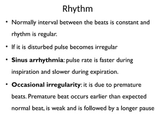
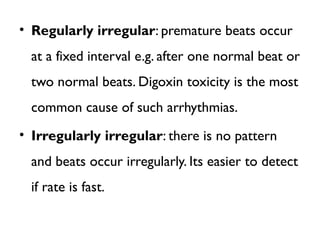
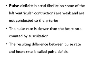
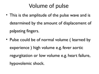
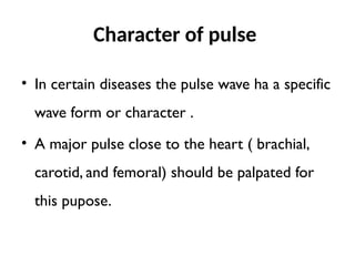
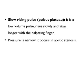
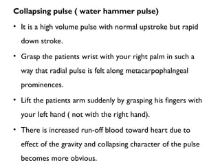
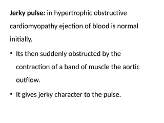
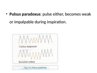
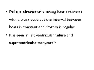
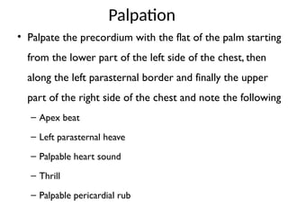
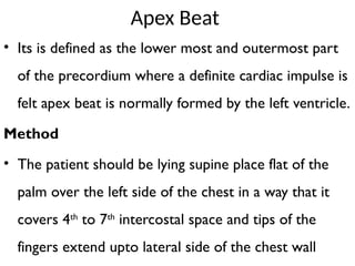
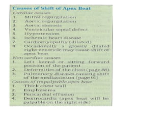
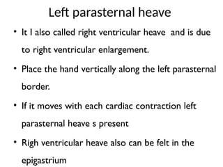
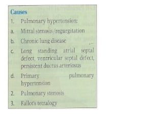
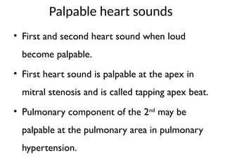
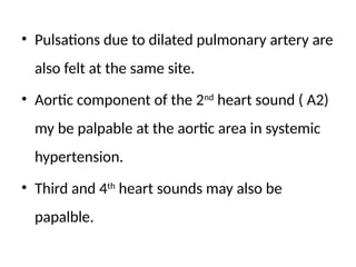
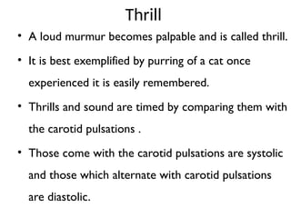
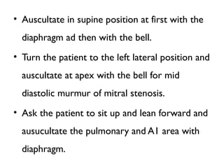
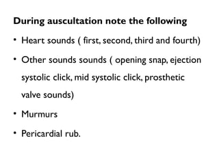
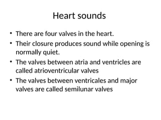
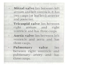
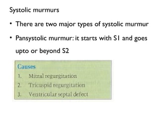
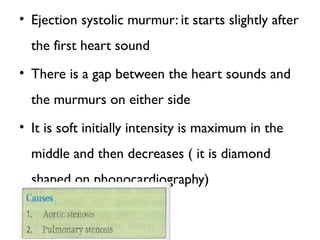
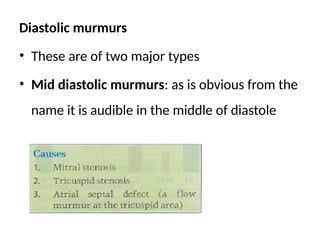
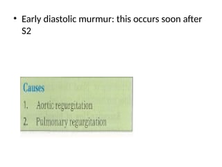
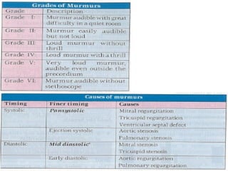
Cardiovascular Physical
Examination
Dr. Mustafe Hussein
MBBS, MMED ( INTERNIST)
introduction
• Clinical examination of the CVS
(cardiovascular system) is particularly
rewarding as it usually leads to an accurate
diagnosis.
• Investigations are carried out either to
confirm the clinical impression or to
differentiate between various possibilities.
Symptoms
• Early diagnosis of important cardiac diseases
like ischemic heart disease and heart failure is
based on carful history taking.
• There are two cardinal symptoms of
cardiovascular disease
– Dyspnea
– Chest pain
Dyspnea
• Dyspnea or breathlessness means difficulty in
breathing it ay occur on exertion or at rest.
Exertion dyspnea
• Its an early symptom of heart failure initially
• It may occur after unaccustomed or strenuous
exertion.
• But as disease progresses patient may become
breathless even on walking a few steps.
Paroxysmal Nocturnal Dyspnea
• The patient wakes up at night due to severe
breathlessness which improves on sitting upright for
several minutes and is usually accompanied by cough
and frothy sputum.
• This is called paroxysmal nocturnal dyspnea.This is
due paroxysmal nocturnal dyspnea.
• This is due to transient pulmonary edema.
• Precipitated by increased venous return to the heart
in recumbent position.
Orthopnea
• In patients of severe heart failure
breathlessness worsens on lying flat, this is
called orthopnea
Pulmonary edema
• There is transudation of fluid into the heart
due to left heart dysfunction.
• Symptoms are persistent severe
breathlessness
• Orthopnea and cough productive of copious,
frothy, watery and blood stained sputum
Chest pain
• It is an important symptom of heart disease its
characteristics vary with the underlying
pathology.
Angina pectoris
• There is transient myocardial ischemia
• The patient develops chest on exertion which is relieved
by rest and sublingual nitroglycerin.
• Pain is retrosternal, across the chest and radiates to the
jaw and left arm.
• Patients describes it as a light band around the chest or
heaviness.
• It my be associated with dyspnea, palpitation and
sweating.
• Total duration of pain is less than 30 minutes
Myocardial Infarction
• There is total occlusion of one or more
branches of the coronary artery and the
dependent myocardium dies.
• Pain is similar to that of angina pectoris but
duration is more than 30 minutes and it is not
relieved by sublingual nitrates or rest.
Pericarditis
• Features are similar to the pain of ischemic
heart disease
• There is no effect of rest or nitrated
• Its relieved by leaning forward and my get
worsen on deep breathing and coughing.
Palpitation
• It is the awareness of the heart beat.
• It’s a common feature of anxiety.
• It also occurs in tachycardia and heart failure.
Examination
• When you are asked to examine a particular
system of patient always start from the general
physical examination except when examiner asks
you to omit it.
• Examination of the cardiovascular system consist of
– Examination of pulse
– Examination blood pressure
– Examination of neck veins
– Examination of pericordium
Examination of the pulse
• The pulse is a wave imparted by the contraction of
the left ventricle to the blood column and travels
10 times faster than the blood it self.
• Pulse is felt where an accessible artery can be
pressed against and underlying bone.
• Commonly felt pulses are radial, branchial, carotid,
femoral, popliteal, posterior tibial and dorsalis
pedis.
Radial pulse
• Its is the most easily accessible and the most
commonly felt pulse.
• The patients hand should be slightly flexed and
pronated.
• Press the radial artery against the head of the
radius.
Branchial pulse
• Flex the patients arm and feel for the tendon
of the biceps press on its medial side with the
thumb of your opposite hand.
Carotid pulse
• Place the thumb or fingers of your opposite
hand along the anterior border of the
sternomastoid at the level of laryngeal
cartilage and press backwards
• Don’t palpate both carotids simultaneously
because blood supply to the brain may be
critically reduced.
• Femoral pulse: Press with the thumb/finger
halfway between the anterior superior iliac
spine and the pubic tubercle along the inguinal
ligament.
• Popliteal pulse: popliteal artery lies deep in
the popliteal fossa and is difficult to palpate.
• Flex the knee at angel of 120 degree and push
fingers of both hands into the popliteal fossa.
• Dorsalis pedis pulse: palpate in the proximal
part of the first intermetatarsal space.
• Posterior tibial pulse: palpate behind the
medial malleolus.
During the examination of pulse note the
following features.
• Rate
• Rhythm
• Volume
• Character
• Comprison with other pulse
• Condition of the vessel wall
• Rate: count the pulse for full minute
• Normal range is between 100 and 60 b/m
• Its equal to the heart rate except in certain
arrhythmias like atrial fibrillation.
• Tachycardia: pulse rate more than 100b/m
• Bradycardia: pulse rate lower than 50b/m
• Relatively bradycardia: normally pulse rise 10
b/m for each degree F or 0.5 C rise in the body
temperature.
Rhythm
• Normally interval between the beats is constant and
rhythm is regular.
• If it is disturbed pulse becomes irregular
• Sinus arrhythmia: pulse rate is faster during
inspiration and slower during expiration.
• Occasional irregularity: it is due to premature
beats. Premature beat occurs earlier than expected
normal beat, is weak and is followed by a longer pause
• Regularly irregular: premature beats occur
at a fixed interval e.g. after one normal beat or
two normal beats. Digoxin toxicity is the most
common cause of such arrhythmias.
• Irregularly irregular: there is no pattern
and beats occur irregularly. Its easier to detect
if rate is fast.
• Pulse deficit: in atrial fibrillation some of the
left ventricular contractions are weak and are
not conducted to the arteries
• The pulse rate is slower than the heart rate
counted by auscultation
• The resulting difference between pulse rate
and heart rate is called pulse deficit.
Volume of pulse
• This is the amplitude of the pulse wave and is
determined by the amount of displacement of
palpating fingers.
• Pulse could be of normal volume ( learned by
experience ) high volume e.g. fever aortic
regurgitation or low volume e.g. heart failure,
hypovolemic shock.
Character of pulse
• In certain diseases the pulse wave ha a specific
wave form or character .
• A major pulse close to the heart ( brachial,
carotid, and femoral) should be palpated for
this pupose.
• Slow rising pulse (pulsus plateau): it is a
low volume pulse, rises slowly and stays
longer with the palpating finger.
• Pressure is narrow it occurs in aortic stenosis.
Collapsing pulse ( water hammer pulse)
• It is a high volume pulse with normal upstroke but rapid
down stroke.
• Grasp the patients wrist with your right palm in such a
way that radial pulse is felt along metacarpophalngeal
prominences.
• Lift the patients arm suddenly by grasping his fingers with
your left hand ( not with the right hand).
• There is increased run-off blood toward heart due to
effect of the gravity and collapsing character of the pulse
becomes more obvious.
Jerky pulse: in hypertrophic obstructive
cardiomyopathy ejection of blood is normal
initially.
• Its then suddenly obstructed by the
contraction of a band of muscle the aortic
outflow.
• It gives jerky character to the pulse.
• Pulsus paradoxus: pulse either, becomes weak
or impalpable during inspiration.
• Pulsus alternant: a strong beat alternates
with a weak beat, but the interval between
beats is constant and rhythm is regular
• It is seen in left ventricular failure and
supraventricular tachycardia
Pulsus bigeminus: it is similar to pulsus
alternans, but interval between beats is variable.
• A strong beat and weak beat occur close to
each other followed by a long pause ( a strong
and weak beats are coupled) and this cycle is
repeated.
• Digoxin toxicity is the most important cause.
Comparison with other pulses
• Palpate corresponding pulses of both sides
simultaneously and compare their volume except
carotids
• Don’t palpate both carotids simultaneously
• Compare radial and femoral pulses in
coarctation of the aorta femoral pulse is weak
and delayed as compared to radial pulse
( radiofemoral delay
Condition of the vessel wall
• Fell the radial pulse with three fingers
• Press with proximal fingers so that the pulse is
occluded and feel the vessel wall with the
middle finger.
• Normally it is not palpable
• In advanced atherosclerosis it can be felt as a
cord between finger and underlying bone.
Neck veins
• The central venous pressure which is the same as the
right atrial pressure is an important guide for the CVS
function.
• Its measured by inserting a catheter into the right
atrium through the internal jugular of subclavian vein.
• A nearly accurate estimation of the right atrial
pressure can be made clinically by observing the upper
limit of venous pulsation in the neck and measuring its
distance from the sternal angle.
• Neck veins are in continuity with the right atrium.
• They become distended when filled with the
blood, otherwise are collapsed.
• Normal mean atrial pressure is 5mmhg equivalent
to 7 cm high column of blood.
• In the upright the proximal 7 to 8cm of veins as
measured from the center of the right atrium
remain distended.
Kussmaul Sign.
• In constrictive pericarditis the JVP instead of
falling rises during inspiration.
• The downward movement of the diaphragm
during inspiration compresses the congested
liver, the venous return is increased but right
atrium cannot expand due to the rigid
pericardium and jvp raised
Examination of Precordium
• Precordium is that part f the chest wall which
overlies the heart examination of the
precordium consists of following steps.
– Inspection
– Palpation
– Percussion
– Auscultation
Inspection
• Looks for the following physical signs
• Chest deformity ( discussed under respiratory
system)
• Bulgin of percordium
• Scars particularly along the sternum of
intercostal spaces ( these indicate past cardiac
surgery)
Pulsation
• Apex beat
• Pulsation along the parasternal border e,g due to
right ventricular hypertrophy.
• Pulsation in the left 2nd
intercostal space e,g due
to dilatation of pulmonary artery
• Pulsations in the right second intercostal space
e,g due to aneurysm of the aorta.
• Pulsations in the suprasternal notch e,g due to
aneurysm of the aorta.
• Pulsations in the epigastrium which are
normally present in thin individuals due to
aorta, the could also be right ventricular
hypertrophy or pulsatile liver in tricuspid
regurgitation.
• Whole of the precordium with each cardiac
beat if the heart is greatly enlarged
Palpation
• Palpate the precordium with the flat of the palm starting
from the lower part of the left side of the chest, then
along the left parasternal border and finally the upper
part of the right side of the chest and note the following
– Apex beat
– Left parasternal heave
– Palpable heart sound
– Thrill
– Palpable pericardial rub
Apex Beat
• Its is defined as the lower most and outermost part
of the precordium where a definite cardiac impulse is
felt apex beat is normally formed by the left ventricle.
Method
• The patient should be lying supine place flat of the
palm over the left side of the chest in a way that it
covers 4th
to 7th
intercostal space and tips of the
fingers extend upto lateral side of the chest wall
Left parasternal heave
• It I also called right ventricular heave and is due
to right ventricular enlargement.
• Place the hand vertically along the left parasternal
border.
• If it moves with each cardiac contraction left
parasternal heave s present
• Righ ventricular heave also can be felt in the
epigastrium
Palpable heart sounds
• First and second heart sound when loud
become palpable.
• First heart sound is palpable at the apex in
mitral stenosis and is called tapping apex beat.
• Pulmonary component of the 2nd
may be
palpable at the pulmonary area in pulmonary
hypertension.
• Pulsations due to dilated pulmonary artery are
also felt at the same site.
• Aortic component of the 2nd
heart sound ( A2)
my be palpable at the aortic area in systemic
hypertension.
• Third and 4th
heart sounds may also be
papalble.
Thrill
• A loud murmur becomes palpable and is called thrill.
• It is best exemplified by purring of a cat once
experienced it is easily remembered.
• Thrills and sound are timed by comparing them with
the carotid pulsations .
• Those come with the carotid pulsations are systolic
and those which alternate with carotid pulsations
are diastolic.
Percussion
• As chest radiograph is a routine investigation
and shows the exact size and shape of the
heart.
• Percussion of the precordium for cardiac
dullness is not performed routinely nowadays
• Increased cardiac dullness due to a large
pericardial effusion may still be detected on
percussion.
Auscultation
• This is the most important step in the
examination of the cardiovascular system.
Method of Auscultation
• auscultate whole of the precordium, either
starting starting from the apex moving up along
the left parasternal border to the pulmonary to
the pulmonary area and then to the A1 area or
starting from the A1 area and moving towards
the apex.
• Auscultate in supine position at first with the
diaphragm ad then with the bell.
• Turn the patient to the left lateral position and
auscultate at apex with the bell for mid
diastolic murmur of mitral stenosis.
• Ask the patient to sit up and lean forward and
ausucultate the pulmonary and A1 area with
diaphragm.
During auscultation note the following
• Heart sounds ( first, second, third and fourth)
• Other sounds sounds ( opening snap, ejection
systolic click, mid systolic click, prosthetic
valve sounds)
• Murmurs
• Pericardial rub.
Heart sounds
• There are four valves in the heart.
• Their closure produces sound while opening is
normally quiet.
• The valves between atria and ventricles are
called atrioventricular valves
• The valves between ventricales and major
valves are called semilunar valves
First and second heart sounds
• The first heart sound is produced by closure
of the mitral and tricuspid valves.
• It marks the beginning of systole
the second heart sound is produced by
closure of the aortic and pulmonary valves.
• It denotes the end of systole and the
beginning of diastole.
• How to differentiate between first and second
heart sound
• Palpate the carotid artery while auscultating:
the sound which comes just before the
caroitd pulsation is S1 and the sound which
comes after carotid pulsation is S2
• At normal heart rate the systolic interval is
shorter than the diastolic interval.
Third Heart Sound
Third Heart Sound
• This is a low pitched sound and occurs in early
diastole at the time of rapid ventricular filling.
Fourth Heart Sound
• This is a low pitched sound and occurs in late
diastole due to atrial contraction if ventricles
are stiff or non complaint due to disease
• Causes of third heart
sound
• Causes of fourth heart
sound
Murmurs
• These are abnormal sounds and are of longer
duration as compared to heart sounds.
• These are produced due to the turbulence of blood
flow and one of three mechanisms is involved.
1. Excessive flow of blood: across a normal valve e.g.
severe anemia or pregnancy, such murmurs are
also called functional or flow murmurs
2. Folow of normal amount of blood across a
narrowed valve e,g mitral stenosis or aortic
stenosis.
3. Flow of blood in abnormal direction
a) Normally a valve allows only unidirectional flow, if it
is abnormal leakage may occur e,g mitral
regurgitation or aortic regurgitation.
b) Abnormal communication within the heart e,g atrial
septal defect, ventricular septal defect or outside
the heart e,g persistent ductus arteriosus
Characteristics of a murmur
If a murmur is audible note the following characteristics
1. Timing
2. Intensity
3. Site of maximum intensity
4. Radiation
5. Character
6. Pitch
7. Effect of respiration
Timing
• For a beginner it will be sufficient if he can
differentiate between a systolic murmur and a
diastolic murmur
• Palpate the carotid artery while auscultating.
• The murmur which comes with the carotid
pulsation is systolic and the murmur which
alternated with it is diastolic.
Systolic murmurs
• There are two major types of systolic murmur
• Pansystolic murmur: it starts with S1 and goes
upto or beyond S2
• Ejection systolic murmur: it starts slightly after
the first heart sound
• There is a gap between the heart sounds and
the murmurs on either side
• It is soft initially intensity is maximum in the
middle and then decreases ( it is diamond
shaped on phonocardiography)
Diastolic murmurs
• These are of two major types
• Mid diastolic murmurs: as is obvious from the
name it is audible in the middle of diastole
• Early diastolic murmur: this occurs soon after
S2
Intensity
• A murmur may be audible with great difficulty
in a quiet room ( grade I) or it may be audible
with out stethoscope (grad Iv)
• A loud murmur without a thrill is grade III and
similar murmur with thrill is grade IV
Site of maximum intensity
• A murmur may be audible all over the
precordium depending upon its loudness but its
intensity is maximum where it is being produced
( with few exceptions )
• e.g. murmur of mitral regurgitation is loudest at
the apex while murmur of tricuspid regurgitation
is of maximum intensity at the tricuspid area
Radiation
• A murmur may be better audible in one
particular direction outside the precordium,
depending upon the direction of flow of blood
• This is called radiation e,g murmur of mitral
regurgitation radiates towards the axilla, murmur
of pulmonary stenosis radiates to the left
shoulder and the murmur of aortic stenosis
radiates to the neck.
Effect of respiration
• Murmur of right heart ( e,g murmur of
tricuspid regurgitation, pulmonary stenosis)
increases in intensity during inspiration.
• Murmurs of left heart (e,g murmur of mitral
regurgitation, aortic stenosis,VSD) increase in
intensity during expiration.
Effect of posture
• Murmur of mitral stenosis is best heard in the
left lateral position while murmurs of
pulmonary of pulmonary and aortic
regurgitation are best audible when the
patient sits up and leans forwards.
Pericardial rub
• This is a superficial scratch sound audible both in
systole and diastole due to rubbing of two
surfaces of pericardium as a result of pericarditis.
• It is best audible at the left lower sternum and
increases in intensity when the patient leans
forward or when the stethoscope is pressed
• Rub usual disappears with the development of
pericardial effusion
• If pleura close to the heart is inflamed, pleura
rub is produced which is also audible along
the left parasternal border.
• Sometimes both rubs are present then it is
called pleuropericardial rub.

cardiovascular (1).pptx teament and pervention

  • 1.
    Cardiovascular Physical Examination Dr. MustafeHussein MBBS, MMED ( INTERNIST)
  • 2.
    introduction • Clinical examinationof the CVS (cardiovascular system) is particularly rewarding as it usually leads to an accurate diagnosis. • Investigations are carried out either to confirm the clinical impression or to differentiate between various possibilities.
  • 3.
    Symptoms • Early diagnosisof important cardiac diseases like ischemic heart disease and heart failure is based on carful history taking. • There are two cardinal symptoms of cardiovascular disease – Dyspnea – Chest pain
  • 4.
    Dyspnea • Dyspnea orbreathlessness means difficulty in breathing it ay occur on exertion or at rest. Exertion dyspnea • Its an early symptom of heart failure initially • It may occur after unaccustomed or strenuous exertion. • But as disease progresses patient may become breathless even on walking a few steps.
  • 5.
    Paroxysmal Nocturnal Dyspnea •The patient wakes up at night due to severe breathlessness which improves on sitting upright for several minutes and is usually accompanied by cough and frothy sputum. • This is called paroxysmal nocturnal dyspnea.This is due paroxysmal nocturnal dyspnea. • This is due to transient pulmonary edema. • Precipitated by increased venous return to the heart in recumbent position.
  • 6.
    Orthopnea • In patientsof severe heart failure breathlessness worsens on lying flat, this is called orthopnea
  • 7.
    Pulmonary edema • Thereis transudation of fluid into the heart due to left heart dysfunction. • Symptoms are persistent severe breathlessness • Orthopnea and cough productive of copious, frothy, watery and blood stained sputum
  • 8.
    Chest pain • Itis an important symptom of heart disease its characteristics vary with the underlying pathology.
  • 9.
    Angina pectoris • Thereis transient myocardial ischemia • The patient develops chest on exertion which is relieved by rest and sublingual nitroglycerin. • Pain is retrosternal, across the chest and radiates to the jaw and left arm. • Patients describes it as a light band around the chest or heaviness. • It my be associated with dyspnea, palpitation and sweating. • Total duration of pain is less than 30 minutes
  • 10.
    Myocardial Infarction • Thereis total occlusion of one or more branches of the coronary artery and the dependent myocardium dies. • Pain is similar to that of angina pectoris but duration is more than 30 minutes and it is not relieved by sublingual nitrates or rest.
  • 11.
    Pericarditis • Features aresimilar to the pain of ischemic heart disease • There is no effect of rest or nitrated • Its relieved by leaning forward and my get worsen on deep breathing and coughing.
  • 12.
    Palpitation • It isthe awareness of the heart beat. • It’s a common feature of anxiety. • It also occurs in tachycardia and heart failure.
  • 13.
    Examination • When youare asked to examine a particular system of patient always start from the general physical examination except when examiner asks you to omit it. • Examination of the cardiovascular system consist of – Examination of pulse – Examination blood pressure – Examination of neck veins – Examination of pericordium
  • 14.
    Examination of thepulse • The pulse is a wave imparted by the contraction of the left ventricle to the blood column and travels 10 times faster than the blood it self. • Pulse is felt where an accessible artery can be pressed against and underlying bone. • Commonly felt pulses are radial, branchial, carotid, femoral, popliteal, posterior tibial and dorsalis pedis.
  • 15.
    Radial pulse • Itsis the most easily accessible and the most commonly felt pulse. • The patients hand should be slightly flexed and pronated. • Press the radial artery against the head of the radius.
  • 16.
    Branchial pulse • Flexthe patients arm and feel for the tendon of the biceps press on its medial side with the thumb of your opposite hand.
  • 18.
    Carotid pulse • Placethe thumb or fingers of your opposite hand along the anterior border of the sternomastoid at the level of laryngeal cartilage and press backwards • Don’t palpate both carotids simultaneously because blood supply to the brain may be critically reduced.
  • 20.
    • Femoral pulse:Press with the thumb/finger halfway between the anterior superior iliac spine and the pubic tubercle along the inguinal ligament. • Popliteal pulse: popliteal artery lies deep in the popliteal fossa and is difficult to palpate. • Flex the knee at angel of 120 degree and push fingers of both hands into the popliteal fossa.
  • 22.
    • Dorsalis pedispulse: palpate in the proximal part of the first intermetatarsal space. • Posterior tibial pulse: palpate behind the medial malleolus.
  • 23.
    During the examinationof pulse note the following features. • Rate • Rhythm • Volume • Character • Comprison with other pulse • Condition of the vessel wall
  • 24.
    • Rate: countthe pulse for full minute • Normal range is between 100 and 60 b/m • Its equal to the heart rate except in certain arrhythmias like atrial fibrillation. • Tachycardia: pulse rate more than 100b/m • Bradycardia: pulse rate lower than 50b/m • Relatively bradycardia: normally pulse rise 10 b/m for each degree F or 0.5 C rise in the body temperature.
  • 25.
    Rhythm • Normally intervalbetween the beats is constant and rhythm is regular. • If it is disturbed pulse becomes irregular • Sinus arrhythmia: pulse rate is faster during inspiration and slower during expiration. • Occasional irregularity: it is due to premature beats. Premature beat occurs earlier than expected normal beat, is weak and is followed by a longer pause
  • 26.
    • Regularly irregular:premature beats occur at a fixed interval e.g. after one normal beat or two normal beats. Digoxin toxicity is the most common cause of such arrhythmias. • Irregularly irregular: there is no pattern and beats occur irregularly. Its easier to detect if rate is fast.
  • 27.
    • Pulse deficit:in atrial fibrillation some of the left ventricular contractions are weak and are not conducted to the arteries • The pulse rate is slower than the heart rate counted by auscultation • The resulting difference between pulse rate and heart rate is called pulse deficit.
  • 28.
    Volume of pulse •This is the amplitude of the pulse wave and is determined by the amount of displacement of palpating fingers. • Pulse could be of normal volume ( learned by experience ) high volume e.g. fever aortic regurgitation or low volume e.g. heart failure, hypovolemic shock.
  • 29.
    Character of pulse •In certain diseases the pulse wave ha a specific wave form or character . • A major pulse close to the heart ( brachial, carotid, and femoral) should be palpated for this pupose.
  • 30.
    • Slow risingpulse (pulsus plateau): it is a low volume pulse, rises slowly and stays longer with the palpating finger. • Pressure is narrow it occurs in aortic stenosis.
  • 31.
    Collapsing pulse (water hammer pulse) • It is a high volume pulse with normal upstroke but rapid down stroke. • Grasp the patients wrist with your right palm in such a way that radial pulse is felt along metacarpophalngeal prominences. • Lift the patients arm suddenly by grasping his fingers with your left hand ( not with the right hand). • There is increased run-off blood toward heart due to effect of the gravity and collapsing character of the pulse becomes more obvious.
  • 32.
    Jerky pulse: inhypertrophic obstructive cardiomyopathy ejection of blood is normal initially. • Its then suddenly obstructed by the contraction of a band of muscle the aortic outflow. • It gives jerky character to the pulse.
  • 33.
    • Pulsus paradoxus:pulse either, becomes weak or impalpable during inspiration.
  • 34.
    • Pulsus alternant:a strong beat alternates with a weak beat, but the interval between beats is constant and rhythm is regular • It is seen in left ventricular failure and supraventricular tachycardia
  • 35.
    Pulsus bigeminus: itis similar to pulsus alternans, but interval between beats is variable. • A strong beat and weak beat occur close to each other followed by a long pause ( a strong and weak beats are coupled) and this cycle is repeated. • Digoxin toxicity is the most important cause.
  • 36.
    Comparison with otherpulses • Palpate corresponding pulses of both sides simultaneously and compare their volume except carotids • Don’t palpate both carotids simultaneously • Compare radial and femoral pulses in coarctation of the aorta femoral pulse is weak and delayed as compared to radial pulse ( radiofemoral delay
  • 37.
    Condition of thevessel wall • Fell the radial pulse with three fingers • Press with proximal fingers so that the pulse is occluded and feel the vessel wall with the middle finger. • Normally it is not palpable • In advanced atherosclerosis it can be felt as a cord between finger and underlying bone.
  • 38.
    Neck veins • Thecentral venous pressure which is the same as the right atrial pressure is an important guide for the CVS function. • Its measured by inserting a catheter into the right atrium through the internal jugular of subclavian vein. • A nearly accurate estimation of the right atrial pressure can be made clinically by observing the upper limit of venous pulsation in the neck and measuring its distance from the sternal angle.
  • 39.
    • Neck veinsare in continuity with the right atrium. • They become distended when filled with the blood, otherwise are collapsed. • Normal mean atrial pressure is 5mmhg equivalent to 7 cm high column of blood. • In the upright the proximal 7 to 8cm of veins as measured from the center of the right atrium remain distended.
  • 40.
    Kussmaul Sign. • Inconstrictive pericarditis the JVP instead of falling rises during inspiration. • The downward movement of the diaphragm during inspiration compresses the congested liver, the venous return is increased but right atrium cannot expand due to the rigid pericardium and jvp raised
  • 41.
    Examination of Precordium •Precordium is that part f the chest wall which overlies the heart examination of the precordium consists of following steps. – Inspection – Palpation – Percussion – Auscultation
  • 42.
    Inspection • Looks forthe following physical signs • Chest deformity ( discussed under respiratory system) • Bulgin of percordium • Scars particularly along the sternum of intercostal spaces ( these indicate past cardiac surgery)
  • 43.
    Pulsation • Apex beat •Pulsation along the parasternal border e,g due to right ventricular hypertrophy. • Pulsation in the left 2nd intercostal space e,g due to dilatation of pulmonary artery • Pulsations in the right second intercostal space e,g due to aneurysm of the aorta.
  • 44.
    • Pulsations inthe suprasternal notch e,g due to aneurysm of the aorta. • Pulsations in the epigastrium which are normally present in thin individuals due to aorta, the could also be right ventricular hypertrophy or pulsatile liver in tricuspid regurgitation. • Whole of the precordium with each cardiac beat if the heart is greatly enlarged
  • 45.
    Palpation • Palpate theprecordium with the flat of the palm starting from the lower part of the left side of the chest, then along the left parasternal border and finally the upper part of the right side of the chest and note the following – Apex beat – Left parasternal heave – Palpable heart sound – Thrill – Palpable pericardial rub
  • 46.
    Apex Beat • Itsis defined as the lower most and outermost part of the precordium where a definite cardiac impulse is felt apex beat is normally formed by the left ventricle. Method • The patient should be lying supine place flat of the palm over the left side of the chest in a way that it covers 4th to 7th intercostal space and tips of the fingers extend upto lateral side of the chest wall
  • 48.
    Left parasternal heave •It I also called right ventricular heave and is due to right ventricular enlargement. • Place the hand vertically along the left parasternal border. • If it moves with each cardiac contraction left parasternal heave s present • Righ ventricular heave also can be felt in the epigastrium
  • 50.
    Palpable heart sounds •First and second heart sound when loud become palpable. • First heart sound is palpable at the apex in mitral stenosis and is called tapping apex beat. • Pulmonary component of the 2nd may be palpable at the pulmonary area in pulmonary hypertension.
  • 51.
    • Pulsations dueto dilated pulmonary artery are also felt at the same site. • Aortic component of the 2nd heart sound ( A2) my be palpable at the aortic area in systemic hypertension. • Third and 4th heart sounds may also be papalble.
  • 52.
    Thrill • A loudmurmur becomes palpable and is called thrill. • It is best exemplified by purring of a cat once experienced it is easily remembered. • Thrills and sound are timed by comparing them with the carotid pulsations . • Those come with the carotid pulsations are systolic and those which alternate with carotid pulsations are diastolic.
  • 54.
    Percussion • As chestradiograph is a routine investigation and shows the exact size and shape of the heart. • Percussion of the precordium for cardiac dullness is not performed routinely nowadays • Increased cardiac dullness due to a large pericardial effusion may still be detected on percussion.
  • 55.
    Auscultation • This isthe most important step in the examination of the cardiovascular system.
  • 56.
    Method of Auscultation •auscultate whole of the precordium, either starting starting from the apex moving up along the left parasternal border to the pulmonary to the pulmonary area and then to the A1 area or starting from the A1 area and moving towards the apex.
  • 57.
    • Auscultate insupine position at first with the diaphragm ad then with the bell. • Turn the patient to the left lateral position and auscultate at apex with the bell for mid diastolic murmur of mitral stenosis. • Ask the patient to sit up and lean forward and ausucultate the pulmonary and A1 area with diaphragm.
  • 58.
    During auscultation notethe following • Heart sounds ( first, second, third and fourth) • Other sounds sounds ( opening snap, ejection systolic click, mid systolic click, prosthetic valve sounds) • Murmurs • Pericardial rub.
  • 59.
    Heart sounds • Thereare four valves in the heart. • Their closure produces sound while opening is normally quiet. • The valves between atria and ventricles are called atrioventricular valves • The valves between ventricales and major valves are called semilunar valves
  • 61.
    First and secondheart sounds • The first heart sound is produced by closure of the mitral and tricuspid valves. • It marks the beginning of systole the second heart sound is produced by closure of the aortic and pulmonary valves. • It denotes the end of systole and the beginning of diastole.
  • 62.
    • How todifferentiate between first and second heart sound • Palpate the carotid artery while auscultating: the sound which comes just before the caroitd pulsation is S1 and the sound which comes after carotid pulsation is S2 • At normal heart rate the systolic interval is shorter than the diastolic interval.
  • 63.
    Third Heart Sound ThirdHeart Sound • This is a low pitched sound and occurs in early diastole at the time of rapid ventricular filling. Fourth Heart Sound • This is a low pitched sound and occurs in late diastole due to atrial contraction if ventricles are stiff or non complaint due to disease
  • 64.
    • Causes ofthird heart sound • Causes of fourth heart sound
  • 65.
    Murmurs • These areabnormal sounds and are of longer duration as compared to heart sounds. • These are produced due to the turbulence of blood flow and one of three mechanisms is involved. 1. Excessive flow of blood: across a normal valve e.g. severe anemia or pregnancy, such murmurs are also called functional or flow murmurs
  • 66.
    2. Folow ofnormal amount of blood across a narrowed valve e,g mitral stenosis or aortic stenosis. 3. Flow of blood in abnormal direction a) Normally a valve allows only unidirectional flow, if it is abnormal leakage may occur e,g mitral regurgitation or aortic regurgitation. b) Abnormal communication within the heart e,g atrial septal defect, ventricular septal defect or outside the heart e,g persistent ductus arteriosus
  • 67.
    Characteristics of amurmur If a murmur is audible note the following characteristics 1. Timing 2. Intensity 3. Site of maximum intensity 4. Radiation 5. Character 6. Pitch 7. Effect of respiration
  • 68.
    Timing • For abeginner it will be sufficient if he can differentiate between a systolic murmur and a diastolic murmur • Palpate the carotid artery while auscultating. • The murmur which comes with the carotid pulsation is systolic and the murmur which alternated with it is diastolic.
  • 69.
    Systolic murmurs • Thereare two major types of systolic murmur • Pansystolic murmur: it starts with S1 and goes upto or beyond S2
  • 70.
    • Ejection systolicmurmur: it starts slightly after the first heart sound • There is a gap between the heart sounds and the murmurs on either side • It is soft initially intensity is maximum in the middle and then decreases ( it is diamond shaped on phonocardiography)
  • 71.
    Diastolic murmurs • Theseare of two major types • Mid diastolic murmurs: as is obvious from the name it is audible in the middle of diastole
  • 72.
    • Early diastolicmurmur: this occurs soon after S2
  • 74.
    Intensity • A murmurmay be audible with great difficulty in a quiet room ( grade I) or it may be audible with out stethoscope (grad Iv) • A loud murmur without a thrill is grade III and similar murmur with thrill is grade IV
  • 75.
    Site of maximumintensity • A murmur may be audible all over the precordium depending upon its loudness but its intensity is maximum where it is being produced ( with few exceptions ) • e.g. murmur of mitral regurgitation is loudest at the apex while murmur of tricuspid regurgitation is of maximum intensity at the tricuspid area
  • 76.
    Radiation • A murmurmay be better audible in one particular direction outside the precordium, depending upon the direction of flow of blood • This is called radiation e,g murmur of mitral regurgitation radiates towards the axilla, murmur of pulmonary stenosis radiates to the left shoulder and the murmur of aortic stenosis radiates to the neck.
  • 77.
    Effect of respiration •Murmur of right heart ( e,g murmur of tricuspid regurgitation, pulmonary stenosis) increases in intensity during inspiration. • Murmurs of left heart (e,g murmur of mitral regurgitation, aortic stenosis,VSD) increase in intensity during expiration.
  • 78.
    Effect of posture •Murmur of mitral stenosis is best heard in the left lateral position while murmurs of pulmonary of pulmonary and aortic regurgitation are best audible when the patient sits up and leans forwards.
  • 79.
    Pericardial rub • Thisis a superficial scratch sound audible both in systole and diastole due to rubbing of two surfaces of pericardium as a result of pericarditis. • It is best audible at the left lower sternum and increases in intensity when the patient leans forward or when the stethoscope is pressed • Rub usual disappears with the development of pericardial effusion
  • 80.
    • If pleuraclose to the heart is inflamed, pleura rub is produced which is also audible along the left parasternal border. • Sometimes both rubs are present then it is called pleuropericardial rub.