Download as PPSX, PPTX

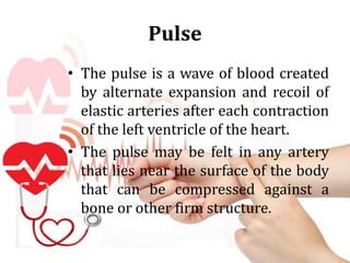
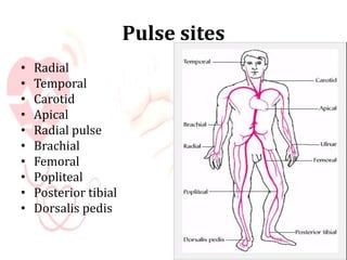
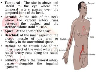
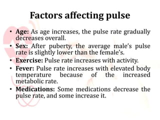
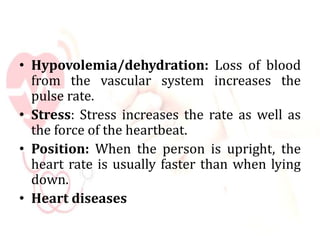
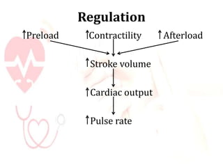
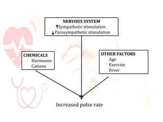


This document discusses the pulse, including its definition, assessment sites on the body, characteristics, factors that affect it, and regulation. The main points are: - The pulse is a wave of blood created by the expansion and recoil of arteries with each heartbeat. It can be felt at various artery sites near the skin. - Common pulse assessment sites include the radial, carotid, femoral, and posterior tibial arteries. The radial site is most commonly used. - A pulse is assessed by palpation or auscultation and expressed in beats per minute, normally 60-100 BPM for adults. Characteristics include rate, rhythm, volume, and arterial wall elasticity. - Fact