…
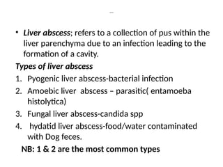
• Liver abscess;refers to a collection of pus within the
liver parenchyma due to an infection leading to the
formation of a cavity.
Types of liver abscess
1. Pyogenic liver abscess-bacterial infection
2. Amoebic liver abscess – parasitic( entamoeba
histolytica)
3. Fungal liver abscess-candida spp
4. hydatid liver abscess-food/water contaminated
with Dog feces.
NB: 1 & 2 are the most common types
4.
Etiology
Pyogenic liver abscess
Biliarytract disease( e.g.
cholangitis,cholecystitis)
Hematogenous spread( e.g. from sepsis)
trauma
Direct extension from adjacent structures
5.
………..
Amoebic liver abscess
Ingestionof cysts from contaminated foods or
water.
• Fungal liver abscess
Immunocompromised state e.g. in patients with
HIV/AIDS, transplant recipients.
Hydatid liver abscess –usually asymptomatic, but
symptoms depend on location, size and mass
effect, symptoms same as pyogenic, CT scan
reveals multiloculated mass; RX; albendazole /
mebendazole.
6.
Pathophysiology
Pyogenic liver abscess
Bacterialinvasion leads to localized inflammation,
tissue necrosis, and pus formation.
It’s the most common type of liver abscess and is
more common in immunocompromised as well
seen commonly in male patients of 5th
to 6th
decades who generally present with complaint of
fever with chills followed by abdominal
pain,anorexia,weight loss, fatigue,
jaundice(25%cases)
M:F 1.5 : 1
7.
……
Most common organismcausing pyogenic
liver abscess in western countries &
worldwide is E.coli,and in Asian countries its
klebsiella while in children with chronic
granulomatous disease its staph.Aureus.
Note; chronic granulomatous disease is
associated with neutrophillic dysfunction
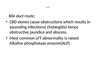
….
Bile duct route;
•CBD stones cause obstructions which results in
ascending infections( cholangitis) hence
obstructive jaundice and abscess.
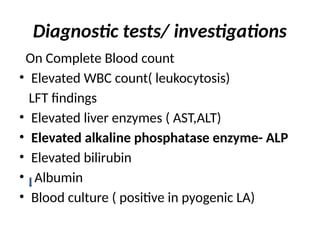
• Most common LFT abnormality is raised
Alkaline phosphatase enzyme(ALP).
10.
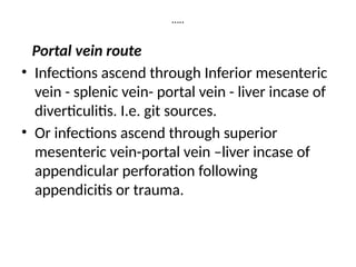
…..
Portal vein route
•Infections ascend through Inferior mesenteric
vein - splenic vein- portal vein - liver incase of
diverticulitis. I.e. git sources.
• Or infections ascend through superior
mesenteric vein-portal vein –liver incase of
appendicular perforation following
appendicitis or trauma.
11.
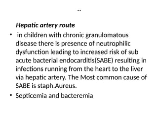
..
Hepatic artery route
•in children with chronic granulomatous
disease there is presence of neutrophilic
dysfunction leading to increased risk of sub
acute bacterial endocarditis(SABE) resulting in
infections running from the heart to the liver
via hepatic artery. The Most common cause of
SABE is staph.Aureus.
• Septicemia and bacteremia
12.
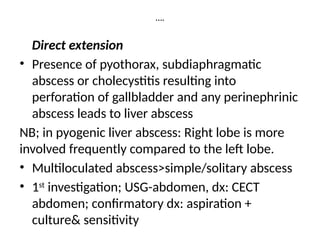
….
Direct extension
• Presenceof pyothorax, subdiaphragmatic
abscess or cholecystitis resulting into
perforation of gallbladder and any perinephrinic
abscess leads to liver abscess
NB; in pyogenic liver abscess: Right lobe is more
involved frequently compared to the left lobe.
• Multiloculated abscess>simple/solitary abscess
• 1st
investigation; USG-abdomen, dx: CECT
abdomen; confirmatory dx: aspiration +
culture& sensitivity
13.
…..
b) Amebic liverabscess
• Its caused by Entamoeba histolytica parasite
• Ingested cysts of the parasite release
trophozoites that invade the intestinal
mucosa, enter the blood stream to reach the
liver which either directly injure hepatocytes
or illicit an immune cascade.
14.
….
• Its morecommon in developing countries, young
patients( 2nd
-3rd
decades) and Alcoholics.
• M>>>F
Rt lobe>> Lt lobe and Mc route of infection- fecal oral route.
The infective stage; Quadrinucleate stage of cyst converts to
octanucleate which finally transforms to the active state in
the liver.
from Amoebic colitis ( flask shaped ulcer seen in amoebic
colitis), the colon amoeba gains entry into smv then via
portal vein into the liver and resultant reddish brown fluid
collection of damaged hepatocytes gives –An chay sauce
appearance of the formed abscess.
15.
…..
• Mc symptomis abdominal pain +/- fever and
jaundice is rare in ALA.
• Mc LFT abnormality is raised PT ( prothrombin
time).
• Simple abscess focus>>> multiple abscess.
• 1st
investigation-USG abdomen.
• Ioc for diagnosis/to confirm the dx is serology
( ELISA testing for Amebic serology or
antibodies)
16.
….
Fungal abscess
• Inthe immune compromised patients the
fungal spores spread hematogenously to the
liver causing abscess
17.
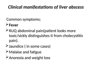
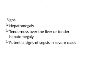
Clinical manifestations ofliver abscess
Common symptoms;
Fever
RUQ abdominal pain(patient looks more
toxic/sickly distinguishes it from cholecystitis
pain).
Jaundice ( in some cases)
Malaise and fatigue
Anorexia and weight loss
…..
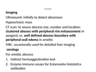
Imaging
Ultrasound: initially todetect abscesses:
Hypoechoeic mass
CT scan: to assess abscess size, number and location:
clustered abscess with peripheral rim enhancement in
pyogenic vs. well defined abscess boundary with
peripheral wall edema in amebic
MRI ; occasionally used for detailed liver imaging
serology
For amebic abscess
1. Indirect hemoagglutination test
2. Enzyme immuno assays for Entamoeba histolytica
antibodies
21.
…
Aspiration and culture
•Percutaneous aspiration of abscess for gram
stain, culture and sensitivity.
• Ideal for pyogenic LA
22.
Management
Medical therapy
Pyogenic abscess
•Broad spectrum antibiotics initially and then
tailored based on culture results.
Amoebic abscess
• Metronidazole or tinidazole followed by luminal
agents to eradicate intestinal cysts e.g. paromycin.
• Metronidazole is the drug of choice; dose 750mg
tds for 10-14 days
• Generally symptoms get resolved within 3-5 days
23.
…..
Indications for Aspiration
1.Symptoms do not improve in 3 to 5 days
2. Abscess size > 5cm
3. Left liver abscess
4. Pregnancy ( metro is not safe in pregnancy}
5. Diagnostic uncertainty
24.
….
Fungal abscess
• Antifungaltherapy e.g.. Amphotericin B,
fluconazole.
Drainage
percutaneous drainage ( guided by USG or CT)
Surgical drainage if percutaneous drainage fails or
if abscess is large and multiloculated.
Supportive care
Pain management with analgesics
Hydration and nutritional support
25.
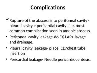
Complications
Rupture of theabscess into peritoneal cavity>
pleural cavity > pericardial cavity ..i.e. most
common complication seen in amebic abscess.
• Peritoneal cavity leakage-do EX-LAP+ lavage
and drainage.
• Pleural cavity leakage- place ICD/chest tube
insertion
• Pericardial leakage- Needle pericardiocentesis.
26.
….
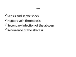
Sepsis and septicshock
Hepatic vein thrombosis
Secondary infection of the abscess
Recurrence of the abscess.
27.
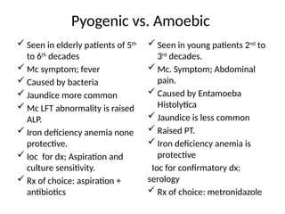
Pyogenic vs. Amoebic
Seen in elderly patients of 5th
to 6th
decades
Mc symptom; fever
Caused by bacteria
Jaundice more common
Mc LFT abnormality is raised
ALP.
Iron deficiency anemia none
protective.
Ioc for dx; Aspiration and
culture sensitivity.
Rx of choice: aspiration +
antibiotics
Seen in young patients 2nd
to
3rd
decades.
Mc. Symptom; Abdominal
pain.
Caused by Entamoeba
Histolytica
Jaundice is less common
Raised PT.
Iron deficiency anemia is
protective
Ioc for confirmatory dx;
serology
Rx of choice: metronidazole
28.
references
• Davidson’s principlesand practices of
medicine
• Sabiston text book of surgery 27th
edn.
• NEET PG quick revision guide on liver abscess
2024


![CASE_PRESENTATION_ON_subdural_hematoma(SDH)[1 FINAL PPT]-1.pptx](https://cdn.slidesharecdn.com/ss_thumbnails/casepresentationonsubduralhematomasdh1finalppt-1-260129172522-d405d375-thumbnail.jpg?width=640&height=640&fit=bounds)


