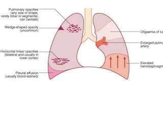
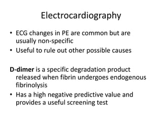
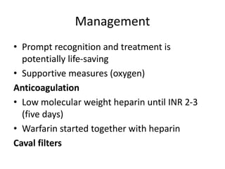
Venous Thromboembolism (VTE).
• Deep venous thrombosis and pulmonary
embolism (PE) can be considered as venous
thromboembolism (VTE).
• Exogenous or endogenous material that
travels to the lungs through the pulmonary
circulation,
• Causes a potential spectrum of consequences.
• The majority (75%) of pulmonary emboli arise
from the propagation of lower limb DVT.
• Amniotic fluid, placenta, air, fat, tumour
(especially choriocarcinoma), and
• septic emboli (from endocarditis ) are rare.
• PE occurs in around 1% of all patients
admitted to hospital
• Accounts for around 5% of in-hospital deaths.
• Common mode of death in patients with
cancer and stroke
• Common cause of death in pregnancy.
RISK FACTORS FOR VENOUS
THROMBOEMBOLISM
Surgery
• Major abdominal/pelvic surgery
• Hip/knee surgery
• Post-operative intensive care
Obstetrics
• Pregnancy/puerperium
Cardiorespiratory disease
• COPD
• Congestive cardiac failure
• Other disabling disease
Lower limb problems
• Fracture
• Varicose veins
• Stroke/spinal cord injury
• Malignant disease
• Miscellaneous
• Increasing age
• Previous proven VTE
• Immobility
• Thrombotic disorders
• Trauma
Pathophysiology
• The obstruction of blood flow creates alveolar
dead space
• High ventilation perfusion ratios as well as
shunting due to perfusion of atelectatic areas.
• This imbalance appears to be the principal
explanation for hypoxemia in acute PE.
Clinical Manifestations
• The history and physical examination are
insensitive and nonspecific for both DVT and
PE
Lower extremity venous thrombosis
• Erythema, warmth, pain, swelling, or
tenderness
• Homans' sign : pain with dorsiflexion of the
foot
CATEGORIES OF PULMONARY
THROMBOEMBOLI
• Acute small/medium PE
• Occlusion of segmental pulmonary artery →
infarction ± effusion
• Pleuritic chest pain, dyspnea, haemoptysis
• Tachycardia ,pleural rub, crackles, effusion
(often blood-stained)
• Low-grade fever
• Chest X-ray
Chronic PE
• Chronic occlusion of pulmonary
microvasculature, right heart failure
• Exertional dyspnoea, late symptoms of
pulmonary hypertension or right heart failure
• RV heave, loud, split P2
terminal-right heart failure
• Chest X –ray. Enlarged pulmonary artery
trunk, enlarged heart, prominent RV
Acute massive PE
• Major haemodynamic effects: ↓ cardiac
output; acute right heart failure
• Faintness or collapse, central chest pain,
apprehension, severe dyspnoea
• Major circulatory collapse: tachycardia,
hypotension, ↑ JVP, right ventricular gallop
rhythm, split P2 Severe cyanosis ↓ Urinary
output
• Usually normal, may be subtle oligaemia
Approch
• Is the clinical presentation consistent with PE?
• Does the patient have risk factors for PE?
• Is there any alternative diagnosis that can
explain the patient's presentation?
Investigations
• Chest radiography : non specific
• Important to exclude other differential
diagnosis
Electrocardiography
• ECG changes in PE are common but are
usually non-specific
• Useful to rule out other possible causes
D-dimer is a specific degradation product
released when fibrin undergoes endogenous
fibrinolysis
• Has a high negative predictive value and
provides a useful screening test
Management
• Prompt recognition and treatment is
potentially life-saving
• Supportive measures (oxygen)
Anticoagulation
• Low molecular weight heparin until INR 2-3
(five days)
• Warfarin started together with heparin
Caval filters
Duration of treatment
• Underlying prothrombotic risk or a history of
previous emboli
• Identifiable and reversible risk factor require
• Idiopathic VTE

venous thromboembolism (VTE).ppt

  • 1.
  • 2.
    • Deep venousthrombosis and pulmonary embolism (PE) can be considered as venous thromboembolism (VTE). • Exogenous or endogenous material that travels to the lungs through the pulmonary circulation, • Causes a potential spectrum of consequences.
  • 3.
    • The majority(75%) of pulmonary emboli arise from the propagation of lower limb DVT. • Amniotic fluid, placenta, air, fat, tumour (especially choriocarcinoma), and • septic emboli (from endocarditis ) are rare.
  • 4.
    • PE occursin around 1% of all patients admitted to hospital • Accounts for around 5% of in-hospital deaths. • Common mode of death in patients with cancer and stroke • Common cause of death in pregnancy.
  • 5.
    RISK FACTORS FORVENOUS THROMBOEMBOLISM Surgery • Major abdominal/pelvic surgery • Hip/knee surgery • Post-operative intensive care Obstetrics • Pregnancy/puerperium
  • 6.
    Cardiorespiratory disease • COPD •Congestive cardiac failure • Other disabling disease Lower limb problems • Fracture • Varicose veins • Stroke/spinal cord injury
  • 7.
    • Malignant disease •Miscellaneous • Increasing age • Previous proven VTE • Immobility • Thrombotic disorders • Trauma
  • 8.
    Pathophysiology • The obstructionof blood flow creates alveolar dead space • High ventilation perfusion ratios as well as shunting due to perfusion of atelectatic areas. • This imbalance appears to be the principal explanation for hypoxemia in acute PE.
  • 9.
    Clinical Manifestations • Thehistory and physical examination are insensitive and nonspecific for both DVT and PE Lower extremity venous thrombosis • Erythema, warmth, pain, swelling, or tenderness • Homans' sign : pain with dorsiflexion of the foot
  • 10.
    CATEGORIES OF PULMONARY THROMBOEMBOLI •Acute small/medium PE • Occlusion of segmental pulmonary artery → infarction ± effusion • Pleuritic chest pain, dyspnea, haemoptysis • Tachycardia ,pleural rub, crackles, effusion (often blood-stained) • Low-grade fever
  • 11.
  • 13.
    Chronic PE • Chronicocclusion of pulmonary microvasculature, right heart failure • Exertional dyspnoea, late symptoms of pulmonary hypertension or right heart failure • RV heave, loud, split P2 terminal-right heart failure • Chest X –ray. Enlarged pulmonary artery trunk, enlarged heart, prominent RV
  • 14.
    Acute massive PE •Major haemodynamic effects: ↓ cardiac output; acute right heart failure • Faintness or collapse, central chest pain, apprehension, severe dyspnoea • Major circulatory collapse: tachycardia, hypotension, ↑ JVP, right ventricular gallop rhythm, split P2 Severe cyanosis ↓ Urinary output
  • 15.
    • Usually normal,may be subtle oligaemia
  • 16.
    Approch • Is theclinical presentation consistent with PE? • Does the patient have risk factors for PE? • Is there any alternative diagnosis that can explain the patient's presentation?
  • 17.
    Investigations • Chest radiography: non specific • Important to exclude other differential diagnosis
  • 19.
    Electrocardiography • ECG changesin PE are common but are usually non-specific • Useful to rule out other possible causes D-dimer is a specific degradation product released when fibrin undergoes endogenous fibrinolysis • Has a high negative predictive value and provides a useful screening test
  • 20.
    Management • Prompt recognitionand treatment is potentially life-saving • Supportive measures (oxygen) Anticoagulation • Low molecular weight heparin until INR 2-3 (five days) • Warfarin started together with heparin Caval filters
  • 21.
    Duration of treatment •Underlying prothrombotic risk or a history of previous emboli • Identifiable and reversible risk factor require • Idiopathic VTE