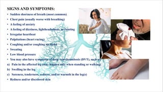
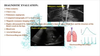
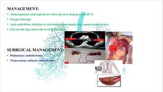
Download as PDF, PPTX

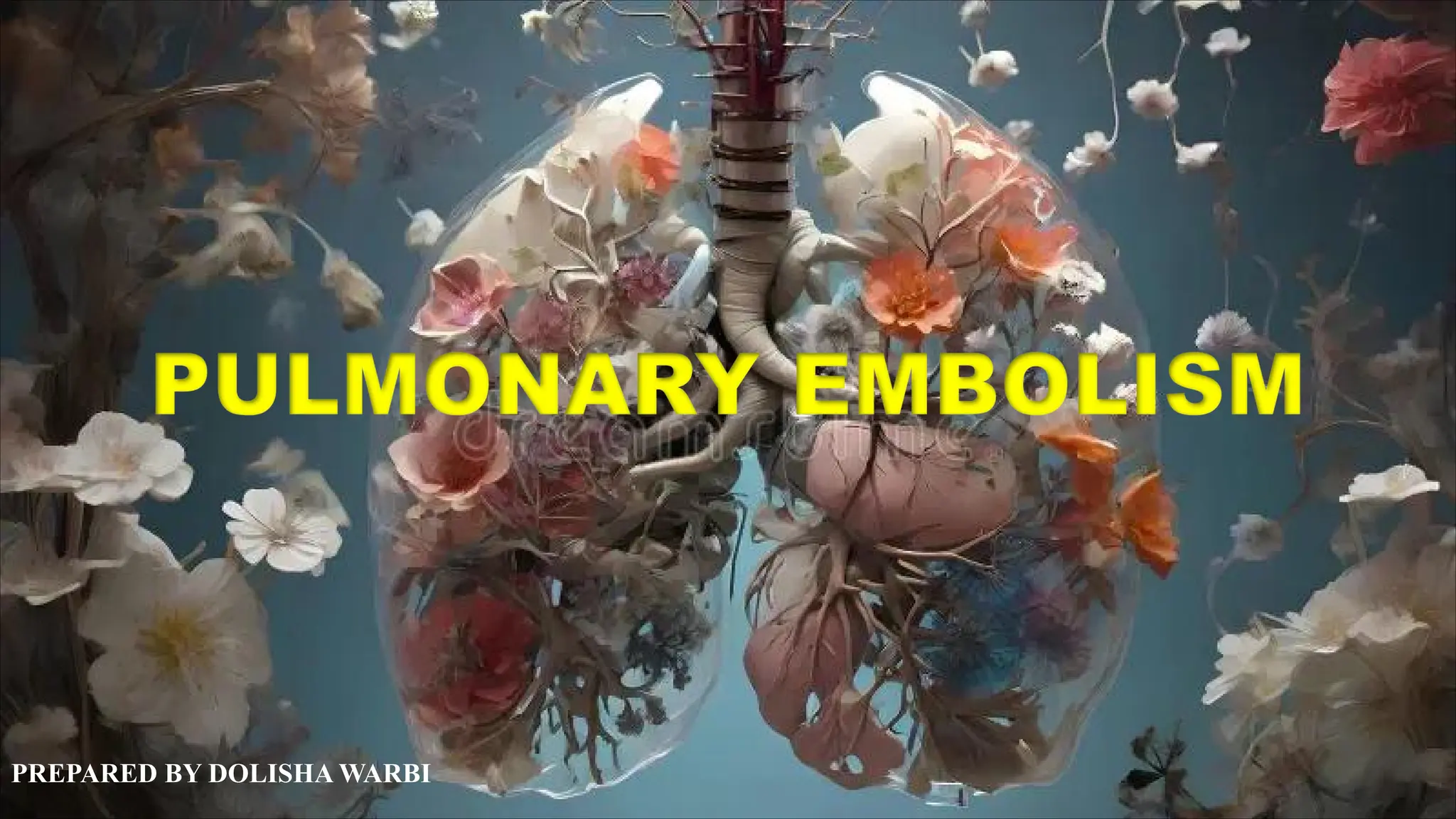
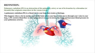
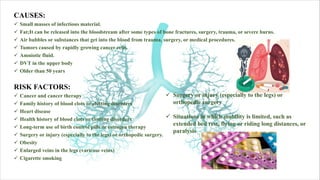
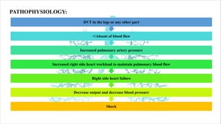


Pulmonary embolism (PE) is a blockage in the pulmonary artery caused by a thrombus, typically originating from the venous system, leading to reduced blood flow and oxygen levels in the lungs. Risk factors include cancer, family history of clots, obesity, and prolonged immobility. Symptoms often include sudden shortness of breath, chest pain, and dizziness, and management strategies involve anticoagulants, oxygen therapy, and possible surgical interventions.


![CTEV [ clubfoot] DR ARUN LAL ,DR MOHAMED ASHRAF travancore medical college k...](https://cdn.slidesharecdn.com/ss_thumbnails/ctevclubfootdrarunlaldrmohamedashraftravancoremedicalcollegekollamkeralaindia-260208063247-18fc466c-thumbnail.jpg?width=640&height=640&fit=bounds)

![PERI-PROSTHETIC FRACTURE NAIL-PLATE CONSTRUCT [NPC].pptx](https://cdn.slidesharecdn.com/ss_thumbnails/drarunkumardrmohamedashrafperiprostheticfrasturenail-plateconstructnpc-260209164459-7e9d15a1-thumbnail.jpg?width=640&height=640&fit=bounds)


