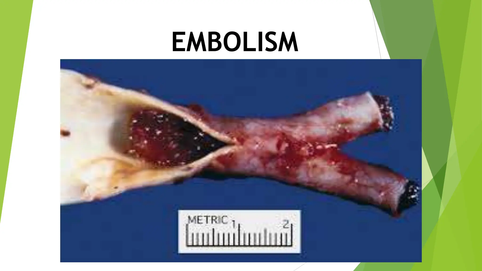
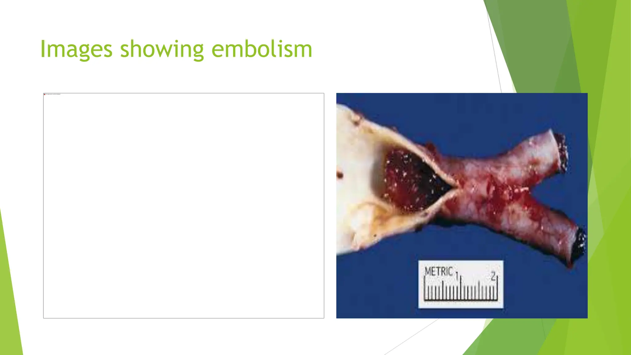
EMBOLISM
DEFINATION
An obstruction of the flow of blood by an
embolus, a particle or aggregate of substance
that is abnormally present in the bloodstream.
The substance may be a blood clot that has
broken loose from its point of formation (while it
is still adherent to the vessel at the point where
it was formed, the clot is called a thrombus); It
may be transferred to another area far from
where it has been formed.
contd
it may be a drop of soluble fat from a
crushing injury of fatty tissue; it may be a
clump of tumor cells, bacteria, or
detached tissue cells; it may be it may be
a drop of amniotic fluid that has entered
the maternal circulation during
childbirth; or it may be an air bubble
(called an air embolism).
TYPES
pulmonary embolism: An embolus, usually formed
in the leg (sometimes known as a deep vein
thrombosis or DVT), lodges in one of the arteries of
the lungs. Many emboli are broken down by the body
and go away by themselves; however, serious
pulmonary embolism may cause death.
brain embolism: If a blood clot travels to the brain,
this causes an ischemic stroke or TIA (transient
ischemic attack).
contd
retinal embolism: Small clots that wouldn't
block a major artery can block the smaller
blood vessels feeding the retina at the back of
the eye. The result is usually sudden blindness
in one eye.
septic embolism: This occurs when particles
created by infection in the body reach the
bloodstream and block blood vessels.
contd
amniotic embolism: Not all emboli are made of
clotted blood. In pregnancy, the womb is filled
with amniotic fluid, which protects the fetus.
Amniotic fluid can embolize and reach the
mother's lungs, causing pulmonary amniotic
embolism.
air embolism: Scuba divers who rise to the
surface too rapidly can generate air embolism,
bubbles in the blood that can block arterial
blood flow.
Images showing embolism
CAUSES
Mostly happen to people who have risk factors
for blood clot formation, such as smoking and
heart disease. Other RF for other types of
emboli include high blood pressure,
atherosclerosis ,high cholesterol, and obesity.
contd
Pulmonary embolism is linked to a lot of causes
and these are the most common:
Trauma. Trauma anywhere in the body could
cause PE especially if a clot is released from the
venous system.
Surgery. Certain surgical procedures such as
orthopedic, major abdominal, pelvic, and
gynecologic surgeries could cause PE.
contd
Hypercoagulable states. A patient with
hypercoagulability disorders would
most likely develop a clot that could
result in PE.
Prolonged immobility. Being unable to
move for a prolonged time predisposes
a person to PE.
SYMPTOMS
 Symptoms are determined by the type of embolism and which organ has been occluded.
Brain storm:
1. BRAIN?
2. KIDNEY?
3. LUNGS?
4. HEART ?
dx
CTPA or a computed tomographic angiography is
a special type of X-ray that is the most common
test used to diagnose PE because it uses
contrast to analyze blood vessels
D-Dimer blood tests to detect clot formation in
your blood.
Chest X-ray of your heart and lungs to rule out
other conditions with similar symptoms
contd
 Pulmonary V/Q scan to show which parts of your lungs are getting airflow and
blood flow
 Ultrasound of the legs to measure blood flow and assess for clots in the veins
 Pulmonary angiography to show the blood clots in the lungs
 Electrocardiogram to record heart activity
 Echocardiogram to measure heart function and assess for elevated pressure in
the pulmonary arteries.
 MRI is usually reserved for pregnant people and individuals that may not be
able to tolerate the contrast used in other imaging tests
Pulmonary embolism
 A pulmonary embolism (PE) is a blood clot in the blood vessels of your lung.
This happens when a clot in another part of your body (often your leg or arm)
moves through the veins to your lung. A PE restricts blood flow to your lungs,
lowers oxygen levels in your lungs and increases blood pressure in your
pulmonary arteries.
symptoms
Sudden shortness of breath — whether you’ve
been active or at rest.
Unexplained sharp pain in your chest, arm,
shoulder, neck or jaw. The pain may also be
similar to symptoms of a heart attack.
contd
Cough with or without bloody mucus.
Pale, clammy or bluish skin.
Rapid heartbeat (pulse).
contd
Excessive sweating.
In some cases, feeling anxious,
lightheaded, faint or passing out.
Wheezing.
Risk for
 Who is at risk for a pulmonary embolism?
 People at risk of developing a PE include those who:
 Have a blood clot in their leg, or deep vein thrombosis (DVT).
 Are inactive for long periods of time while traveling via motor
vehicle, train or plane (such as a long, cross-country car ride).
 Have recently had trauma or injury to a vein, possibly after a
recent surgery, fracture or from varicose veins.
 Are taking birth control pills (oral contraceptives) or hormone
replacement therapy.
contd
Currently smoke.
Have a history of heart failure or stroke.
Have overweight (a Body Mass Index or BMI greater
than 25)/obesity (a BMI greater than 30).
Are pregnant or have given birth in the previous six
weeks.
Received a central venous catheter through their
arm or leg.
dx
Blood tests (including the D-dimer test).
Computed tomography (CT) angiogram.
Doppler ultra sound: (This helps identify blood
clots in people’s legs, or deep vein thrombosis,
which can move to the lungs and become a PE
and cause more damage.)
contd
A ventilation/perfusion (V/Q) scan, if
you’re unable to get contrast for a CT
scan. (This is a nuclear scan that can
detect clots in your lung.)
Pulmonary angiogram.
Chest X-ray.
Mnx
Anticoagulants
Thrombolytic therapy: TPA
Surgery: Surgical embolectomy, Transvenous
catheter embolectomy
Nursing dx
Ineffective peripheral tissue perfusion related
to obstructed pulmonary artery.
Risk for shock related to increased workload of
the right ventricle.
Acute pain related to pleuritic origin.
Nursing mnx
A key role of the nurse is to identify
the patient at high risk for pulmonary
embolism and to minimize the risk of
PE in all patients.
Nursing mnx
Planning and goals for a patient with pulmonary
embolism include the following:
Increase perfusion
Verbalize understanding of condition, therapy
regimen, and medication side effects.
Display hemodynamic stability.
Report pain is relieved or controlled.
Follow prescribed pharmacologic regimen.
Intervention
 Prevent venous stasis. Encourage ambulation and
active and passive leg exercises to prevent venous
stasis.
 Monitor thrombolytic therapy. Monitoring
thrombolytic and anticoagulant therapy through INR
or PTT.
 Manage pain. Turn patient frequently and reposition
to improve ventilation-perfusion ratio.
contd
Manage oxygen therapy. Assess for
signs of hypoxemia and monitor the
pulse oximetry values.
Relieve anxiety. Encourage the patient
to talk about any fears or concerns
related to this frightening episode.
Evaluation
Success of the treatment plan will be evaluated with the following:
Increased perfusion.
Verbalized understanding of condition, therapy
regimen, and medication side effects.
Displayed hemodynamic stability.
Reported pain is relieved or controlled.
Followed prescribed pharmacologic regimen.

Jaundice in adults

  • 1.
  • 2.
    DEFINATION An obstruction ofthe flow of blood by an embolus, a particle or aggregate of substance that is abnormally present in the bloodstream. The substance may be a blood clot that has broken loose from its point of formation (while it is still adherent to the vessel at the point where it was formed, the clot is called a thrombus); It may be transferred to another area far from where it has been formed.
  • 3.
    contd it may bea drop of soluble fat from a crushing injury of fatty tissue; it may be a clump of tumor cells, bacteria, or detached tissue cells; it may be it may be a drop of amniotic fluid that has entered the maternal circulation during childbirth; or it may be an air bubble (called an air embolism).
  • 4.
    TYPES pulmonary embolism: Anembolus, usually formed in the leg (sometimes known as a deep vein thrombosis or DVT), lodges in one of the arteries of the lungs. Many emboli are broken down by the body and go away by themselves; however, serious pulmonary embolism may cause death. brain embolism: If a blood clot travels to the brain, this causes an ischemic stroke or TIA (transient ischemic attack).
  • 5.
    contd retinal embolism: Smallclots that wouldn't block a major artery can block the smaller blood vessels feeding the retina at the back of the eye. The result is usually sudden blindness in one eye. septic embolism: This occurs when particles created by infection in the body reach the bloodstream and block blood vessels.
  • 6.
    contd amniotic embolism: Notall emboli are made of clotted blood. In pregnancy, the womb is filled with amniotic fluid, which protects the fetus. Amniotic fluid can embolize and reach the mother's lungs, causing pulmonary amniotic embolism. air embolism: Scuba divers who rise to the surface too rapidly can generate air embolism, bubbles in the blood that can block arterial blood flow.
  • 7.
  • 8.
  • 9.
    CAUSES Mostly happen topeople who have risk factors for blood clot formation, such as smoking and heart disease. Other RF for other types of emboli include high blood pressure, atherosclerosis ,high cholesterol, and obesity.
  • 10.
    contd Pulmonary embolism islinked to a lot of causes and these are the most common: Trauma. Trauma anywhere in the body could cause PE especially if a clot is released from the venous system. Surgery. Certain surgical procedures such as orthopedic, major abdominal, pelvic, and gynecologic surgeries could cause PE.
  • 11.
    contd Hypercoagulable states. Apatient with hypercoagulability disorders would most likely develop a clot that could result in PE. Prolonged immobility. Being unable to move for a prolonged time predisposes a person to PE.
  • 12.
    SYMPTOMS  Symptoms aredetermined by the type of embolism and which organ has been occluded. Brain storm: 1. BRAIN? 2. KIDNEY? 3. LUNGS? 4. HEART ?
  • 13.
    dx CTPA or acomputed tomographic angiography is a special type of X-ray that is the most common test used to diagnose PE because it uses contrast to analyze blood vessels D-Dimer blood tests to detect clot formation in your blood. Chest X-ray of your heart and lungs to rule out other conditions with similar symptoms
  • 14.
    contd  Pulmonary V/Qscan to show which parts of your lungs are getting airflow and blood flow  Ultrasound of the legs to measure blood flow and assess for clots in the veins  Pulmonary angiography to show the blood clots in the lungs  Electrocardiogram to record heart activity
  • 15.
     Echocardiogram tomeasure heart function and assess for elevated pressure in the pulmonary arteries.  MRI is usually reserved for pregnant people and individuals that may not be able to tolerate the contrast used in other imaging tests
  • 16.
    Pulmonary embolism  Apulmonary embolism (PE) is a blood clot in the blood vessels of your lung. This happens when a clot in another part of your body (often your leg or arm) moves through the veins to your lung. A PE restricts blood flow to your lungs, lowers oxygen levels in your lungs and increases blood pressure in your pulmonary arteries.
  • 17.
    symptoms Sudden shortness ofbreath — whether you’ve been active or at rest. Unexplained sharp pain in your chest, arm, shoulder, neck or jaw. The pain may also be similar to symptoms of a heart attack.
  • 18.
    contd Cough with orwithout bloody mucus. Pale, clammy or bluish skin. Rapid heartbeat (pulse).
  • 19.
    contd Excessive sweating. In somecases, feeling anxious, lightheaded, faint or passing out. Wheezing.
  • 20.
    Risk for  Whois at risk for a pulmonary embolism?  People at risk of developing a PE include those who:  Have a blood clot in their leg, or deep vein thrombosis (DVT).  Are inactive for long periods of time while traveling via motor vehicle, train or plane (such as a long, cross-country car ride).  Have recently had trauma or injury to a vein, possibly after a recent surgery, fracture or from varicose veins.  Are taking birth control pills (oral contraceptives) or hormone replacement therapy.
  • 21.
    contd Currently smoke. Have ahistory of heart failure or stroke. Have overweight (a Body Mass Index or BMI greater than 25)/obesity (a BMI greater than 30). Are pregnant or have given birth in the previous six weeks. Received a central venous catheter through their arm or leg.
  • 22.
    dx Blood tests (includingthe D-dimer test). Computed tomography (CT) angiogram. Doppler ultra sound: (This helps identify blood clots in people’s legs, or deep vein thrombosis, which can move to the lungs and become a PE and cause more damage.)
  • 23.
    contd A ventilation/perfusion (V/Q)scan, if you’re unable to get contrast for a CT scan. (This is a nuclear scan that can detect clots in your lung.) Pulmonary angiogram. Chest X-ray.
  • 24.
    Mnx Anticoagulants Thrombolytic therapy: TPA Surgery:Surgical embolectomy, Transvenous catheter embolectomy
  • 25.
    Nursing dx Ineffective peripheraltissue perfusion related to obstructed pulmonary artery. Risk for shock related to increased workload of the right ventricle. Acute pain related to pleuritic origin.
  • 26.
    Nursing mnx A keyrole of the nurse is to identify the patient at high risk for pulmonary embolism and to minimize the risk of PE in all patients.
  • 27.
    Nursing mnx Planning andgoals for a patient with pulmonary embolism include the following: Increase perfusion Verbalize understanding of condition, therapy regimen, and medication side effects. Display hemodynamic stability. Report pain is relieved or controlled. Follow prescribed pharmacologic regimen.
  • 28.
    Intervention  Prevent venousstasis. Encourage ambulation and active and passive leg exercises to prevent venous stasis.  Monitor thrombolytic therapy. Monitoring thrombolytic and anticoagulant therapy through INR or PTT.  Manage pain. Turn patient frequently and reposition to improve ventilation-perfusion ratio.
  • 29.
    contd Manage oxygen therapy.Assess for signs of hypoxemia and monitor the pulse oximetry values. Relieve anxiety. Encourage the patient to talk about any fears or concerns related to this frightening episode.
  • 30.
    Evaluation Success of thetreatment plan will be evaluated with the following: Increased perfusion. Verbalized understanding of condition, therapy regimen, and medication side effects. Displayed hemodynamic stability. Reported pain is relieved or controlled. Followed prescribed pharmacologic regimen.