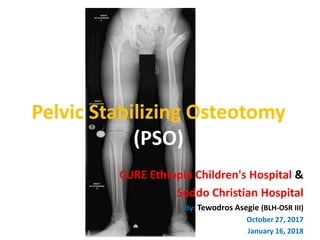
Pelvic stabilizing osteotomy (PSO) is a surgical procedure used to treat hip instability. It involves performing a valgus osteotomy of the proximal femur and sometimes a varus osteotomy of the distal femur. This realigns the hip to improve biomechanics and stability. For patient Abonesh, who has a dislocated left hip, previous girdlestone procedure, and limb length discrepancy, a PSO may be a suitable option to reduce pain, equalize limb length, and improve hip function and gait. Careful preoperative planning including measurement of adduction contracture and overcorrection angle is required to determine the degree of valgus angulation needed at the proximal osteotomy
a simplified version of periprosthetic fractures, easy to learn and understand with lots of images and classification. It includes hip, shaft of femur, knee, shoulder
Deformity: It’s the position of a limb/Joint, from which it cannot be brought back to its normal anatomical position.
Described as abnormalities of :
Length
Angulation
Rotation
Translation
Combination
Deformity: It’s the position of a limb/Joint, from which it cannot be brought back to its normal anatomical position.
Described as abnormalities of :
Length
Angulation
Rotation
Translation
Combination
This presentation goes in depth about Primary and Recurrent Patellar dislocation. Its cause, clinical and radiographic evaluation and various modalities of management with update from latest literature.
Hip dysplasia in adults, types, radiographs and management!
Useful for Orthopaedic residents and Surgeons.
Include most of the basics from reliable sources, pardon for any mistakes. Contact at singh_prabhjeet@yahoo.com for any corrections.
The hip joint is a ball and socket joint consisting of the femoral head and acetabulum. This articulation provides multiple planes of movement and is highly congruent. Articular cartilage, consisting of type II collagen, covers the majority of the femoral head. The acetabulum peripherally consists of articular cartilage while the central floor is non-articular and filled with a fatty layer termed the pulvinar. The ligamentum teres arises from both the transverse acetabular ligament and the central non-articular layer of the acetabulum and attaches to the central femoral head. It may play a role in stabilizing the hip joint.
Knee replacement in a recurvatum deformity is one of the most challenging scenarios. This uncommon surgery is based on a few simple principles which are explained in this ppt. Hyperextension can be a part of neurological disorder, primary ligament laxity, mal-alignment around the joint, post traumatic conditions etc. a careful dissection and minimal bone cuts are essential points in this surgery. Contrary to regular principle we end up stuffing the joint to balance the posterior capsular hammock.
Hope this ppt helps you with your surgery planning.
Ethanol (CH3CH2OH), or beverage alcohol, is a two-carbon alcohol
that is rapidly distributed in the body and brain. Ethanol alters many
neurochemical systems and has rewarding and addictive properties. It
is the oldest recreational drug and likely contributes to more morbidity,
mortality, and public health costs than all illicit drugs combined. The
5th edition of the Diagnostic and Statistical Manual of Mental Disorders
(DSM-5) integrates alcohol abuse and alcohol dependence into a single
disorder called alcohol use disorder (AUD), with mild, moderate,
and severe subclassifications (American Psychiatric Association, 2013).
In the DSM-5, all types of substance abuse and dependence have been
combined into a single substance use disorder (SUD) on a continuum
from mild to severe. A diagnosis of AUD requires that at least two of
the 11 DSM-5 behaviors be present within a 12-month period (mild
AUD: 2–3 criteria; moderate AUD: 4–5 criteria; severe AUD: 6–11 criteria).
The four main behavioral effects of AUD are impaired control over
drinking, negative social consequences, risky use, and altered physiological
effects (tolerance, withdrawal). This chapter presents an overview
of the prevalence and harmful consequences of AUD in the U.S.,
the systemic nature of the disease, neurocircuitry and stages of AUD,
comorbidities, fetal alcohol spectrum disorders, genetic risk factors, and
pharmacotherapies for AUD.
Report Back from SGO 2024: What’s the Latest in Cervical Cancer?bkling
Are you curious about what’s new in cervical cancer research or unsure what the findings mean? Join Dr. Emily Ko, a gynecologic oncologist at Penn Medicine, to learn about the latest updates from the Society of Gynecologic Oncology (SGO) 2024 Annual Meeting on Women’s Cancer. Dr. Ko will discuss what the research presented at the conference means for you and answer your questions about the new developments.
- Video recording of this lecture in English language: https://youtu.be/lK81BzxMqdo
- Video recording of this lecture in Arabic language: https://youtu.be/Ve4P0COk9OI
- Link to download the book free: https://nephrotube.blogspot.com/p/nephrotube-nephrology-books.html
- Link to NephroTube website: www.NephroTube.com
- Link to NephroTube social media accounts: https://nephrotube.blogspot.com/p/join-nephrotube-on-social-media.html
Anti ulcer drugs and their Advance pharmacology ||
Anti-ulcer drugs are medications used to prevent and treat ulcers in the stomach and upper part of the small intestine (duodenal ulcers). These ulcers are often caused by an imbalance between stomach acid and the mucosal lining, which protects the stomach lining.
||Scope: Overview of various classes of anti-ulcer drugs, their mechanisms of action, indications, side effects, and clinical considerations.
Couples presenting to the infertility clinic- Do they really have infertility...Sujoy Dasgupta
Dr Sujoy Dasgupta presented the study on "Couples presenting to the infertility clinic- Do they really have infertility? – The unexplored stories of non-consummation" in the 13th Congress of the Asia Pacific Initiative on Reproduction (ASPIRE 2024) at Manila on 24 May, 2024.
These lecture slides, by Dr Sidra Arshad, offer a quick overview of physiological basis of a normal electrocardiogram.
Learning objectives:
1. Define an electrocardiogram (ECG) and electrocardiography
2. Describe how dipoles generated by the heart produce the waveforms of the ECG
3. Describe the components of a normal electrocardiogram of a typical bipolar leads (limb II)
4. Differentiate between intervals and segments
5. Enlist some common indications for obtaining an ECG
Study Resources:
1. Chapter 11, Guyton and Hall Textbook of Medical Physiology, 14th edition
2. Chapter 9, Human Physiology - From Cells to Systems, Lauralee Sherwood, 9th edition
3. Chapter 29, Ganong’s Review of Medical Physiology, 26th edition
4. Electrocardiogram, StatPearls - https://www.ncbi.nlm.nih.gov/books/NBK549803/
5. ECG in Medical Practice by ABM Abdullah, 4th edition
6. ECG Basics, http://www.nataliescasebook.com/tag/e-c-g-basics
Factory Supply Best Quality Pmk Oil CAS 28578–16–7 PMK Powder in Stockrebeccabio
Factory Supply Best Quality Pmk Oil CAS 28578–16–7 PMK Powder in Stock
Telegram: bmksupplier
signal: +85264872720
threema: TUD4A6YC
You can contact me on Telegram or Threema
Communicate promptly and reply
Free of customs clearance, Double Clearance 100% pass delivery to USA, Canada, Spain, Germany, Netherland, Poland, Italy, Sweden, UK, Czech Republic, Australia, Mexico, Russia, Ukraine, Kazakhstan.Door to door service
Hot Selling Organic intermediates
ARTIFICIAL INTELLIGENCE IN HEALTHCARE.pdfAnujkumaranit
Artificial intelligence (AI) refers to the simulation of human intelligence processes by machines, especially computer systems. It encompasses tasks such as learning, reasoning, problem-solving, perception, and language understanding. AI technologies are revolutionizing various fields, from healthcare to finance, by enabling machines to perform tasks that typically require human intelligence.
Title: Sense of Taste
Presenter: Dr. Faiza, Assistant Professor of Physiology
Qualifications:
MBBS (Best Graduate, AIMC Lahore)
FCPS Physiology
ICMT, CHPE, DHPE (STMU)
MPH (GC University, Faisalabad)
MBA (Virtual University of Pakistan)
Learning Objectives:
Describe the structure and function of taste buds.
Describe the relationship between the taste threshold and taste index of common substances.
Explain the chemical basis and signal transduction of taste perception for each type of primary taste sensation.
Recognize different abnormalities of taste perception and their causes.
Key Topics:
Significance of Taste Sensation:
Differentiation between pleasant and harmful food
Influence on behavior
Selection of food based on metabolic needs
Receptors of Taste:
Taste buds on the tongue
Influence of sense of smell, texture of food, and pain stimulation (e.g., by pepper)
Primary and Secondary Taste Sensations:
Primary taste sensations: Sweet, Sour, Salty, Bitter, Umami
Chemical basis and signal transduction mechanisms for each taste
Taste Threshold and Index:
Taste threshold values for Sweet (sucrose), Salty (NaCl), Sour (HCl), and Bitter (Quinine)
Taste index relationship: Inversely proportional to taste threshold
Taste Blindness:
Inability to taste certain substances, particularly thiourea compounds
Example: Phenylthiocarbamide
Structure and Function of Taste Buds:
Composition: Epithelial cells, Sustentacular/Supporting cells, Taste cells, Basal cells
Features: Taste pores, Taste hairs/microvilli, and Taste nerve fibers
Location of Taste Buds:
Found in papillae of the tongue (Fungiform, Circumvallate, Foliate)
Also present on the palate, tonsillar pillars, epiglottis, and proximal esophagus
Mechanism of Taste Stimulation:
Interaction of taste substances with receptors on microvilli
Signal transduction pathways for Umami, Sweet, Bitter, Sour, and Salty tastes
Taste Sensitivity and Adaptation:
Decrease in sensitivity with age
Rapid adaptation of taste sensation
Role of Saliva in Taste:
Dissolution of tastants to reach receptors
Washing away the stimulus
Taste Preferences and Aversions:
Mechanisms behind taste preference and aversion
Influence of receptors and neural pathways
Impact of Sensory Nerve Damage:
Degeneration of taste buds if the sensory nerve fiber is cut
Abnormalities of Taste Detection:
Conditions: Ageusia, Hypogeusia, Dysgeusia (parageusia)
Causes: Nerve damage, neurological disorders, infections, poor oral hygiene, adverse drug effects, deficiencies, aging, tobacco use, altered neurotransmitter levels
Neurotransmitters and Taste Threshold:
Effects of serotonin (5-HT) and norepinephrine (NE) on taste sensitivity
Supertasters:
25% of the population with heightened sensitivity to taste, especially bitterness
Increased number of fungiform papillae
Pulmonary Thromboembolism - etilogy, types, medical- Surgical and nursing man...VarunMahajani
Disruption of blood supply to lung alveoli due to blockage of one or more pulmonary blood vessels is called as Pulmonary thromboembolism. In this presentation we will discuss its causes, types and its management in depth.
Pulmonary Thromboembolism - etilogy, types, medical- Surgical and nursing man...
Pso t. asegie
1. Pelvic Stabilizing Osteotomy
(PSO)
CURE Ethiopia Children's Hospital &
Soddo Christian Hospital
By: Tewodros Asegie (BLH-OSR III)
October 27, 2017
January 16, 2018
2. Introduction
• PSO: hip bypass procedure where upper femur is
bent at an angle that stabilizes the connection
between the femur and pelvis to allow walking
with less limp
• Salvage of damaged hips for whom arthrodesis or
arthroplasty not appropriate
• Successful PSO:
– Reduces limp,
– Equalizes LL and,
– Facilitates a more Energy-Efficient gait
3. Basic Principle and Goal
• Enhance femoro-pelvic stability by PF valgus
osteotomy,
– Increase weight-bearing surface between
periacetabular region and residual upper femur and
– Minimize excessive pelvic drop during single limb
stance
• Improve hip biomechanics by displacing the
center of gravity medially, which results in an
improvement in the mechanical efficiencies of
Abductor Muscles
4. History
• Bouvier in 1838 described the technique of PSO
for CDH
• Lorenz:
• Valgus osteotomy + med and prox displacement of the shaft
of femur
– Limitation of movement
• Schanz:
• Valgus, and sometimes extension, position to the distal
femoral segment
– Increased pelvic stability
• Both techniques induced limitation of movement
5. • Milch: expanded the concept and
popularized the PSO in the United
States during the mid-20th century
– (β)- postosteotomy angle
predicts abutment to pelvis
when its greater than pelvic
inclination angle (α)
– Excessive difference
restriction of movement
and secondary disability
• Recommended post-
osteotomy angle should lie
210°-240°
6. Limitations Of Traditional PSO
• Optimal extent of angulation is difficult to
achieve:
– If the angle is too large:
• Excessive genu valgum,
• Fixed pelvic obliquity, and
• Impingement pain on adduction of the lower extremity
to the neutral position may ensue
– If the angle is too small:
• Insufficient improvement in hip biomechanics, and
• Most importantly, Remaining LLD cannot be addressed
7. Ilizarov Hip Reconstruction
• Early mid 1970s
• Modified form of PSO, which combines distal
femoral osteotomy for concomitant
lengthening and varus angulation
8. • Ilizarov provided a solution through a second,
more distal osteotomy
– Enable a proximal valgus osteotomy large enough
to eradicate any degree of adduction in the hip
(and thereby eliminate the T-gait) but, through
the distal varus osteotomy, to achieve parallelism
of both legs
9. Indications
• Hip Instability: Severe Dysplastic Acetabulum
• DDH: neglected, unsuccessfully treated
• Traumatic hip dislocation with instability
• Paralytic or spastic dislocation (postpoliomyelitis, cerebral
palsy, muscular dystrophy)
• Femoral head and neck absence: partial or total
• Severe sequelae of septic arthritis (Choi type IV)
• Skeletal dysplasia (SED, Morquio, etc.)
• Severe AVN
• Post-Girdlestone resection arthroplasty
10. Relative Contraindications
• NOT performed in young children (Wolff’s law)
– Young children <12 yrs, . Rozbruch SR et al, 2005
• Rapid remodeling in 1 yr
• LLD
• Older patients when THR is better alternative
– No Specific recommendations for a cut-off age
not possible
• Chronic paralytic hip dislocations in non-
ambulating patients
12. A. Clinical Planning
1. Adduction contracture, Arc of
abduction/adduction, Maximum adduction?
• Flexing the hip over the unaffected or Contralateral hip
abducted over the edge of the examining couch
(unilateral)- better
• Unstable hip or abductor insufficiency pelvic obliquity
is equivalent to an adduction of that hip
• Measuring maximum adduction is to provide a basis
for the size of valgus osteotomy
13. 2. Direction and degree of rotation when
maximally adducted?
• Movement of limb as it maximally adducted, foot
progression angle as pt walks
i. Normal foot progression angle but the limb externally
rotates as it is adducted-Needs to compensate during
osteotomy
ii. Abnormal foot progression angle – Calculate derotation
angle
14. 3. FFD and arc of flexion/extension?
– FFD can be addressed at proximal osteotomy site
4. Contralateral hip?
15. B. X-ray Planning
• Three radiographs:
• Standing AP view of both lower limbs (preferably a
parallel beam scanogram),
• Pelvis AP view with
– Maximum abduction affected hip and
– Fully adducted affected pelvis
16. • Document:
• Presence of any deformities of
femur and tibia
• Hip pathology
• Estimate of LLD
19. • Position of the pelvis and
of the weight-bearing
limb in single stance that
serves as a reference in
planning for a pelvic
support osteotomy
• Horizontal line to the
pelvis with the knee and
ankle joint inclinations
parallel to it and to the
ground are the reference
positions
20.
21. C. Translating Findings
1. Proximal femoral osteotomy:
• Level,
• Degree and
• Direction of osteotomy
2. Distal femoral osteotomy:
• Level,
• Degree and
• Direction of osteotomy
22. Level of PFO
• Fully adduct femur
• Level situated between the
infracotyloid recess and the
ischial tuberosity
• Level can vary:
– Undiagnosed hip dislocations:
may be at a level coincident
with the superior border of
obturator foramen;
– Other scenarios: Lies with
projection of the ischial
tuberosity
• Long prox segment
advantage:abductor
mechanical benefits
23. Amount of Valgus
• Highly controversial
• Milch: Traditonal PSO recommends
• Postosteotomy angle 210-230 degree
• Mean PFO angle in different literature 35-60
degree
• IHR: irrespective of PF valgus overcorrection,
abduction deformity can be compensated by
DF osteotomy if it restores knee joint
inclination parallel to horizontal
24. • Paley: recommends
overcorrection 15-
25 degree in
addition to pelvic
drop angle
– Overcorrection
offsets the loss of
the valgus from
remodelling at the
osteotomy site
25. • Pafilas and Nayagam:
• Valgus Adduction = Max
Addn + Addn Contract + +
9 degree + Overcorrection
(30-40deg or 21-31
extravalgus)
• Choi et al:
• Recommend at least 25
degrees
overcorrection, and
>30 degrees in
preadolescents
26. • Overcorrection is entirely empirical in
anticipation of remodeling at the valgus
osteotomy and some atrophy of the soft
tissue interposed between the femur and
lateral pelvic wall
27. Amount of Extension
• Extension should be adjusted to correct hip
flexion contracture and pelvic tilt, and the
sacrofemoral angle
• FFC can be compensated for in the osteotomy
and this usually amounts to 20 degree
28. Amount of Derotation
• Clinicaly assessed
• Foot progression angle when walking and the
amount and direction of rotation when the
femur was fully adducted
• The sum of the two will be incorporated as a
derotation osteotomy either at the PF or DF
29. DFO Level
• Addresses:
– Excessive valgus of PFO
– Allows for derotation, and
– Lengthening as well
30. • Paley: CORA method which
utilizes an imaginary
proximal mechanical axis line
• proximal MAL corresponds
to a line perpendicular to the
horizontal pelvic line, passing
through the point of 1/3 to
1/2 the distance lateral to
the medial edge of the
proximal fragment
• Too proximally it can
medialise the entire limb
and vice versa
31. • IHR: presented a
formula that took into
consideration the
amount of PF valgus
and DF varus
angulation
32. • This level can be determined using image
manipulation software or using simple trigonometry
33. Amount of Varus
• Serves to bring the inclination of the knee
joint parallel with that of the horizontal line of
the pelvis
• Mean DF varus angulations varied: 10 and 22
degrees
• To maintain some overcorrection, we suggest
that the distal osteotomy is undercorrected
(the knee joint line left in some valgus created
by the proximal osteotomy);
35. Amount of Lengthening
• Most reliable measurement is after PSO
• Woodblock or scanogram to obtain a level
pelvis
• Overlengthening is poorly tolerated
36. Where Is the Weight-bearing Fulcrum?
• Not absolute, nor a true joint or false
articulation
• The center of rotation seems to vary with the
position direction of motion of the lower
limbs and depends on the soft-tissue
interpositional weight-bearing surface
between the PF osteotomy and the pelvis
37. • When determining the
center of rotation during
adduction/ abduction
motion, the main fulcrum
is seemingly located
around the lesser
trochanter and not
around the apex of the
valgus angulation
adjacent to the ischial
tuberosity during passive
abduction and adduction
38. T-Gait after PSO
• 30.3% (0-62.5%)
– Abductor Insufficiency (preop atrophy or loss of
fulcrum during follow up)
• Inan et al.: correction of Trendelenburg sign
depended on the age at the time of operation
and the volume of the gluteus medius muscle
39. PSO to THR
• Still possible, although it is
a technically demanding
procedure due to the PF
deformity
• More complications are
present for patients with
hip instability due to septic
reasons
– Chen et al. : 14% rate of
infection after hip
arthroplasty in patients with
coxarthrosis due to previous
septic arthritis, and a 36%
rate of total complications
41. • Retrospective, 21 hips of 20 pts, 13 neglected hip dislocation, 7
septic, 1 failed THR, age 12- 34(22.6), follow up 33.45months(16-45)
• Result:
• Harris score 48.3 t0 80.1
• Preop LLD 53.3 mm and mean proximal migration was 42.9 mm, reduced to 16
mm after an average lengthening of 63.3mm
• Trendelenburg gait disappeared completely in 13 of 21 hips and was improved
in 8 hips
• Conclusion: Pelvic support osteotomy is a good treatment option to
overcome hip instability asit improves pain and equalizes limb length
47. Abonesh Lemi, 17 F, ET34277
• Subj:
– Left hip deformity since 10 yrs old
– Severe pain in the right hip, lay in bed for the last 3 yrs
– Had no hx of previous treatment
• Obj:
– Hip;
• Gait: Severely antalgic,
• LLD = 4 cm,
• FFD of hip = 10 degree,
• Flexion = 90 degree,
• Minimal rotation,
• Fixed adduction = 15-20 degree,
– Knee: fine
• Ass: Lt degenerate/dislocated hip
51. 12/10/2017
• In the wards put on traction
• Wound healed without complication
• Done:
– Lt Schanz Osteotomy
– Approach: Lateral vastus elevating
– Prebent plate with 60 degrees applied
– Predrilled with 3 proximal screws
– Osteotomy at the level of ischium with 60 degree abduction of
hip
– Wedge excised and plated distally
– Anterior 4 hole recon plate applied
– Hip stable
– Wound closed in layers
59. Options of treatment for Abonesh in
Our Setup
• Treatment of hip instability in adolescents and
young adults:
– Greater trochanteric arthroplasty,
– THR
– Hip arthrodesis,
– Femoral osteotomy, and
– Girdlestone operation---unstable hip, crutches
for life
– PSO
60. Summary
• The aim in the treatment of patients with hip
instability is overcoming:
– hip pain,
– equalizing limb length deficiency,
– preventing limping,
– improving range of motion of the hip and thus
restoring hip stability whilst preserving the
biomechanical alignment of the extremity
• Milch: femoral head resection is an improvement
of PSO
• PSO is a possible option in Ethiopian set up too
61. References
• In Ho Choi, Tae-Joon Cho, etal, Recurrent
dislocations and complete necrosis: the role
of pelvic support osteotomy. J Pediatr
Orthop july/august,2013
• Dimitros Pafilas, Selvadurai Nayagam, The
pelvic support osteotomy: Indications and
preoperative planning. Strat Traum Limb
Recon, August 2008
65. • Loss of parallelism was in effect an ‘abduction
contracture’
– Compensate with eversion of the foot and with
tilting of the pelvis (relative adduction of
contralateral hip)
66. Valgus Osteotomy
= Max Addn + Addn Contracture
• Brings shaft to vertical with 9 deg
overcorrection(a)
– Does not lateralise shaft or knee joint
sufficiently
• Overcorrection in the region of 30
deg is preferable to allow a shift of
the limb from the midline
– Produces an ‘abduction contracture’,
i.e. in order to stand with both legs
parallel, the patient has to tilt the
pelvis
67. • If a varus correction is produced at the distal
osteotomy, then the effect of this ‘abduction
contracture’ is lost (and so will any
overcorrection at the proximal osteotomy)
• overcorrection factor should be added to the
valgus correction at the proximal osteotomy
because a failure to do so will leave the entire
limb medialised and much closer to the
midline than the contralateral side
68. • overcorrection of 9 deg will leave the shaft of the
femur parallel to the vertical axis of the pelvis
• In view of this, we suggest that the
overcorrection performed at the proximal
osteotomy level should be greater than the 25
suggested by Choi et al to enable an abduction of
the shaft of the femur away from the midline
• An overcorrection of 30–40 brings the
postosteotomy angle of Milch close to the
recommended 240 deg
69. • X-ray parameters:
1. Distance of the centre of the contralateral knee to the
midline axis of the body (from the standing AP film of
both lower limbs)
2. Distance of the proposed proximal osteotomy site from
the midline axis of the body (from the AP film of the
pelvis with hip in maximum adduction
3. Proposed overcorrection—we recommend adding 30 of
extra valgus to the sum of the adduction range and
measured adduction contracture
• Nine degrees of this overcorrection will bring the femur parallel
to the vertical axis, and the remaining 21 will take the femur
away from the midline
Editor's Notes
Double level femoral osteotomy with the objective of eliminating a Trendelenburg and short limb gait
Increase weight-bearing surface between periacetabular region and residual upper femur and
Minimize excessive pelvic drop during single limb stance
impingement occurred when
the patient attempted to bring the leg into parallel with the
contralateral side Milch described a
post-osteotomy angle (b) which
predicted abutment against the
lateral wall of the pelvis when it
exceeded the angle of pelvic
inclination (a). When the
difference was excessive,
restriction of movement was
significant and created a
secondary disability
IHR is considered a breakthrough in terms of resolving
the inherent problems of PSO, as the treatment
goals for normal gait are to obtain stability by reconstructing
a stable fulcrum, to improve energy efficiency
by restoring abductor mechanism, and to improve
cosmetic appearance by eliminating shortening/joint
contracture-related problems
Even when performed at the age of 12 years, a
repeat procedure is likely to be needed at skeletal maturity time of surgery
LLD even if adressed at th
chronic paralytic hip dislocations
(from neuromuscular disorders, e.g. cerebral
palsy, myelomeningocele, poliomyelitis) in non-ambulating
patients
this obliquity can be prevented by eradicating of
any adduction at the hip in the single stance phase of gait.
This is achieved through the proximal femoral osteotomy by
placing the involved hip in maximum adduction
The
standing radiograph was used to measure the angle
between the adducted femur’s anatomical axis and
either a line connecting the upper ends of the iliac
crest or a line connecting the lower ends of the
sacroiliac joints. A valgus angulation with a similar
angle plus a 15 degree overcorrection was made. In
order to detect the level of the osteotomy site, radiographs
with the instable hip in maximum adduction
were used
Schematic
representation of relationships
between the pelvis, knee and
femur in bipedal and single limb
stance. a In bipedal stance with
the feet at shoulder’s width, the
knee joint is at slight valgus to
the horizontal plane (38). Both
knees are equidistant to the
midline vertical axis (x1 = x2). b
In single stance, the knee and
ankle joint of the weightbearing
limb are horizontal and
parallel to the pelvic line. This
is accomplished through a slight
adduction at the hip. The
femoral shaft subtends an angle
of 98 to the midline vertical
axis. The ground reaction force
moves closer to the standing
leg, x1 not equal to x2
When femoral shaft is fully adducted against the lateral wall of the pelvis, an AP X-ray of the pelvis gives a projection of abutment and this depicts the level of the proximal osteotomy
Level can vary:
Undiagnosed hip dislocations: may be at a level coincident with the superior border of obturator foramen;
Other scenarios: Lies with projection of the ischial tuberosity
In undiagnosed hip dislocations, where there can be greater proximal migration of the femur, this may be located at a level coincident with the superior border of the obturator foramen;
In other scenarios the level of proposed osteotomy lies coincident with part of the projection of the ischial tuberosity
firstly it can be difficult to obtain a
single stance pelvic drop angle without the patient requiring
some form of additional support to achieve balance
and, secondly, the measured range of adduction does not
account for an adduction contracture that may commonly
Exis
Abduction angle that is equal to the single stance pelvic drop angle or measured range of adduction, plus an overcorrection factor of 15–25 deg……difficult to assess
Measured range of adduction plus size of adduction contracture (If present) plus overcorrection
Max Addn + Addn Contract + + 9 degree bring the femur parallel to perp axis of pelvic line while overcorrection brings the distal femur away from midline
Overcorrection at the proximal level leaves the patient with an ‘abduction contracture
Based on FFD, sacrofemoral angle
Full correction of FFD without consideration of hip flexion arc is disadvantageous
If the arc is small, some consideration to the loss of maximum hip flexion is needed; whilst patients may benefit from a better standing posture (having reduced or eliminated their lumbar lordosis), they may complain from being unable to fasten on their shoes
Extension overcomes FFC and pelvic tilt
The second osteotomy removes the ‘abduction contracture’
and allows both limbs to be parallel, with the knee, ankle and the
pelvis horizontal. The treated side remains in maximum adduction at
its articulation with the pelvis, and therefore prevents a Trendelenburg
gait. Lengthening at the second osteotomy removes limb length
inequality
the site of the distal osteotomy will vary
depending on the amount of valgus overcorrection introduced;
similarly, it will also vary if the position of the
proximal osteotomy is in line with the medial edge of the
ischial tuberosity as compared with its lateral edgeperformed at the intersection of two lines: a vertical axis that is dropped from the horizontal line of the pelvis which traverses through the proximal osteotomy site and the mechanical axis of the tibia extrapolated proximally
The distal osteotomy was made at a level
between the proximal osteotomy site and the knee
joint line
The level of the second osteotomy is to allow the limb to
return to a near parallel alignment to the contralateral normal side but
leave a small residual overcorrection of valgus. The position of this
osteotomy can be worked out as part of the preoperative plan using
the equation described in the text where y = (x1 - x2)/sine h
To maintain some overcorrection, we suggest that the distal osteotomy is undercorrected (the knee joint line left in some valgus created by the proximal osteotomy); a convenient method is to leave the femoral shaft parallel to the vertical axis of the pelvis, thereby producing a valgus overcorrection of 9–10
This explains the trigonometric method above which aims to bring the femoral shaft parallel to the vertical midline axis, thereby leaving a valgus inclination at the knee of 9 which is equivalent to an ‘abduction contracture’ of the same amount
Fulcrum at LT cineradiography in a patient who underwent Shanz-type
PSO and managed to figure out that the center of rotation
was located around the lesser trochanter and not around
the apex of the valgus angulation adjacent to the ischial
tuberosity during passive abduction and adduction
34 yrs old, neglected DDH at 24 , IHR, THR at 34
1 Final orthoroentgenogram of the patient with no limb length
discrepancy. Lengthening was not made and the osteotomy
sites were fixed using locking plates
2 Pelvic support osteotomy using monolateral external fixator
in a patient with hip instability due to congenital dysplasia
of the hip
The mean Harris hip score in the final follow-up was
80.1 (range: 60 to 93), whilst it was 48.3 (range: 28
to 79) prior to surgical treatment (Table 1). The
increase in the Harris Hip Score was statistically significant
(p<0.05
PSO=53 degree, near the planned 60 degree
Schanz or Lorenz Osteotomies, exceeded pelvic inclination,
impingement occurred when the patient attempted to bring the leg into parallel with the contralateral side
Eliminating the Trendelenburg gait some surgeons erroneously increased the abduction angle (and consequently the post-osteotomy angulation) to such an excessive degree that it became a disability
Performing a valgus osteotomy equal in size to the maximum
range of adduction plus any adduction contracture will bring the
femoral shaft to its normal inclination of 9 to the vertical. Bringing
the shaft to vertical therefore overcorrects by 9 (valgus correction a).
This does not lateralise the shaft or knee joint sufficiently. Therefore
an overcorrection in the region of 30 is preferable to allow a shift of
the limb from the midline (valgus correction b). This overcorrection
in effect produces an ‘abduction contracture’, i.e. in order to stand
with both legs parallel, the patient has to tilt the pelvis