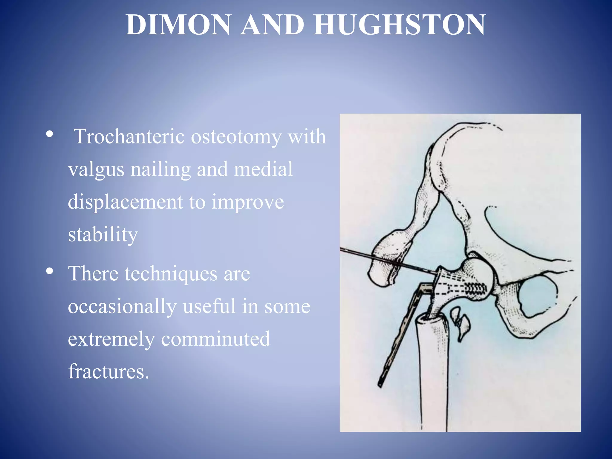
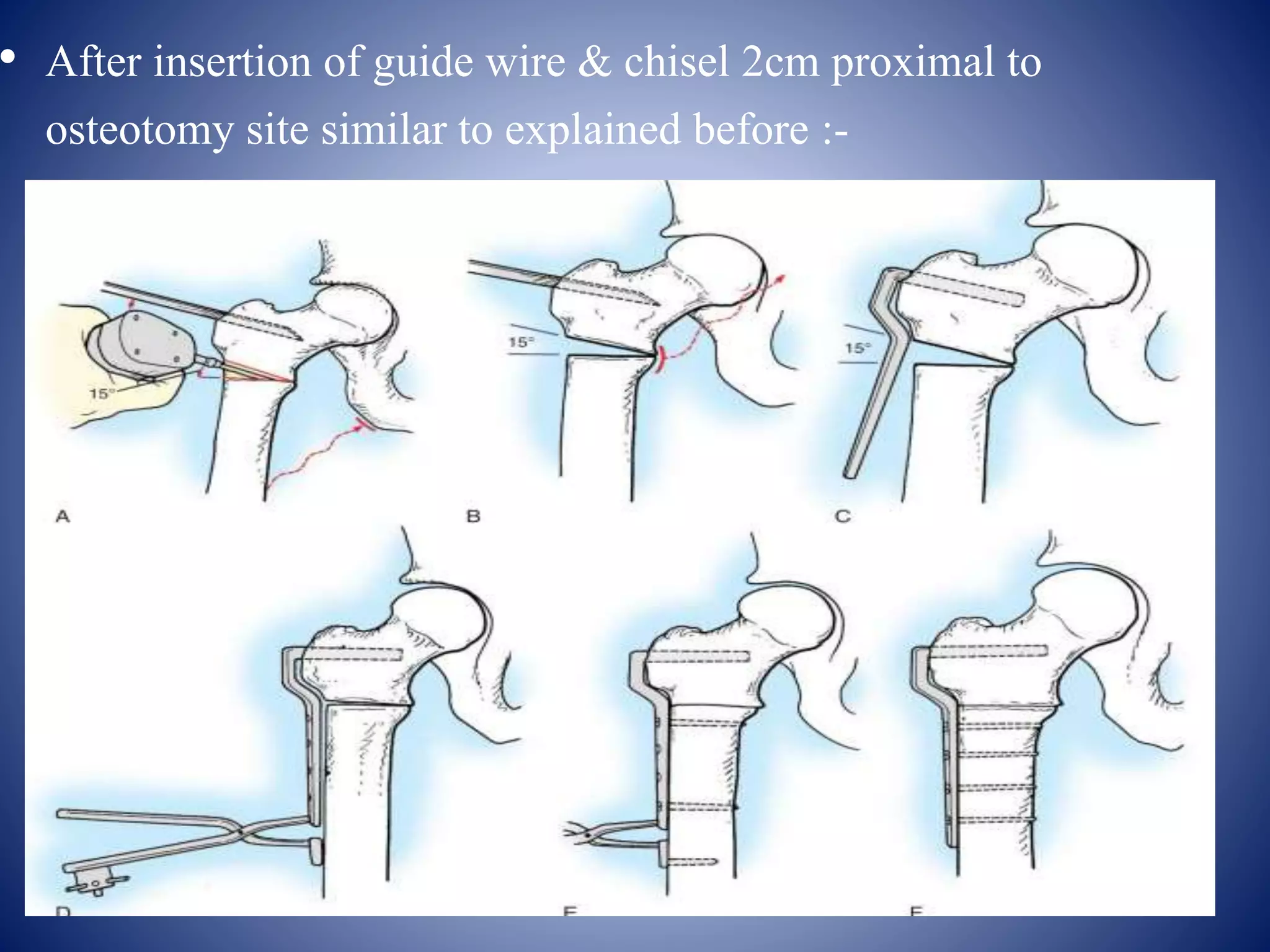
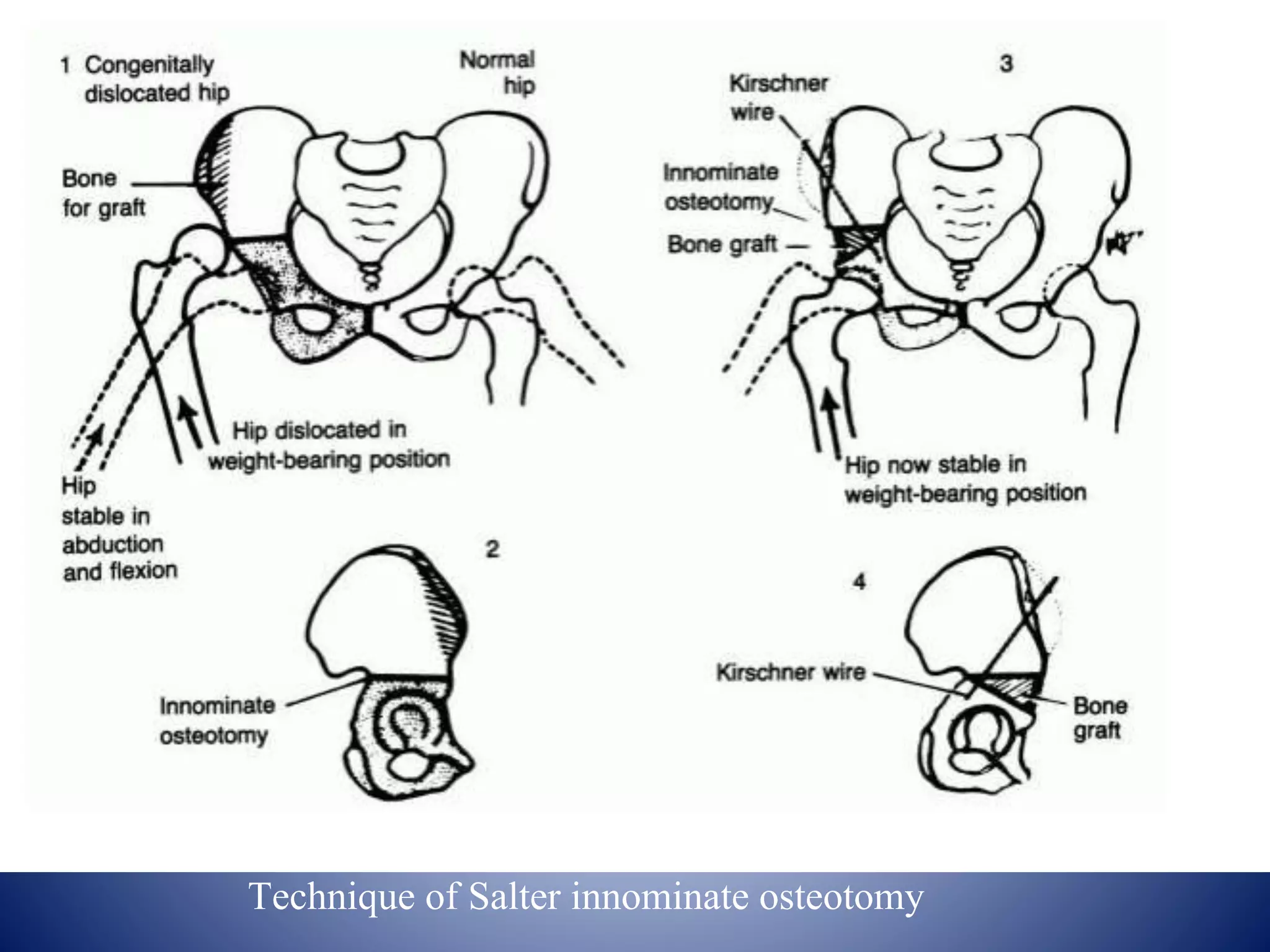
An osteotomy around the hip is a surgical procedure used to correct biomechanical alignment and load transmission. There are various types of osteotomies of the proximal femur classified by displacement of the distal fragment, anatomical location, and indication. Common osteotomies include McMurray's displacement osteotomy, Pauwel's varus/valgus osteotomy, and Salter's innominate osteotomy. The goal of osteotomies is to improve joint congruity, relieve pain, and correct deformities around the hip joint.
Deformity: It’s the position of a limb/Joint, from which it cannot be brought back to its normal anatomical position.
Described as abnormalities of :
Length
Angulation
Rotation
Translation
Combination
Deformity: It’s the position of a limb/Joint, from which it cannot be brought back to its normal anatomical position.
Described as abnormalities of :
Length
Angulation
Rotation
Translation
Combination
Presentation contain etiology, blood supply of femoral head & neck,pathogenesis ,classification system ,clinical features,diagnosis,managment, pelvic & femoral osteotomies in detail
Medial patellofemoral ligament reconstruction ---- an update on techniques used. This lecture was taken by me at Trinity Arthroscopy Course, Chandigarh.
a simplified version of periprosthetic fractures, easy to learn and understand with lots of images and classification. It includes hip, shaft of femur, knee, shoulder
GWT COMPENSATION PLAN 5 Source of income
60 to 100% profit on retail sales,Wholesale Profit
of up to $50 for each new VDMlauched , Package Sales
Earn up to 8% Payout to INFINITY, Website Purchases Business Development Bonus (BDB), Business Development Bonus (BDB) Elite, and more if you need more information about this business just contact me @ joannebiz@outlook.com i will be Glad to help you
Includes detailed description of BIOMECHANICS & PATHOMECHANICS OF KNEE JOINT AND PATELLOFEMORAL JOINT with recent evidences . Hope you find it useful!!
this slideshow describes about the hip joint anatomy, biomechanics and its pathomechanics along with angles of hip joint. the slide show also briefs about the pelvic femoral rhythm in daily activities
These simplified slides by Dr. Sidra Arshad present an overview of the non-respiratory functions of the respiratory tract.
Learning objectives:
1. Enlist the non-respiratory functions of the respiratory tract
2. Briefly explain how these functions are carried out
3. Discuss the significance of dead space
4. Differentiate between minute ventilation and alveolar ventilation
5. Describe the cough and sneeze reflexes
Study Resources:
1. Chapter 39, Guyton and Hall Textbook of Medical Physiology, 14th edition
2. Chapter 34, Ganong’s Review of Medical Physiology, 26th edition
3. Chapter 17, Human Physiology by Lauralee Sherwood, 9th edition
4. Non-respiratory functions of the lungs https://academic.oup.com/bjaed/article/13/3/98/278874
HOT NEW PRODUCT! BIG SALES FAST SHIPPING NOW FROM CHINA!! EU KU DB BK substit...GL Anaacs
Contact us if you are interested:
Email / Skype : kefaya1771@gmail.com
Threema: PXHY5PDH
New BATCH Ku !!! MUCH IN DEMAND FAST SALE EVERY BATCH HAPPY GOOD EFFECT BIG BATCH !
Contact me on Threema or skype to start big business!!
Hot-sale products:
NEW HOT EUTYLONE WHITE CRYSTAL!!
5cl-adba precursor (semi finished )
5cl-adba raw materials
ADBB precursor (semi finished )
ADBB raw materials
APVP powder
5fadb/4f-adb
Jwh018 / Jwh210
Eutylone crystal
Protonitazene (hydrochloride) CAS: 119276-01-6
Flubrotizolam CAS: 57801-95-3
Metonitazene CAS: 14680-51-4
Payment terms: Western Union,MoneyGram,Bitcoin or USDT.
Deliver Time: Usually 7-15days
Shipping method: FedEx, TNT, DHL,UPS etc.Our deliveries are 100% safe, fast, reliable and discreet.
Samples will be sent for your evaluation!If you are interested in, please contact me, let's talk details.
We specializes in exporting high quality Research chemical, medical intermediate, Pharmaceutical chemicals and so on. Products are exported to USA, Canada, France, Korea, Japan,Russia, Southeast Asia and other countries.
Factory Supply Best Quality Pmk Oil CAS 28578–16–7 PMK Powder in Stockrebeccabio
Factory Supply Best Quality Pmk Oil CAS 28578–16–7 PMK Powder in Stock
Telegram: bmksupplier
signal: +85264872720
threema: TUD4A6YC
You can contact me on Telegram or Threema
Communicate promptly and reply
Free of customs clearance, Double Clearance 100% pass delivery to USA, Canada, Spain, Germany, Netherland, Poland, Italy, Sweden, UK, Czech Republic, Australia, Mexico, Russia, Ukraine, Kazakhstan.Door to door service
Hot Selling Organic intermediates
Title: Sense of Taste
Presenter: Dr. Faiza, Assistant Professor of Physiology
Qualifications:
MBBS (Best Graduate, AIMC Lahore)
FCPS Physiology
ICMT, CHPE, DHPE (STMU)
MPH (GC University, Faisalabad)
MBA (Virtual University of Pakistan)
Learning Objectives:
Describe the structure and function of taste buds.
Describe the relationship between the taste threshold and taste index of common substances.
Explain the chemical basis and signal transduction of taste perception for each type of primary taste sensation.
Recognize different abnormalities of taste perception and their causes.
Key Topics:
Significance of Taste Sensation:
Differentiation between pleasant and harmful food
Influence on behavior
Selection of food based on metabolic needs
Receptors of Taste:
Taste buds on the tongue
Influence of sense of smell, texture of food, and pain stimulation (e.g., by pepper)
Primary and Secondary Taste Sensations:
Primary taste sensations: Sweet, Sour, Salty, Bitter, Umami
Chemical basis and signal transduction mechanisms for each taste
Taste Threshold and Index:
Taste threshold values for Sweet (sucrose), Salty (NaCl), Sour (HCl), and Bitter (Quinine)
Taste index relationship: Inversely proportional to taste threshold
Taste Blindness:
Inability to taste certain substances, particularly thiourea compounds
Example: Phenylthiocarbamide
Structure and Function of Taste Buds:
Composition: Epithelial cells, Sustentacular/Supporting cells, Taste cells, Basal cells
Features: Taste pores, Taste hairs/microvilli, and Taste nerve fibers
Location of Taste Buds:
Found in papillae of the tongue (Fungiform, Circumvallate, Foliate)
Also present on the palate, tonsillar pillars, epiglottis, and proximal esophagus
Mechanism of Taste Stimulation:
Interaction of taste substances with receptors on microvilli
Signal transduction pathways for Umami, Sweet, Bitter, Sour, and Salty tastes
Taste Sensitivity and Adaptation:
Decrease in sensitivity with age
Rapid adaptation of taste sensation
Role of Saliva in Taste:
Dissolution of tastants to reach receptors
Washing away the stimulus
Taste Preferences and Aversions:
Mechanisms behind taste preference and aversion
Influence of receptors and neural pathways
Impact of Sensory Nerve Damage:
Degeneration of taste buds if the sensory nerve fiber is cut
Abnormalities of Taste Detection:
Conditions: Ageusia, Hypogeusia, Dysgeusia (parageusia)
Causes: Nerve damage, neurological disorders, infections, poor oral hygiene, adverse drug effects, deficiencies, aging, tobacco use, altered neurotransmitter levels
Neurotransmitters and Taste Threshold:
Effects of serotonin (5-HT) and norepinephrine (NE) on taste sensitivity
Supertasters:
25% of the population with heightened sensitivity to taste, especially bitterness
Increased number of fungiform papillae
Couples presenting to the infertility clinic- Do they really have infertility...Sujoy Dasgupta
Dr Sujoy Dasgupta presented the study on "Couples presenting to the infertility clinic- Do they really have infertility? – The unexplored stories of non-consummation" in the 13th Congress of the Asia Pacific Initiative on Reproduction (ASPIRE 2024) at Manila on 24 May, 2024.
Lung Cancer: Artificial Intelligence, Synergetics, Complex System Analysis, S...Oleg Kshivets
RESULTS: Overall life span (LS) was 2252.1±1742.5 days and cumulative 5-year survival (5YS) reached 73.2%, 10 years – 64.8%, 20 years – 42.5%. 513 LCP lived more than 5 years (LS=3124.6±1525.6 days), 148 LCP – more than 10 years (LS=5054.4±1504.1 days).199 LCP died because of LC (LS=562.7±374.5 days). 5YS of LCP after bi/lobectomies was significantly superior in comparison with LCP after pneumonectomies (78.1% vs.63.7%, P=0.00001 by log-rank test). AT significantly improved 5YS (66.3% vs. 34.8%) (P=0.00000 by log-rank test) only for LCP with N1-2. Cox modeling displayed that 5YS of LCP significantly depended on: phase transition (PT) early-invasive LC in terms of synergetics, PT N0—N12, cell ratio factors (ratio between cancer cells- CC and blood cells subpopulations), G1-3, histology, glucose, AT, blood cell circuit, prothrombin index, heparin tolerance, recalcification time (P=0.000-0.038). Neural networks, genetic algorithm selection and bootstrap simulation revealed relationships between 5YS and PT early-invasive LC (rank=1), PT N0—N12 (rank=2), thrombocytes/CC (3), erythrocytes/CC (4), eosinophils/CC (5), healthy cells/CC (6), lymphocytes/CC (7), segmented neutrophils/CC (8), stick neutrophils/CC (9), monocytes/CC (10); leucocytes/CC (11). Correct prediction of 5YS was 100% by neural networks computing (area under ROC curve=1.0; error=0.0).
CONCLUSIONS: 5YS of LCP after radical procedures significantly depended on: 1) PT early-invasive cancer; 2) PT N0--N12; 3) cell ratio factors; 4) blood cell circuit; 5) biochemical factors; 6) hemostasis system; 7) AT; 8) LC characteristics; 9) LC cell dynamics; 10) surgery type: lobectomy/pneumonectomy; 11) anthropometric data. Optimal diagnosis and treatment strategies for LC are: 1) screening and early detection of LC; 2) availability of experienced thoracic surgeons because of complexity of radical procedures; 3) aggressive en block surgery and adequate lymph node dissection for completeness; 4) precise prediction; 5) adjuvant chemoimmunoradiotherapy for LCP with unfavorable prognosis.
- Video recording of this lecture in English language: https://youtu.be/lK81BzxMqdo
- Video recording of this lecture in Arabic language: https://youtu.be/Ve4P0COk9OI
- Link to download the book free: https://nephrotube.blogspot.com/p/nephrotube-nephrology-books.html
- Link to NephroTube website: www.NephroTube.com
- Link to NephroTube social media accounts: https://nephrotube.blogspot.com/p/join-nephrotube-on-social-media.html
Ozempic: Preoperative Management of Patients on GLP-1 Receptor Agonists Saeid Safari
Preoperative Management of Patients on GLP-1 Receptor Agonists like Ozempic and Semiglutide
ASA GUIDELINE
NYSORA Guideline
2 Case Reports of Gastric Ultrasound
micro teaching on communication m.sc nursing.pdfAnurag Sharma
Microteaching is a unique model of practice teaching. It is a viable instrument for the. desired change in the teaching behavior or the behavior potential which, in specified types of real. classroom situations, tends to facilitate the achievement of specified types of objectives.
TEST BANK for Operations Management, 14th Edition by William J. Stevenson, Ve...kevinkariuki227
TEST BANK for Operations Management, 14th Edition by William J. Stevenson, Verified Chapters 1 - 19, Complete Newest Version.pdf
Tom Selleck Health: A Comprehensive Look at the Iconic Actor’s Wellness Journeygreendigital
Tom Selleck, an enduring figure in Hollywood. has captivated audiences for decades with his rugged charm, iconic moustache. and memorable roles in television and film. From his breakout role as Thomas Magnum in Magnum P.I. to his current portrayal of Frank Reagan in Blue Bloods. Selleck's career has spanned over 50 years. But beyond his professional achievements. fans have often been curious about Tom Selleck Health. especially as he has aged in the public eye.
Follow us on: Pinterest
Introduction
Many have been interested in Tom Selleck health. not only because of his enduring presence on screen but also because of the challenges. and lifestyle choices he has faced and made over the years. This article delves into the various aspects of Tom Selleck health. exploring his fitness regimen, diet, mental health. and the challenges he has encountered as he ages. We'll look at how he maintains his well-being. the health issues he has faced, and his approach to ageing .
Early Life and Career
Childhood and Athletic Beginnings
Tom Selleck was born on January 29, 1945, in Detroit, Michigan, and grew up in Sherman Oaks, California. From an early age, he was involved in sports, particularly basketball. which played a significant role in his physical development. His athletic pursuits continued into college. where he attended the University of Southern California (USC) on a basketball scholarship. This early involvement in sports laid a strong foundation for his physical health and disciplined lifestyle.
Transition to Acting
Selleck's transition from an athlete to an actor came with its physical demands. His first significant role in "Magnum P.I." required him to perform various stunts and maintain a fit appearance. This role, which he played from 1980 to 1988. necessitated a rigorous fitness routine to meet the show's demands. setting the stage for his long-term commitment to health and wellness.
Fitness Regimen
Workout Routine
Tom Selleck health and fitness regimen has evolved. adapting to his changing roles and age. During his "Magnum, P.I." days. Selleck's workouts were intense and focused on building and maintaining muscle mass. His routine included weightlifting, cardiovascular exercises. and specific training for the stunts he performed on the show.
Selleck adjusted his fitness routine as he aged to suit his body's needs. Today, his workouts focus on maintaining flexibility, strength, and cardiovascular health. He incorporates low-impact exercises such as swimming, walking, and light weightlifting. This balanced approach helps him stay fit without putting undue strain on his joints and muscles.
Importance of Flexibility and Mobility
In recent years, Selleck has emphasized the importance of flexibility and mobility in his fitness regimen. Understanding the natural decline in muscle mass and joint flexibility with age. he includes stretching and yoga in his routine. These practices help prevent injuries, improve posture, and maintain mobilit
2. BIOMECHANICS OF THE HIP JOINT
• The acetabulum and femoral head form a multiaxial spheroidal
(‘ball and socket') joint
• Allows relatively unhindered motion in three degrees of
freedom-flexon/extenson
-abduction/adduction
-internal/external rotation
• This articulation is innately limited in its capacity for
translational motion in anteroposterior, transverse and vertical
planes
3. • In the coronal plane, the femoral neck is inclined obliquely to
the shaft at an angle of 135° (range 120–145°).
• The centre of the neck in the coronal plane is at the level of the
apex of the greater trochanter.
• In the axial plane the femoral neck is anteverted, i.e. rotated
anteriorly relative to the posterior surfaces of the femoral
condyles: in the adult, this angle is 10–15
4. The angle (NSA) between the long axis of the femoral shaft (S) and the axis of the femoral neck (N) is on
average 135° (range 125–140°). In addition, in most hips, a line perpendicular to S from the tip of the
greater trochanter (B) passes through the centre of the femoral head. This approximation can be utlilized in
judging the position of the femoral osteotomy in hip arthroplasty
5. ACETABULUM
• The acetabulum consists of the confluence of the ilium, ischium
and pubis at the triradiate cartilage.
• By itself, the acetabulum covers an area slightly less than a
hemisphere: it is deepened by the acetabular labrum.
• The degree of acetabular anteversion in the erect position is 14°
(men) and 19° (women).
• In the coronal plane, the acetabular axis is inclined
approximately 45° from the horizontal
6. FORCES ACTING ON THE HIP
• Under bipedal loading conditions, the femoral heads support
the weight of the body minus the weight of both legs
(approximately one-third body weight) and the resultant vectors
are vertical.
• When viewed in the sagittal plane, minimal muscle forces are
required to maintain equilibrium and balance when the weight
of the upper body is directly over the femoral heads
• During normal gait the hip is subjected to->
one third of body weight-: double-leg support phase
four times body weight-: single-leg support phase
7. In single-leg stance, 5/6 of total body weight (W) passes just lateral to the midline, exerting a clockwise
moment on the pelvis; this is counteracted by the pull of the abductors, whose lever arm (a) is approximately
half that of the body centre (b). Thus the abductor force (Ab) required to maintain equilibrium is
approximately twice that of body weight, resulting in a joint reaction force (JRF) approximately 3 to 4 times
that of body weight.
8. DEFINITION
An OSTEOTOMY AROUND THE HIP is a
surgical corrective procedure used to obtain a
correct biomechanical alignment of the extremity
so as to achieve equivocal load transmission,
performed with or without removal of a portion of
the bone.
9. OSTEOTOMY AROUND THE HIP work….
as it
• Increases the contact area / congruency.
• Improves coverage of head.
• Moves normal articular cartilage into weight bearing zone.
• Restore biomechanical advantage.
10. OSTEOTOMIES AROUND HIP JOINT CLASSIFIED
AS –
• OSTEOTOMIES OF PROXIMAL FEMUR
• OSTEOTOMIES OF PELVIS
11. OSTEOTOMIES OF PROXIMAL FEMUR ARE
CLASSIFIED ACCORDING TO:
• DISPLACEMENT OF DISTAL FRAGMENT.
• ANATOMICAL LOCATION OF OSTEOTOMY.
• ACCORDING TO INDICATION.
12. I) DISPLACEMENT OF DISTAL
FRAGMENT
1.TRANSPOSITIONAL OSTEOTOMY:
• Longitudinal axis of distal fragment
remains parallel to the longitudinal
terminal axis of proximal fragment.
• Used in : Fracture neck of femur and OA.
• Eg: McMurray osteotomy, Pauwel’s
osteotomy & Putti osteotomy.
13. 2. ANGULATION OSTEOTOMY :
• Longitudinal axis of distal fragment
forms an angle with that of proximal
fragment . It is done in
• Sagittal plane – Extension osteotomy for
FFD.
• Coronal plane - Adduction osteotomy
- Abduction osteotomy
17. • 7.AVN
-Sugioka – Trans trochanteric osteotomy
-Varus de-rotation osteotomy
-Girdle stone osteotomy
• 8.SLIPPED CAPITAL FEMORAL EPIPHYSIS.
A) Closing wedge osteotomy of neck:
a. The technique of Fish
b. Technique of Dunn just distal to slip
c. Base of neck technique by Kramer et al d.
d. Technique of Abraham et al
B) Compensatory osteotomies:
a. Ball and socket osteotomy
b. Biplanar IT osteotomy (Southwick)
• 9. OSTEOTOMIES IN PARALYTIC DISORDER OF HIP
-Varus osteotomy
-Rotation osteotomy
-Extension osteotomy.
18. OSTEOTOMIES OF PELVIS DIVIDED INTO :
• a) SINGLE INNOMINATE - Salter osteotomy
• b) DOUBLE INNOMINATE - Sutherland
• c) TRIPLE INNOMINATE - Steel osteotomy
- Tonnis
• d) PERI-ACETABULAR - Wagner osteotomy
- Ganz osteotomy.
19. McMURRAY’S DISPLACEMENT OSTEOTOMY
• Described as medial displacement linear oblique inter-trochantric
pelvic supporting osteotomy
• INDICATIONS:
-Nonunion of femoral neck fracture
-Advanced osteoarthritis .
• Extends from lateral aspect of shaft at level just below the lower
border of lesser trochanter and terminates medially between lesser
trochanter and lower border of neck.
• Shaft is displaced medially
• AIM :
-Line of weight bearing is shifted medially
-Shearing force at the nonunion is decreased, because the fracture
surface has become more horizontal
20.
21. DICKSON’S HIGH GEOMETRIC OSTEOTOMY
• Line of osteotomy is changed from vertical (shearing) force
to a horizontal (impacting) force. This osteotomy is done just
below the greater trochanter, the distal fragment is abducted
60° and fixed with plate.
• Gives high rate of union
• Improves abductor strength
• Increases limb length
22. SCHANZ ANGULATION OSTEOTOMY
AIM :
• To turn the shaft from the adducted to
abducted position, so that the shearing
stress of weight bearing and muscle
retraction becomes an impaction force.
INDICATIONS:
• Non-union fracture neck of femur
• Congenital dislocation of hip
23. • The femur is cut transversely at ischial tuberosity level & the
proximal fragment is adducted until it rests against the side wall
of the pelvis.
• This lengthens the distance of the gluteus medius and provides a
fulcrum so that adequate leverage of the muscle is obtained.
• A plate is prepared and angulated sufficiently.
24. • This is a post op radiograph after SCHANZ
OSTEOTOMY for neglected CDH…
25. GIRDLE STONE OSTEOTOMY
In this head & neck of femur are excised at Inter trochanteric
level to create pseudo arthrosis in order to improve stability.
Angulations Osteotomy is added.
INDICATION
• T.B. Hip
• Pyogenic Hip
• Non union #.neck femur [in elderly pt.]
• AVN of femoral head.
Advantage :-
• Painless mobile hip joint
26. LORENZ (BIFURCATION OSTEOTOMY)
In this upper end of the lower fragment is abducted and
inserted in to the acetabulum after making on
intertrochanteric osteotomy “plane of osteotomy” below &
outward to above & inward.
DISADVANTAGE :
• Increased shortening.
• Less mobility and arthritic pain.
27. Limb is Abducted and extended so proximal end of distal
fragment directed medially and anteriorly in acetabulum
LORENZ (BIFURCATION OSTEOTOMY)
28. DIMON AND HUGHSTON
• Trochanteric osteotomy with
valgus nailing and medial
displacement to improve
stability
• There techniques are
occasionally useful in some
extremely comminuted
fractures.
30. OSTEOARTHRITIS OF HIP
AIM OF OSTEOTOMY:
• 1. RELIEF OF PAIN:
-Mechanical : reducing the ratio between abductor and body weight,
lever, relaxing capsule.
-Haemodynamic: Also by decreasing the intra osseous pressure.
• 2. CORRECTION OF DEFORMITY: flexion, adduction, external
rotation.
• 3. REVERSAL OF DEGENERATIVE PROCESS: helped by
increase in joint space.
31. CLINICALLY THE FOLLOWING SHOULD BE
RECORDED :
• Limp – antalgic or trendelenberg
• Position in which hip is least painful.
• Amount of lengthening or shortening.
• Fixed abduction/flexion and rotation deformity.
• Degree of both active and passive movement of joint.
32. PAUWEL’S VARUS OSTEOTOMY
AIM :
Varus intertrochanteric femoral osteotomies are designed
to elevate the greater trochanter and move it laterally, while moving the
abductor and psoas muscles medially, to :
• Restore joint congruity
• Decrease the force acting on the edge of the acetabulum moves to the
middle of weight bearing surface.
INDICATIONS:
• Antalgic abductor limb
• Abduction deformity
• Painful adduction
• Neck shaft angle > 135°
33. After insertion of guide wire & chisel 2cm proximal to osteotomy
site
Oblique cut is made parallel to
the chisel inserted
Proximal fragment is
rotated
36. • CONTRAINDICATIONS:
-Fixed external rotation of > 25°
-Flexion of 70° or less.
• DISADVANTAGES:
-Shortens the limb to some degrees.
-Creates a trendelenberg gait.
-Increases the prominence of greater trochanter.
-Overloading of the medial compartment of knee.
37. PAUWEL’S VALGUS OSTEOTOMY
AIM:
• Valgus intertrochanteric femoral osteotomies transfer the
center of hip rotation medially from the superior aspect of the
acetabulum to decrease the weight bearing area of femoral head .
• Normally 15° of correction is required.
INDICATIONS:
• Trendelenburg Limb
• Adduction deformity
• Motion in adduction beyond adduction deformity
• Painful abduction
CONTRAINDICATIONS:
• Flexion of less than 60°
• Knock knees as this will increase the deformity at knee.
38. • After insertion of guide wire & chisel 2cm proximal to
osteotomy site similar to explained before :-
39. UNREDUCED CDH
IN CDH, THE BASIC PATHOLOGY IS:
• A dysplastic acetabulum that is shallow and vertical. This
permits the femoral head to slip out when the limb is in
extension and adduction.
• A displaced head rests against the lateral wall of ilium. This
constant pressure on the femoral head increases the degree of
anteversion. An osteotomy in CDH is thus aimed at correcting
these defects.
AIM:
• To contain the femoral head within the acetabulum.
• To improve the dynamic and static forces maintaining reduction
40. SALTER'S INNOMINATE OSTEOTOMY:
AIM :
• In this, the entire acetabulum together with pubis and ischium
is rotated as a unit.
INDICATIONS:
• CDH in children from 18 months to 6 years of age and in
congenital subluxation upto early adult life.
• Before the osteotomy, femoral head should be positioned
opposite the level of the acetabulum achieved by period of
traction.
• Contractures of iliopsoas and adductor muscles must be
released.
42. ADVANTAGES:
• Relatively simple procedure.
• No change in acetabular configuration.
DISADVANTAGES:
• Relatively unstable needs internal fixation.
• Second surgery for pin removal.
• Possibility of joint penetration by pins
43. Subluxation in 4 yrs old girl of DDH 1yrs post op after SALTERS
osteotomy
44. PEMBERTON ACETABULOPLASTY
AIM:
• This operation redirects the inclination of the acetabular roof
by an osteotomy of the ilium, superior to the acetabulum
followed by levering of the roof inferiorly.
INDICATION:
• In dysplastic hips between the age of 1 year and the age when
the tri-radiate cartilage became too inflexible to serve as a
hinge (about 12 years in girls and 14 years in boys).
45.
46. ADVANTAGES:
• Osteotomy is incomplete, therefore more stable
• Internal fixation is not required
• Greater degree of correction can be achieved with less rotation
of the acetabulum.
DISADVANTAGES:
• Technically more difficult
• It alters the configuration and capacity of the acetabulum and
can result in an incongruence relationship between it and
femoral head.
47. Acetabular dysplasia in 8-year-old girl after treatment of
CDH with Pemberton acetabuloplasty on Right side.
48. TRIPLE INNOMINATE OSTEOTOMY BY STEEL
• INDICATIONS- Adolescents &
skeletally mature adults with residual
dysplasia & subluxation in whom
remodelling of acetabulum is no
longer anticipated.
• ADVANTAGE - Better coverage of
femoral head by articular cartilage
[chiari- fibrous cartilage], Better hip
joint stability, no need of spica cast.
50. DISADVANTAGES:
• Difficult to perform.
• Does not change the size of the acetabulum.
• It distorts the pelvis so natural child birth is impossible in
adulthood.
MODIFIED BY LIPTON & BOWEN
• Resecting 1-1.5 cm bone from ischial tuberosity to favor
medialization.
• To resect a triangular wedge from outer part of ilium which
favors slot formation which serves as abutment.
• Use 7.3mm cannulated screws instead of steinmann pins.
51. SHELF OSTEOTOMY BY STAHELI
• Have commonly been performed to enlarge the volume of
the acetabulum.
• The objective is to create a shelf, the size of which is
decided by measuring the “width of augmentation (WA)”
using the CE angle of wiberg.
• Best to do after 5 years of age.
52. • CE angle is measured in
standing AP radiograph ..
• Graft length(gl)= WA +
slot depth
• An acetabular slot is
created exactly at the
acetabular margin by
drilling a series of holes
with 4.5mm drill bit. Slot
should be 1 cm deep.
• Place the graft in the slot
53. The rectus femoris is sutured for stability of the graft .
Postoperative: hip spica can be applied in 15 deg of
abduction and 20° of flexion.
54. CONTRA-INDICATIONS:
• DDH with spherical congruity suited for re-directional
osteotomy.
• Hips requiring concurrent open reduction that must have
supplementary stability.
• Patients un-suited for spica cast application
55. CHIARI OSTEOTOMY
This is a capsular interposition osteotomy as the capsule is interposed
between the newly formed acetabular roof and femoral head.
INDICATIONS:
• Congenital subluxation in patients 4 to 6 years or older, including
adults.
• Dysplastic hip with osteoarthritis
• For Coxa magna after Perthes disease or avascular necrosis after
treatment of congenital dysplasia.
• For paralytic dislocation caused by muscular weakness or
spasticity.
56. • The osteotomy is made precisely
between the insertion of the capsule
and reflected head of rectus femoris.
• Ending distal to the AIIS anteriorly
and in sciatic notch posteriorly.
• on lateral table with plane directed
20° superiorly towards inner table.
57. • The distal fragment is displaced medially
by forcing the limb into abduction hinging
at symphysis pubis.
• It is displaced enough medially so that the
proximal fragment completely covers the
femoral head
• If necessary the fragments may be
transfixed by screw driven obliquely.
59. GANZ OSTEOTOMY: (BERNESE)
PRIACETUBULAR OSTEOTOMY
• This triplaner osteotomy is for adolescent and adult
dysplastic hip that required correction of congruency &
containment of the femoral head with little or no arthritis.
• If significant degenerative changes are presents a
proximal femoral osteotomy can be added.
• Approach Smith Peterson approach.
60.
61. ADVANTAGES :
• Only one approach is used.
• A large amount of correction can be obtained in all directions,
including the medial and lateral planes.
• Blood supply to the acetabulum is preserved.
• The posterior column of the hemipelvis remains mechanically
intact, allowing immediate crutch walking with minimal
internal fixation.
• The shape of the true pelvis is unaltered, permitting a normal
child delivery.
• Can be combined with trochanteric osteotomy if needed
62.
63. LEGG CALVE PERTHES DISEASE
PATHOLOGY:
• Self limited disease of avascular necrosis of ossification
center of the capital epiphysis, resulting in variable degree
of deformity of femoral head.
AIM:
• To prevent or minimize residual deformity of femoral head
by creating the biomechanical environment which is not
detrimental to normal growth and remodeling of epiphysis.
• This is achieved by containing the femoral head within the
acetabulum.
64. VARUS DE-ROTATION OSTEOTOMY
AIM :
• By reducing the ante-version and neck shaft angle to obtain
maximum coverage of the femoral head.
• This osteotomy is done before 4 years of age, as after this age,
there are less chances of Acetabular remodeling.
DISADVANTAGES:
• Excessive varus angulation that may not correct with growth
• Further shortening of already shortened extremity
• Possibility of a gluteus lurch produced by decreasing the length of
the lever arm of the gluteus musculature.
65. • The degree of de roration is estimated with the amount of
internal rotation but furthur adjustments can be made
during the surgery.
• If the internal rotation is severely limited even after 4
weeks of bed rest with traction: Varus osteotomy is done
along with extension by giving slight backward tilt to the
proximal segment.
66. Using the side plate and screws firmly
join the proximal and distal fragments
Insert the barrel guide into the back of
the implanted lag screw.
Make the osteotomy cut & tilt the
head into varus
67. OTHER OSTEOTOMIES IN PERTHES
DISEASE
• SALTER Innominate osteotomy:
• SHELF procedure (Staheli): If the hip is congruous, it can be
performed for coxa magna and lack of acetabular coverage for
the femoral head.
• CHIARI Osteotomy: It is used as a salvage procedure to
accomplish coverage of large flattened femoral head.
• VALGUS EXTENSION osteotomy: Indicated in malformed
femoral head in residual Perthe's disease with hinge abduction.
68. AVASCULAR NECROSIS OF FEMORAL HEAD
AIM :
• To reposition the necrotic part of the femoral head
to a non-weight bearing area.
INDICATIONS:
• Osteotomy is done in FICAT'S stage I and
II of AVN.
69. PAUWEL'S `Y' OSTEOTOMY
• A guide pin is inserted from the greater
trochanter to head of femur.
• One limb of osteotomy is made from the
base of greater trochanter towards the
base of neck medially and inferiorly.
• The distal limb of the Y then passes
upwards and medially to reach the
proximal limb and a wedge of bone with
the required correction is removed
from the proximal aspect of distal
fragment with its base directed laterally.
70. • The trochanter head segment is levered
into valgus.
• The two fragments are apposed by
displacing the proximal end of the shaft
medially and abducting the limb.
• The nail is then attached by a plate to
the shaft
71. SUGIOKA TRANSTROCHANTRIC
ROTATIONAL OSTEOTOMY
• This is done for osteonecrosis to prevent progressive collapse
of the articular surface and to improve the congruity of hip
joint.
• To do this the femoral head and neck segment is rotated
anteriorly around its longitudinal axis, though a trans-
trochantric osteotomy.
• So that the weight bearing force is transmitted to the
posterior articular surface of femoral head, which is not
involved in the ischemic process
72. • Reflect it proximally along with the attached tendon of Gluteus
medius, minimus and Piriformis Incise the joint capsule
circumferentially.
• Carefully protect the posterior branch of medial circumflex femoral
artery at inferior edge of Quadratus femoris
TECHNIQUE :
• Through lateral approach
expose the capsule,
osteotomize the greater
trochanter.
73. • Place two pins in greater
trochanter from lateral to medial.
in plane perpendicular to
femoral neck.
• Make a trans-trochantric
osteotomy and a second
osteotomy at right angle to the
first, at superior edge of lesser
trochanter, to leave the lesser
trochanter with distal fragment.
74. After completing second
osteotomy use the
proximal pin to rotate
proximal fragment 45-90°
depending on the size of
necrotic area.
Fix the osteotomy internally with large screws and washer.
Re-attach the greater trochanter to proximal and distal
fragment with screws.
75. Post op after one yr
Postoperative: skin traction is given for 2-3 weeks
active range of motion exercises of hip are begun at 10-14 days.
76. SLIPPED CAPITAL FEMORAL EPIPHYSIS
In this condition, the epiphysis slowly displaces inferiorly and
causing adduction and external rotation deformity of the limb.
AIM:
• Osteotomy is performed here to reposition the
femoral head concentrically within the acetabulum.
INDICATIONS:
• Chronic slip with moderate to severe displacement.
• Malunited slip
77. TWO BASIC TYPES:
• CLOSING WEDGE OSTEOTOMY OF NECK: Usually associated with
serious complications of AVN and chondrolysis, therefore these osteotomies
are not recommended. These are of four types.
a. The technique of Fish
b. Technique of Dunn just distal to slip
c. Base of neck technique by Kramer et al d.
d. Technique of Abraham et al
• COMPENSATORY OSTEOTOMIES THROUGH THE TROCHANTRIC
REGION: These osteotomies produce a deformity in the opposite direction. It
includes
a. Ball and socket osteotomy
b. Biplane intertrochanteric osteotomy (Southwick)
78. CUNEIFORM OSTEOTOMY OF
FEMORAL NECK (FISH):
• Fish recommended this in moderate to
severe slips of more than 30°.
• Watson-Jones approach
• Capsule is incised & femoral neck is
exposed.
• Locate the physis.
• Determine the size of wedge to be
removed by noting the degree of slip.
79. • Adjacent to the epiphyseal plate, a
wedge shaped piece of bone is
removed with its base directed
anteriorly and superiorly with apex
psotero-inferiorly.
• Take care that osteotome does not
penetrate the intact posterior
periosteum, damaging retinacular
vessels.
80. • Reduce the epiphysis by flexion,
abduction and internal rotation of
limb, taking care to put much
tension on the posterior
periosteum, capsule and vessels.
• After reduction fix the epiphysis to
neck with 2-3 pins six inches long
threaded on one half of their
lengths with a nut on the thread.
• Do not penetrate articular
cartilage.
81. 2. CUNIEFORM OSTEOTOMY OF FEMORAL NECK
(DUNN):
• Dunn described an osteotomy for severe chronic slips in
children with open physis.
• This procedure should not be done if the physis is closed.
82. TECHNIQUE :
Through a lateral approach
A. Greater trochanter is
detached.
B. Synovium is elevated from
anterior and postero-lateral
surface of femoral neck with
periosteum elevator.
83. C. Head is free of all fibrocartilage and callus.
D. Osteotomy line on upper end of femoral neck is made for
excision of trapezoid segment.
84. E. Head of femur is replaced on femoral neck and three
threaded Steinmann pins are used for fixation of shaft, head,
and neck of femur.
F. Two cancellous screws are used to fix greater trochanter
in normal position.
85. CONTRAINDICATIONS OF OSTEOTOMY
• NEUROPATHIC ARTHROPATHY
• INFLAMMATORY ARTHROPATHY
• ACTIVE INFECTIONS
• SEVERE OSTEOPENIA
• ADVANCED ARTHRITIS/ANKYLOSIS
• ADVANCED AGE
• SMOKING, OBESITY
86. REFERANCES
• GRAY’S ANATOMY-40th edition
• CAMPBELL’S OPERATIVE ORTHOPAEDICS, 12th edition.
• TEXT BOOK OF ORTHOPAEDICS – G.S. KULKARNI.
• TUREK ORTHOPAEDICS-4th edition
• STANDARD ORTHOPAEDICS OPERATIONS– ADAMS.
• OPERATIVE ORTHOPAEDICS – CHAPMAN’S.
• INTERNET