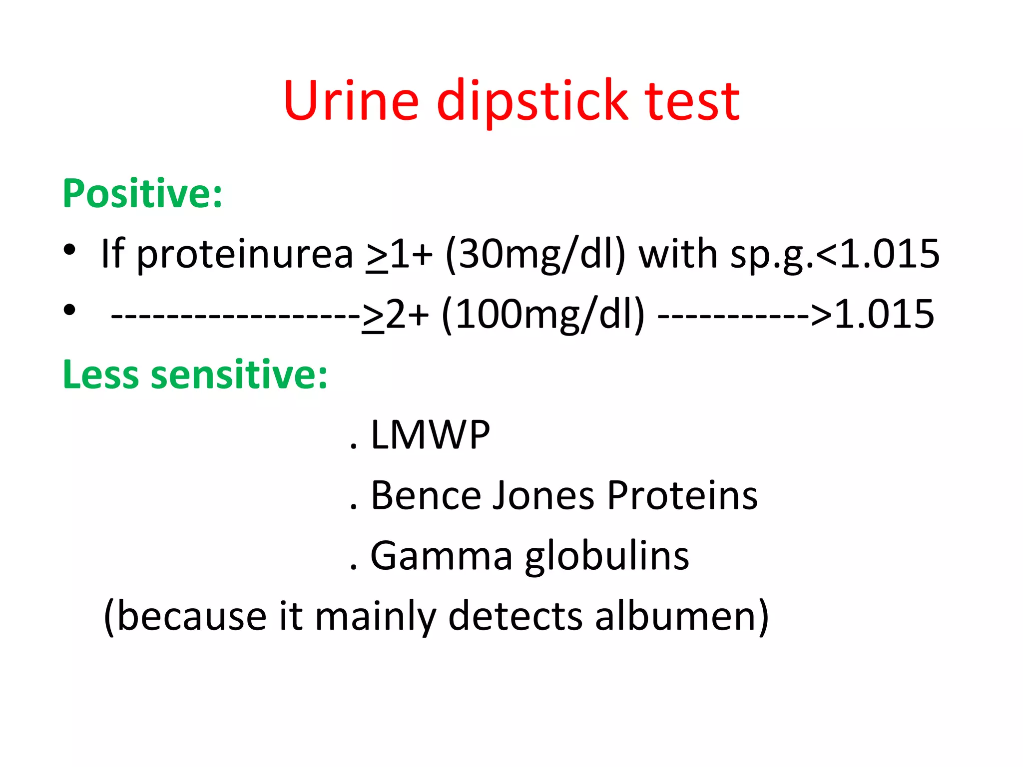
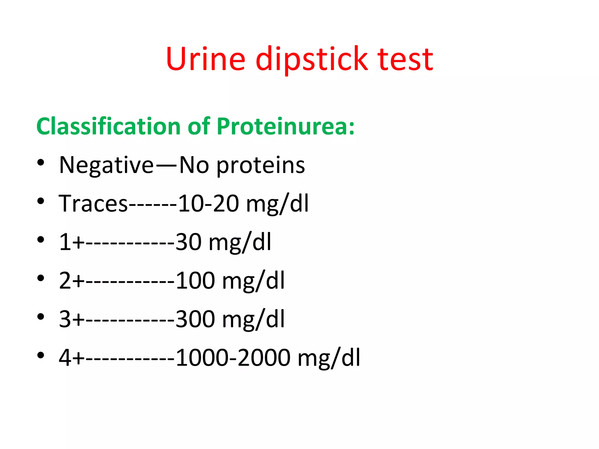
Proteinurea
&
Nephrotic Syndrome
By
Dr. Muhammad Rafique
Assist. Prof. Paediatrics
Diagnosis:
1- Urine dipstick
2- Urinary pr./cr. ratio
3- 24 hours urine collection
Urine dipstick test
Positive:
• If proteinurea >1+ (30mg/dl) with sp.g.<1.015
• ------------------>2+ (100mg/dl) ----------->1.015
Less sensitive:
. LMWP
. Bence Jones Proteins
. Gamma globulins
(because it mainly detects albumen)
Urine dipstick test
Classification of Proteinurea:
• Negative—No proteins
• Traces------10-20 mg/dl
• 1+-----------30 mg/dl
• 2+-----------100 mg/dl
• 3+-----------300 mg/dl
• 4+-----------1000-2000 mg/dl
Urine dipstick test
False Negative:
-Diluted urine(sp.g.<1.005)
-Diseases in which predominant
urinary protein is not albumen
False Positive:
- Concentrated urine
- Urine PH>7
- Gross hematurea
- Antiseptic contamination
Urinary protein/creatinine ratio
• First morning voided urine should be tested.
• This ratio is age dependent:
< 2years <0.5
>2years <0.2
• Nephrotic range ------------ >3
24 Hour Urine Collection
• Start and stop after voiding.
• Normal proteinurea:
<4mg/m2/h or <150mg/d
• Abnormal proteinurea:
>4mg/m2/hour
• Nephrotic range proteinurea:
>40mg/m2/hour
Types of protein urea
• Transient protein urea
• Postural protein urea
• Persistent protein urea
Transient protein urea
Causes:
High fever, exercise, dehydration, exposure
to cold, stress, seizures, heart failure
Note:
• Usually does not exceed 2+
• It is benign
• No need of any evaluation
Postural Protein urea
• Morning voided urine- absent/traces of protein
• Urine pr./cr. <0.2 for 3 consecutive days.
• Most common cause :
In(60%) of school age and adolescent children.
• In standing position may increase up to 10-folds
• Usually asymptomatic
• In supine position no or mild only
• Usually not exceed 1 g/m2/day
• No BP increase,azotemia,odema,hypoalbumenemia
Persistent protein urea
• In it necessary to R/O postural protein urea
• Exact cause unknown.
• Possible causes:
-altered renal hemodynamic
-partial renal vein obstruction.
• If persists:
-long term follow-up for renal
disease detection.
Fixed Protein urea
• Significant protein urea in first morning
voided urine for 3 consecutive days
• >1+ on dipstick
• >0.2 Upr./U cr.
• It usually indicates renal disease
• Classified on the basis of site of origin:
- Glomerulous
- Tubular
Glomerular Protein urea
• Due to increase glomerular capillary
permeability
• Range is <1g - >30 g/day
• It is always selective
• Selective protein urea:
-Excretory protein molecular wt.<albumen.
• Non selective pritein urea:
-Excretary protein, molecular wt.>albumen
Glomerular Protein urea
Suspect if :
- U pr./ Ucr. Ratio>1.0
- Associated with hypertention, azotemia,
oedema, hematurea
Causes are:
FSGS, SCA, PSGN, IgA nephropathy, HSPN,
Alport Synd; Obesity related G nephropathy
• Evaluation is necessary including renal biopsy
Tubular protein urea
• MW <albumen, proteins filter- reabsorb
• Reabsorption reduce in proximal tubular
injury diseases.
• Low MW proteins
• <1g/d
• Urinary pr./cr. <1.0
• Oedema absent
• Asymptomatic before detection
Tubular proteinurea
Causes:
Cystinosis, willson’s disease, Lowe syndrome,
ATN, polycystic K D, reflux nephropathy,
• May be associated with:
glucosurea, phosphateurea, NaHCo3
wasting, amino acid urea
• D/D between glomerular and tubular cause by
urine electrophoresis is possible.
Nephrotic Syndrome
• Incidence : 2-3/100,000 children /year
• 15 times more common in children.
• Characterized by:
-Heavy protein urea >40mg/m2/hour
-Hypoalbumenemia <2.5 g/dl
-Oedema
-Hypercholesterolemia/hyperlipidemia>200mg/dl
Periorbital oedema in nephrotic
Syndrome
Classification of Nephrotic Syndrome
1. Idiopathic: (90%)
-Minimal change 85% (95 % respond to steroids)
-Mesengial proliferative 5%(50% respond to
steroids)
-FSGS10% (20% respond to steroids)
2. Secondary: (10%)
-Membranous nephropathy
-Membranoproliferative glomerulonephritis
3. Congenital
Pathophysiology of MCNS
• Increase G. capillary permeability due to:
- loss of – ve charge glycoproteins.
• Odema :
- low oncotic pressure ,high aldosterone and ADH
• Hyperlipidemia due to:
- more lipoproteins production
- urinary loss of lipase
• Hypoproteinemia due to loss of urinary albumen
Signs/ Symptoms of Idiopathic N S
• Periorbital oedema - marked in the morning.
• Pedal (lower limbs) oedema
• Ascites and pleural effusion
• Genetal (labial/scrotal) oedema
• Generalised body swelling (anasarca)
• Increased BP in few cases(10-20%)
• Hematurea in 10% cases
PROTEINUREA
MCNS(Lipoid Nephrosis)
• Commonest cause of nephrotic syndrome(70%)
• Common in Males M:F 2:1
• Common age 2-7 years
• 15 times more common in children
• Oedema, annorexia, irritability, abdominal pain,
diarrhoea
• Blood pressure is normal
• Hypertention and gross hematuea is uncommon
Diagnosis of MCNS
• Urine analysis: Protein urea 3-4+ (selective)
• Microscopic hematurea 10-20%
• Spot urine, Urinary pr./ cr. >2
• 24 h urine collection: >40mg/m2/h protein urea
• S. albumen <2.5g/dl
• High s. cholesterole & triglyceride >200mg/dl
• S. urea, creatinine, C3, C4 levels normal
• Renal biopsy E/M fusion of epith.cells foot process
• -ve immunoflourescence on microscopy.
Differential Diagnosis of MCNS
D/D of generalized oedema:
• Protein losing enteropathy.
• Hepatic failure
• Protein energy malnutrion (kwashiorkor)
• Acute and chronich glomerulonephritis
• Angioedema
Managemen of MCNS
• Hospitalization for 1st
time for Dx./serious cases.
• Fluid chart (intake/ output)
• I.V. 5% albumen if necessary, 0.5g/kg/dose Q 12h
• Diuretics in severe symptoms.
-Chlorthiazoids 10mg/kg/dose Q 12h/ or
- metrolozone 0.1mg/kg/ dose Q 12 h followed by
- furosemide 30 min. later, 1-2mg/kg/dose Q 12h
Managemen of MCNS
• Diet:
No added salt, fluid restriction if hyponatremia
• Steroids therapy
• Scrotal support
• Other lines of treatment.
• Treatment of complications
Steroids Therapy
• 1-8 years pts. Start without renal biopsy
• Prednisone 60mg/m2 or 2mg/kg/day in 2-3 DD
with antacids, for at least 6 weeks.
• After absence of protein urea/1+ taper to
40mg/m2 on AD single morning dose.
• Slowly taper and discontinue in 2-3 months.
• If age<1 or >8ys, hypertension, hematurea,
azotemia, hypocomplementemia
hypoalbumenemia , do renal biopsy before
starting steroids.
Treatment of Relapse
• Relapse rate mentioned is 60-80%.
• It is 30-40% if first episode is treated prolong.
• Many children have at least one relapse(3-4+
protein urea with oedema)
• Give daily DD steroids until remission(urine
protein traces/- ve for 3 consecutive days)
then change to AD dose and taper in 1-2 mo
NS Definations
Steroid Dependant: Relapse while on AD
therapy /within 28 days after stopping steroids.
Frequent Relapser: Respond well to steroids but
relapse >4 times in one year.
Steroid Resistant: Persistent protein urea >2+
continues after 8 weeks DD steroid therapy.
-Diagnostic renal biopsy is must.
-Mostly FSGS(80%), MCNS(20%), and rarely MP
Alternate Management Agents
• Indications:
-Steroid Resistant
-Steroid Dependant
-Frequent relapser.
• These are:
Pulse steroids, cyclophophosphamide,
cyclosporin A, tacrolimus, microphenolate,
ACE inhibitors, levamisole and imuron.
Complication of N S
Infection: Increase susceptibility due to
-Urinary loss of Ig. and properdon factor B
-Defective cell mediated immunity
-Immunosuppressive therapy (steroids etc.)
-Odematous fluid is a good culture media
-Malnutrition
• Spontaneous bacterial peritonitis
• Strept. Pneumoniae and E-coli are more common
• High index of suspicion, early evaluation and Mx.
Including antibiotics is essential.
Complication of NS---Con.
• During steroid treatment fever and other
physiological findings are minimal.
• Vaccines:
-Polyvalent pneumococcal:
When pts. are in remission and off daily DD
steroid therapy.
-Varicella:
If varicella titer is -ve
Complication of NS--Cont
Thrombo embolic events:
• Risk 2-5% (much less than adults)
• Arterial & venous thrombosis, RVT, pulm.
embolism, saggital sinus and A & V catheters
thrombosis.
• Risk increased due to :
• High level of thrombotic factors (fibrinogen,
thrombocytosis, hemoconcentration)
• Low fibrinolytic factos (Urinary loss of antithrombin
III , protein C& S)
• Note: Avoid overaggressive diuretics and
indwelling catheters
Compaclications of NS—Cont.
C V Diseases:
• Hyperlipidemia may be a risk factor.
• Use of 3-hydroxy 3-methylglutaryl Co-enzyme
A (HMG-CoA) reductase inhibiting drugs to
reduce it.
• In MCNS no need because spontaneously
reduces
• Needed in familial verities like FSGS etc.
Prognosis of MCNS
• Steroid responders--Relapses decrease with
age – spontaneous cure in 2nd
decade of life.
• Rapid response to steroids, if no relapse in 1st
6 months after Dx.- infrequent relapser course
• Counsel parents:
• Non heriditary disease, non ch. Renal disease,
no infertility.
• In remission consider normal. no restriction of
diet and activity, no test for proteinurea etc.
Prognosis of N S
• Steroid Resistant like FSGS:
• Poor prognosis
• Use of cyclophosphamide leads to infertility
• Proggressive renal disease- ESRD
• Require dialysis/ renal transplant
• 30-50 % renal transplant develop recurrence
of NS
Secondary N S
• Suspect when age <1 & >8 ys, hypertention,
hematurea, azotemia, hypocomplementemia &
extra renal S/S like (fever, rash, arthralgia etc.)
Causes:
• Infections:
HBV, HCV, Malaria
• Drugs:
pencillamine, NSAID, captopril
• Systemic diseases:
SLE, HSP
Congenital N S
• Develope with in 3 months of life.
• FINNISH type:
Causes:
Congenital infections like HIV, HBV, TORCHS
Treatment:
• ACE inhibitors, indomethacin and Unilatemal
nephrectomy etc.
• Poor prognosis

Proteinurea & nephrotic syndrome

  • 1.
    Proteinurea & Nephrotic Syndrome By Dr. MuhammadRafique Assist. Prof. Paediatrics
  • 3.
    Diagnosis: 1- Urine dipstick 2-Urinary pr./cr. ratio 3- 24 hours urine collection
  • 7.
    Urine dipstick test Positive: •If proteinurea >1+ (30mg/dl) with sp.g.<1.015 • ------------------>2+ (100mg/dl) ----------->1.015 Less sensitive: . LMWP . Bence Jones Proteins . Gamma globulins (because it mainly detects albumen)
  • 8.
    Urine dipstick test Classificationof Proteinurea: • Negative—No proteins • Traces------10-20 mg/dl • 1+-----------30 mg/dl • 2+-----------100 mg/dl • 3+-----------300 mg/dl • 4+-----------1000-2000 mg/dl
  • 9.
    Urine dipstick test FalseNegative: -Diluted urine(sp.g.<1.005) -Diseases in which predominant urinary protein is not albumen False Positive: - Concentrated urine - Urine PH>7 - Gross hematurea - Antiseptic contamination
  • 10.
    Urinary protein/creatinine ratio •First morning voided urine should be tested. • This ratio is age dependent: < 2years <0.5 >2years <0.2 • Nephrotic range ------------ >3
  • 11.
    24 Hour UrineCollection • Start and stop after voiding. • Normal proteinurea: <4mg/m2/h or <150mg/d • Abnormal proteinurea: >4mg/m2/hour • Nephrotic range proteinurea: >40mg/m2/hour
  • 12.
    Types of proteinurea • Transient protein urea • Postural protein urea • Persistent protein urea
  • 13.
    Transient protein urea Causes: Highfever, exercise, dehydration, exposure to cold, stress, seizures, heart failure Note: • Usually does not exceed 2+ • It is benign • No need of any evaluation
  • 14.
    Postural Protein urea •Morning voided urine- absent/traces of protein • Urine pr./cr. <0.2 for 3 consecutive days. • Most common cause : In(60%) of school age and adolescent children. • In standing position may increase up to 10-folds • Usually asymptomatic • In supine position no or mild only • Usually not exceed 1 g/m2/day • No BP increase,azotemia,odema,hypoalbumenemia
  • 15.
    Persistent protein urea •In it necessary to R/O postural protein urea • Exact cause unknown. • Possible causes: -altered renal hemodynamic -partial renal vein obstruction. • If persists: -long term follow-up for renal disease detection.
  • 16.
    Fixed Protein urea •Significant protein urea in first morning voided urine for 3 consecutive days • >1+ on dipstick • >0.2 Upr./U cr. • It usually indicates renal disease • Classified on the basis of site of origin: - Glomerulous - Tubular
  • 17.
    Glomerular Protein urea •Due to increase glomerular capillary permeability • Range is <1g - >30 g/day • It is always selective • Selective protein urea: -Excretory protein molecular wt.<albumen. • Non selective pritein urea: -Excretary protein, molecular wt.>albumen
  • 18.
    Glomerular Protein urea Suspectif : - U pr./ Ucr. Ratio>1.0 - Associated with hypertention, azotemia, oedema, hematurea Causes are: FSGS, SCA, PSGN, IgA nephropathy, HSPN, Alport Synd; Obesity related G nephropathy • Evaluation is necessary including renal biopsy
  • 19.
    Tubular protein urea •MW <albumen, proteins filter- reabsorb • Reabsorption reduce in proximal tubular injury diseases. • Low MW proteins • <1g/d • Urinary pr./cr. <1.0 • Oedema absent • Asymptomatic before detection
  • 20.
    Tubular proteinurea Causes: Cystinosis, willson’sdisease, Lowe syndrome, ATN, polycystic K D, reflux nephropathy, • May be associated with: glucosurea, phosphateurea, NaHCo3 wasting, amino acid urea • D/D between glomerular and tubular cause by urine electrophoresis is possible.
  • 21.
    Nephrotic Syndrome • Incidence: 2-3/100,000 children /year • 15 times more common in children. • Characterized by: -Heavy protein urea >40mg/m2/hour -Hypoalbumenemia <2.5 g/dl -Oedema -Hypercholesterolemia/hyperlipidemia>200mg/dl
  • 24.
    Periorbital oedema innephrotic Syndrome
  • 26.
    Classification of NephroticSyndrome 1. Idiopathic: (90%) -Minimal change 85% (95 % respond to steroids) -Mesengial proliferative 5%(50% respond to steroids) -FSGS10% (20% respond to steroids) 2. Secondary: (10%) -Membranous nephropathy -Membranoproliferative glomerulonephritis 3. Congenital
  • 28.
    Pathophysiology of MCNS •Increase G. capillary permeability due to: - loss of – ve charge glycoproteins. • Odema : - low oncotic pressure ,high aldosterone and ADH • Hyperlipidemia due to: - more lipoproteins production - urinary loss of lipase • Hypoproteinemia due to loss of urinary albumen
  • 29.
    Signs/ Symptoms ofIdiopathic N S • Periorbital oedema - marked in the morning. • Pedal (lower limbs) oedema • Ascites and pleural effusion • Genetal (labial/scrotal) oedema • Generalised body swelling (anasarca) • Increased BP in few cases(10-20%) • Hematurea in 10% cases
  • 31.
  • 32.
    MCNS(Lipoid Nephrosis) • Commonestcause of nephrotic syndrome(70%) • Common in Males M:F 2:1 • Common age 2-7 years • 15 times more common in children • Oedema, annorexia, irritability, abdominal pain, diarrhoea • Blood pressure is normal • Hypertention and gross hematuea is uncommon
  • 33.
    Diagnosis of MCNS •Urine analysis: Protein urea 3-4+ (selective) • Microscopic hematurea 10-20% • Spot urine, Urinary pr./ cr. >2 • 24 h urine collection: >40mg/m2/h protein urea • S. albumen <2.5g/dl • High s. cholesterole & triglyceride >200mg/dl • S. urea, creatinine, C3, C4 levels normal • Renal biopsy E/M fusion of epith.cells foot process • -ve immunoflourescence on microscopy.
  • 34.
    Differential Diagnosis ofMCNS D/D of generalized oedema: • Protein losing enteropathy. • Hepatic failure • Protein energy malnutrion (kwashiorkor) • Acute and chronich glomerulonephritis • Angioedema
  • 35.
    Managemen of MCNS •Hospitalization for 1st time for Dx./serious cases. • Fluid chart (intake/ output) • I.V. 5% albumen if necessary, 0.5g/kg/dose Q 12h • Diuretics in severe symptoms. -Chlorthiazoids 10mg/kg/dose Q 12h/ or - metrolozone 0.1mg/kg/ dose Q 12 h followed by - furosemide 30 min. later, 1-2mg/kg/dose Q 12h
  • 36.
    Managemen of MCNS •Diet: No added salt, fluid restriction if hyponatremia • Steroids therapy • Scrotal support • Other lines of treatment. • Treatment of complications
  • 37.
    Steroids Therapy • 1-8years pts. Start without renal biopsy • Prednisone 60mg/m2 or 2mg/kg/day in 2-3 DD with antacids, for at least 6 weeks. • After absence of protein urea/1+ taper to 40mg/m2 on AD single morning dose. • Slowly taper and discontinue in 2-3 months. • If age<1 or >8ys, hypertension, hematurea, azotemia, hypocomplementemia hypoalbumenemia , do renal biopsy before starting steroids.
  • 38.
    Treatment of Relapse •Relapse rate mentioned is 60-80%. • It is 30-40% if first episode is treated prolong. • Many children have at least one relapse(3-4+ protein urea with oedema) • Give daily DD steroids until remission(urine protein traces/- ve for 3 consecutive days) then change to AD dose and taper in 1-2 mo
  • 40.
    NS Definations Steroid Dependant:Relapse while on AD therapy /within 28 days after stopping steroids. Frequent Relapser: Respond well to steroids but relapse >4 times in one year. Steroid Resistant: Persistent protein urea >2+ continues after 8 weeks DD steroid therapy. -Diagnostic renal biopsy is must. -Mostly FSGS(80%), MCNS(20%), and rarely MP
  • 41.
    Alternate Management Agents •Indications: -Steroid Resistant -Steroid Dependant -Frequent relapser. • These are: Pulse steroids, cyclophophosphamide, cyclosporin A, tacrolimus, microphenolate, ACE inhibitors, levamisole and imuron.
  • 42.
    Complication of NS Infection: Increase susceptibility due to -Urinary loss of Ig. and properdon factor B -Defective cell mediated immunity -Immunosuppressive therapy (steroids etc.) -Odematous fluid is a good culture media -Malnutrition • Spontaneous bacterial peritonitis • Strept. Pneumoniae and E-coli are more common • High index of suspicion, early evaluation and Mx. Including antibiotics is essential.
  • 43.
    Complication of NS---Con. •During steroid treatment fever and other physiological findings are minimal. • Vaccines: -Polyvalent pneumococcal: When pts. are in remission and off daily DD steroid therapy. -Varicella: If varicella titer is -ve
  • 44.
    Complication of NS--Cont Thromboembolic events: • Risk 2-5% (much less than adults) • Arterial & venous thrombosis, RVT, pulm. embolism, saggital sinus and A & V catheters thrombosis. • Risk increased due to : • High level of thrombotic factors (fibrinogen, thrombocytosis, hemoconcentration) • Low fibrinolytic factos (Urinary loss of antithrombin III , protein C& S) • Note: Avoid overaggressive diuretics and indwelling catheters
  • 45.
    Compaclications of NS—Cont. CV Diseases: • Hyperlipidemia may be a risk factor. • Use of 3-hydroxy 3-methylglutaryl Co-enzyme A (HMG-CoA) reductase inhibiting drugs to reduce it. • In MCNS no need because spontaneously reduces • Needed in familial verities like FSGS etc.
  • 46.
    Prognosis of MCNS •Steroid responders--Relapses decrease with age – spontaneous cure in 2nd decade of life. • Rapid response to steroids, if no relapse in 1st 6 months after Dx.- infrequent relapser course • Counsel parents: • Non heriditary disease, non ch. Renal disease, no infertility. • In remission consider normal. no restriction of diet and activity, no test for proteinurea etc.
  • 47.
    Prognosis of NS • Steroid Resistant like FSGS: • Poor prognosis • Use of cyclophosphamide leads to infertility • Proggressive renal disease- ESRD • Require dialysis/ renal transplant • 30-50 % renal transplant develop recurrence of NS
  • 48.
    Secondary N S •Suspect when age <1 & >8 ys, hypertention, hematurea, azotemia, hypocomplementemia & extra renal S/S like (fever, rash, arthralgia etc.) Causes: • Infections: HBV, HCV, Malaria • Drugs: pencillamine, NSAID, captopril • Systemic diseases: SLE, HSP
  • 49.
    Congenital N S •Develope with in 3 months of life. • FINNISH type: Causes: Congenital infections like HIV, HBV, TORCHS Treatment: • ACE inhibitors, indomethacin and Unilatemal nephrectomy etc. • Poor prognosis