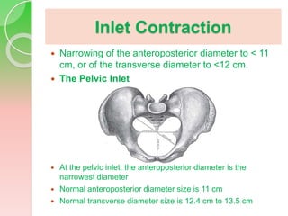
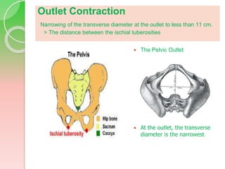
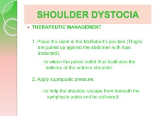
Problems with the
Passageway
Inlet Contraction
 Narrowing of the anteroposterior diameter to < 11
cm, or of the transverse diameter to <12 cm.
 The Pelvic Inlet
 At the pelvic inlet, the anteroposterior diameter is the
narrowest diameter
 Normal anteroposterior diameter size is 11 cm
 Normal transverse diameter size is 12.4 cm to 13.5 cm
Inlet Contraction
 Causes:
1. Rickets
2. Inherited small pelvis
 In primigravidas, the fetal head normally
engages between week 36-38 of pregnancy.
- If this not occur at expected gestational age, either a fetal
abnormality or a pelvic abnormality should be suspected.
 In multigravidas, engagement does not occur
until labor begins. (normal)
Inlet Contraction
 Assessment Findings:
1. prolonged labor
2. arrest of descent
 Complications:
1. Fetal malposition (with CPD)
2. Premature rupture of membranes
3. Cord prolapse
 Therapeutic management:
1. Pelvic measurements should be taken and recorded in
every primigravida before week 24 of pregnancy so as a
birth decision can be made.
2. CS for inadequate inlet measurements and the fetal lie and
position are poor.
Outlet Contraction
Narrowing of the transverse diameter at the outlet to less than 11 cm.
> The distance between the ischial tuberosities
 The Pelvic Outlet
 At the outlet, the transverse
diameter is the narrowest
Cephalopelvic Disproportion
 A condition in which the fetal head size is too large
to fit through the maternal pelvis.
 Risk Factors:
1. Gestational diabetes
2. Multiparity
3. Fetal malformation
4. Shape or size of maternal pelvis
5. Shape or position of the fetus’ head
Cephalopelvic Disproportion
 Signs and Symptoms
1. General lack of cervical change or fetal descent during the
active phase of first stage of labor
2. Dystocia – abnormal labor or failure of labor to progress
3. Uncontrollable pushing before complete dilation of cervix
 Diagnostic Tests
1. Trial labor – attempt at vaginal delivery (when
measurements indicate borderline CPD)
2. Utrasound estimation of fetal size compared to manual
pelvic measurements (prior to labor) and computed
tomography
3. X-ray pelvimetry – to visualize pelvic measures
Cephalopelvic Disproportion
 THERAPEUTIC MANAGEMENT
1. Assist client to cope emotionally with cesarean delivery.
2. Stress the importance of the safety of the baby.
3. Monitor FHR, uterine contractions, and cervical dilatation.
4. Nursing care during “trial of labor” is similar to that of any labor,
except that assessments of cervical dilation and fetal descent
are done more often.
- If progress ceases, a cesarean birth is necessary.
5. Report any signs of fetal distress to the caregiver immediately.
6. Provide emotional support by keeping the woman/couple
informed and explaining procedures
7. Help the woman assume different positions to increase the
pelvic diameters (e.g., sitting or squatting, changing from one
side to the other, hands-and-knees position)
Cephalopelvic Disproportion
 COMPLICTIONS to
the Mother
1. Exhaustion
2. Postpartum hemorrhage
3. Infection
 COMPLICATIONS to
the Baby
1. Cord prolapse
2. Birth trauma
3. Fractured clavicle
4. Erb’s palsy
5. Anoxia
SHOULDER DYSTOCIA
 DESCRIPTION
- the anterior shoulder of the baby is
unable to pass under the maternal pubic
arch after the head is born.
 ETIOLOGY
1. advanced maternal age
2. diabetes
3. multiparity
4. post-date pregnancy
SHOULDER DYSTOCIA
 PATHOPHYSIOLOGY
- The wide anterior shoulder locks beneath the symphysis
pubis.
- The problem occurs at the second stage of labor and often is
not identified until the head has already been born.
 ASSESSMENT FINDINGS
1. Associated findings
1.1 birth process in the second stage is prolonged
1.2 arrest of descent
2. Clinical manifestations
2.1 The fetal head retracts against the mother’s perineum
instead of protruding with each contraction as soon as the
head appears on the perineum/is delivered. (a turtle sign)
2.2 External rotation does not occur.
SHOULDER DYSTOCIA
 COMPLICATIONS to
the Mother
1. Vaginal or cervical
tears
2. Bleeding
 COMPLICATIONS to
the Baby
1. Cord compression
2. Brachial plexus
injury or fractured
clavicle
SHOULDER DYSTOCIA
 THERAPEUTIC MANAGEMENT
1. Place the client in the McRobert’s position (Thighs
are pulled up against the abdomen with hips
abducted).
- to widen the pelvic outlet thus facilitates the
delivery of the anterior shoulder.
2. Apply suprapubic pressure.
- to help the shoulder escape from beneath the
symphysis pubis and be delivered.

Problems-with-the-passage.pptx

  • 1.
  • 2.
    Inlet Contraction  Narrowingof the anteroposterior diameter to < 11 cm, or of the transverse diameter to <12 cm.  The Pelvic Inlet  At the pelvic inlet, the anteroposterior diameter is the narrowest diameter  Normal anteroposterior diameter size is 11 cm  Normal transverse diameter size is 12.4 cm to 13.5 cm
  • 3.
    Inlet Contraction  Causes: 1.Rickets 2. Inherited small pelvis  In primigravidas, the fetal head normally engages between week 36-38 of pregnancy. - If this not occur at expected gestational age, either a fetal abnormality or a pelvic abnormality should be suspected.  In multigravidas, engagement does not occur until labor begins. (normal)
  • 4.
    Inlet Contraction  AssessmentFindings: 1. prolonged labor 2. arrest of descent  Complications: 1. Fetal malposition (with CPD) 2. Premature rupture of membranes 3. Cord prolapse  Therapeutic management: 1. Pelvic measurements should be taken and recorded in every primigravida before week 24 of pregnancy so as a birth decision can be made. 2. CS for inadequate inlet measurements and the fetal lie and position are poor.
  • 5.
    Outlet Contraction Narrowing ofthe transverse diameter at the outlet to less than 11 cm. > The distance between the ischial tuberosities  The Pelvic Outlet  At the outlet, the transverse diameter is the narrowest
  • 6.
    Cephalopelvic Disproportion  Acondition in which the fetal head size is too large to fit through the maternal pelvis.  Risk Factors: 1. Gestational diabetes 2. Multiparity 3. Fetal malformation 4. Shape or size of maternal pelvis 5. Shape or position of the fetus’ head
  • 7.
    Cephalopelvic Disproportion  Signsand Symptoms 1. General lack of cervical change or fetal descent during the active phase of first stage of labor 2. Dystocia – abnormal labor or failure of labor to progress 3. Uncontrollable pushing before complete dilation of cervix  Diagnostic Tests 1. Trial labor – attempt at vaginal delivery (when measurements indicate borderline CPD) 2. Utrasound estimation of fetal size compared to manual pelvic measurements (prior to labor) and computed tomography 3. X-ray pelvimetry – to visualize pelvic measures
  • 8.
    Cephalopelvic Disproportion  THERAPEUTICMANAGEMENT 1. Assist client to cope emotionally with cesarean delivery. 2. Stress the importance of the safety of the baby. 3. Monitor FHR, uterine contractions, and cervical dilatation. 4. Nursing care during “trial of labor” is similar to that of any labor, except that assessments of cervical dilation and fetal descent are done more often. - If progress ceases, a cesarean birth is necessary. 5. Report any signs of fetal distress to the caregiver immediately. 6. Provide emotional support by keeping the woman/couple informed and explaining procedures 7. Help the woman assume different positions to increase the pelvic diameters (e.g., sitting or squatting, changing from one side to the other, hands-and-knees position)
  • 9.
    Cephalopelvic Disproportion  COMPLICTIONSto the Mother 1. Exhaustion 2. Postpartum hemorrhage 3. Infection  COMPLICATIONS to the Baby 1. Cord prolapse 2. Birth trauma 3. Fractured clavicle 4. Erb’s palsy 5. Anoxia
  • 10.
    SHOULDER DYSTOCIA  DESCRIPTION -the anterior shoulder of the baby is unable to pass under the maternal pubic arch after the head is born.  ETIOLOGY 1. advanced maternal age 2. diabetes 3. multiparity 4. post-date pregnancy
  • 11.
    SHOULDER DYSTOCIA  PATHOPHYSIOLOGY -The wide anterior shoulder locks beneath the symphysis pubis. - The problem occurs at the second stage of labor and often is not identified until the head has already been born.  ASSESSMENT FINDINGS 1. Associated findings 1.1 birth process in the second stage is prolonged 1.2 arrest of descent 2. Clinical manifestations 2.1 The fetal head retracts against the mother’s perineum instead of protruding with each contraction as soon as the head appears on the perineum/is delivered. (a turtle sign) 2.2 External rotation does not occur.
  • 12.
    SHOULDER DYSTOCIA  COMPLICATIONSto the Mother 1. Vaginal or cervical tears 2. Bleeding  COMPLICATIONS to the Baby 1. Cord compression 2. Brachial plexus injury or fractured clavicle
  • 13.
    SHOULDER DYSTOCIA  THERAPEUTICMANAGEMENT 1. Place the client in the McRobert’s position (Thighs are pulled up against the abdomen with hips abducted). - to widen the pelvic outlet thus facilitates the delivery of the anterior shoulder. 2. Apply suprapubic pressure. - to help the shoulder escape from beneath the symphysis pubis and be delivered.