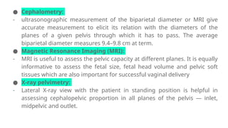
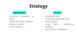
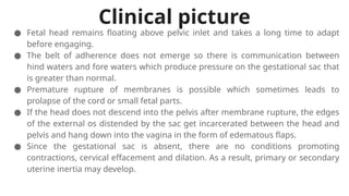
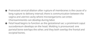
CLINICALLY
CONTRACTED
PELVIS
XABA REFILWE GROUP:75
Definition
Functionally, or clinically contracted
pelvis (cephalopelvic disproportion)
is the case when anatomical
dimensions of particular pelvis
preclude the passage of particular
fetus.
● Contracted inlet :
- when the obstetric conjugate is < 10 cm or the greatest transverse diameter
is < 12 cm or diagonal conjugate is < 11 cm.
● Contracted Midpelvis :
- when the sum of the inter-ischial spinous and posterior sagittal diameters
of the mid pelvis (normal: 10.0 + 5 = 15.0 cm) is 13.0 cm or below.
● Contracted outlet :
- when the inter ischial tuberous diameter is 8 cm or less.
Diagnosis
● Clinical:
- In multigravida, a previous history of spontaneous delivery of an average
size baby, reasonably rules out contracted pelvis. But in a primigravida
with non engagement of the head even at labor, one should rule out
disproportion.
CPD can be diagnosed when labor is in progress:
● with satisfactory contractions;
● after rupture of membranes;
● almost complete cervical dilation;
● the head pressed to the inlet;
● signs of disproportion between the head and pelvic inlet.
Abdominal method
- The patient is placed in dorsal position with
the thighs slightly flexed and separated.
- The head is grasped by the left hand.
- Two fingers (index and middle) of the right
hand are placed above the symphysis pubis
to note the degree of overlapping when the
head is pushed downwards and backwards
Abdominovaginal method
(Muller-Munro Kerr)
is bimanual method as the pelvic assessment can be
done simultaneously.
- The patient is placed in lithotomy position and
the internal examination is done taking all
aseptic precautions.
- Two fingers of the right hand are introduced
into the vagina with the fingertips placed at the
level of ischial spines and thumb is placed over
the symphysis pubis.
- The head is grasped by the left hand and is
pushed in a downward and backward direction
into the pelvis
Vasten’s sign
● with fingertips, perform motions from symphysis pubis upwards to the
head pressed to pelvic inlet determining the proportions between the
fetal head and mother’s pelvis.
● If the head and pelvis are proportionate to each other, the anterior
symphysis surface is higher than the anterior surface of the pressed
head — negative Vasten’s sign.
● If the anterior symphysis surface is on the level with the anterior head
surface — Vasten’s sign at the same level.
● If the anterior head surface is above the symphysis, it is a positive
Vatsen’s sign: cephalopelvic disproportion is present
Zangemeister’s sign,
● measures the external conjugate with a pelvimeter and then shifts the
anterior end of pelvimeter to the most prominent point of the head (the
other end of pelvimeter remains in the same place).
● If the obtained value is less than the external conjugate, it is a negative
Zangemeister’s sign.
● If the obtained value is greater than the external conjugate, this indicates
CPD: positive Zangemeister’s sign.
● If the obtained values are equal, there is a relative CPD
● Cephalometry:
- ultrasonographic measurement of the biparietal diameter or MRI give
accurate measurement to elicit its relation with the diameters of the
planes of a given pelvis through which it has to pass. The average
biparietal diameter measures 9.4–9.8 cm at term.
● Magnetic Resonance Imaging (MRI):
- MRI is useful to assess the pelvic capacity at different planes. It is equally
informative to assess the fetal size, fetal head volume and pelvic soft
tissues which are also important for successful vaginal delivery
● X-ray pelvimetry:
- Lateral X-ray view with the patient in standing position is helpful in
assessing cephalopelvic proportion in all planes of the pelvis — inlet,
midpelvic and outlet.
Etiology
MATERNAL’S FETUS’S
- structural reduction in
pelvis
- Abnormal shape of pelvis :
disease, accident
- Tumor of bone
- large fetal head
- Presenting extended head
- Fetal position
- Large baby : hereditary,
diabetes
- Post mature (>42weeks)
Clinical picture
● Fetal head remains floating above pelvic inlet and takes a long time to adapt
before engaging.
● The belt of adherence does not emerge so there is communication between
hind waters and fore waters which produce pressure on the gestational sac that
is greater than normal.
● Premature rupture of membranes is possible which sometimes leads to
prolapse of the cord or small fetal parts.
● If the head does not descend into the pelvis after membrane rupture, the edges
of the external os distended by the sac get incarcerated between the head and
pelvis and hang down into the vagina in the form of edematous flaps.
● Since the gestational sac is absent, there are no conditions promoting
contractions, cervical effacement and dilation. As a result, primary or secondary
uterine inertia may develop.
● Protracted cervical dilation after rupture of membranes is the cause of a
long rupture to delivery interval; there is communication between the
vagina and uterine cavity where microorganisms can enter.
Chorioamnionitis can develop during labor.
● Fetal head begins to function as the gestational sac: a prominent caput
succedaneum develops on the head. Molding is pronounced: one
parietal bone overlaps the other, and they both overlap the frontal and
occipital bones.
● the fetal head progress through all parts of pelvis is slowed down which often
causes fetal hypoxia;
● protracted course of the first stage leads to the woman’s nervous exhaustion
and physical fatigue and in the expulsion stage secondary uterine inertia may
develop;
● if the head persists in one of pelvic planes due to its reduced size, there is a
risk of cervical incarceration and compression of adjacent organs which can
result in later emergence of genitourinary, cervicovaginal and intestino
genital fistulas;
● in case of powerful contractions the bones of pubic and/or iliac symphysis
can separate, the cervix and perineum can tear;
● in some cases labor is complicated by powerful contractions and even uterine
tetany; excessive labor can result in uterine rupture, placental abruption,
fetal demise;
● in case of protracted labor (18 hours and more) and a prolonged rupture to
delivery interval there is a risk of an ascending infection (chorioamnionitis
during labor).
TRIAL ON LABOR
is the conduction of spontaneous labor in a moderate degree of cephalopelvic
disproportion, in an institution under supervision with watchful expectancy,
hoping for a vaginal delivery.
Aims: A trial labor aims at avoiding an unnecessary cesarean section and at
delivering a healthy baby.
Contraindications :
● Associated midpelvic and outlet contraction;
● Presence of complicating factors like elderly primigravida, malpresentation,
postmaturity, post cesarean pregnancy, pre-eclampsia, medical disorders like
heart disease, diabetes, tuberculosis, etc.
● Where facilities for cesarean section is not available round the clock.
The methods Of termination of trial labor:
● Spontaneous delivery with or without episiotomy (30%).
● Forceps or ventouse (30%)—Difficult forceps delivery is to be avoided.
● Cesarean section (40%)—Judicious and timely decision for cesarean
delivery is to be taken.
However,in significant cases, the section is done even before full dilatation of
the cervix, the indication being uterine inertia or fetal distress.
Cephalopelvic disproportion at the outlet is defined as one where the biparietal-
suboccipitobregmatic plane fails to pass through the bispinous and
anteroposterior planes of the outlet.
● Elective cesarean section: Contraction of both the transverse and
anteroposterior diameters of the midpelvic plane or minor contraction
associated with other complicating factors is dealt by elective cesarean
section.
● To allow vaginal delivery: In uncomplicated cases with minor contraction,
vaginal delivery is allowed under supervision with watchful expectancy.
Moulding and adaptation of the head and “give” of the pelvis may allow the
head to pass through the contracted zone.
If there is no dilatation of cervix or descent of the fetal head after a period of 2
hours in the active phase of labor, arrest of labor is considered. Once arrest
disorder is diagnosed, cesarean delivery is the option.

CLINICALLY CONTRACTED PELVIS (group 75) xaba.pptx

  • 1.
  • 2.
    Definition Functionally, or clinicallycontracted pelvis (cephalopelvic disproportion) is the case when anatomical dimensions of particular pelvis preclude the passage of particular fetus.
  • 3.
    ● Contracted inlet: - when the obstetric conjugate is < 10 cm or the greatest transverse diameter is < 12 cm or diagonal conjugate is < 11 cm. ● Contracted Midpelvis : - when the sum of the inter-ischial spinous and posterior sagittal diameters of the mid pelvis (normal: 10.0 + 5 = 15.0 cm) is 13.0 cm or below. ● Contracted outlet : - when the inter ischial tuberous diameter is 8 cm or less.
  • 4.
    Diagnosis ● Clinical: - Inmultigravida, a previous history of spontaneous delivery of an average size baby, reasonably rules out contracted pelvis. But in a primigravida with non engagement of the head even at labor, one should rule out disproportion. CPD can be diagnosed when labor is in progress: ● with satisfactory contractions; ● after rupture of membranes; ● almost complete cervical dilation; ● the head pressed to the inlet; ● signs of disproportion between the head and pelvic inlet.
  • 5.
    Abdominal method - Thepatient is placed in dorsal position with the thighs slightly flexed and separated. - The head is grasped by the left hand. - Two fingers (index and middle) of the right hand are placed above the symphysis pubis to note the degree of overlapping when the head is pushed downwards and backwards
  • 6.
    Abdominovaginal method (Muller-Munro Kerr) isbimanual method as the pelvic assessment can be done simultaneously. - The patient is placed in lithotomy position and the internal examination is done taking all aseptic precautions. - Two fingers of the right hand are introduced into the vagina with the fingertips placed at the level of ischial spines and thumb is placed over the symphysis pubis. - The head is grasped by the left hand and is pushed in a downward and backward direction into the pelvis
  • 7.
    Vasten’s sign ● withfingertips, perform motions from symphysis pubis upwards to the head pressed to pelvic inlet determining the proportions between the fetal head and mother’s pelvis. ● If the head and pelvis are proportionate to each other, the anterior symphysis surface is higher than the anterior surface of the pressed head — negative Vasten’s sign. ● If the anterior symphysis surface is on the level with the anterior head surface — Vasten’s sign at the same level. ● If the anterior head surface is above the symphysis, it is a positive Vatsen’s sign: cephalopelvic disproportion is present
  • 8.
    Zangemeister’s sign, ● measuresthe external conjugate with a pelvimeter and then shifts the anterior end of pelvimeter to the most prominent point of the head (the other end of pelvimeter remains in the same place). ● If the obtained value is less than the external conjugate, it is a negative Zangemeister’s sign. ● If the obtained value is greater than the external conjugate, this indicates CPD: positive Zangemeister’s sign. ● If the obtained values are equal, there is a relative CPD
  • 10.
    ● Cephalometry: - ultrasonographicmeasurement of the biparietal diameter or MRI give accurate measurement to elicit its relation with the diameters of the planes of a given pelvis through which it has to pass. The average biparietal diameter measures 9.4–9.8 cm at term. ● Magnetic Resonance Imaging (MRI): - MRI is useful to assess the pelvic capacity at different planes. It is equally informative to assess the fetal size, fetal head volume and pelvic soft tissues which are also important for successful vaginal delivery ● X-ray pelvimetry: - Lateral X-ray view with the patient in standing position is helpful in assessing cephalopelvic proportion in all planes of the pelvis — inlet, midpelvic and outlet.
  • 11.
    Etiology MATERNAL’S FETUS’S - structuralreduction in pelvis - Abnormal shape of pelvis : disease, accident - Tumor of bone - large fetal head - Presenting extended head - Fetal position - Large baby : hereditary, diabetes - Post mature (>42weeks)
  • 12.
    Clinical picture ● Fetalhead remains floating above pelvic inlet and takes a long time to adapt before engaging. ● The belt of adherence does not emerge so there is communication between hind waters and fore waters which produce pressure on the gestational sac that is greater than normal. ● Premature rupture of membranes is possible which sometimes leads to prolapse of the cord or small fetal parts. ● If the head does not descend into the pelvis after membrane rupture, the edges of the external os distended by the sac get incarcerated between the head and pelvis and hang down into the vagina in the form of edematous flaps. ● Since the gestational sac is absent, there are no conditions promoting contractions, cervical effacement and dilation. As a result, primary or secondary uterine inertia may develop.
  • 13.
    ● Protracted cervicaldilation after rupture of membranes is the cause of a long rupture to delivery interval; there is communication between the vagina and uterine cavity where microorganisms can enter. Chorioamnionitis can develop during labor. ● Fetal head begins to function as the gestational sac: a prominent caput succedaneum develops on the head. Molding is pronounced: one parietal bone overlaps the other, and they both overlap the frontal and occipital bones.
  • 14.
    ● the fetalhead progress through all parts of pelvis is slowed down which often causes fetal hypoxia; ● protracted course of the first stage leads to the woman’s nervous exhaustion and physical fatigue and in the expulsion stage secondary uterine inertia may develop; ● if the head persists in one of pelvic planes due to its reduced size, there is a risk of cervical incarceration and compression of adjacent organs which can result in later emergence of genitourinary, cervicovaginal and intestino genital fistulas; ● in case of powerful contractions the bones of pubic and/or iliac symphysis can separate, the cervix and perineum can tear; ● in some cases labor is complicated by powerful contractions and even uterine tetany; excessive labor can result in uterine rupture, placental abruption, fetal demise; ● in case of protracted labor (18 hours and more) and a prolonged rupture to delivery interval there is a risk of an ascending infection (chorioamnionitis during labor).
  • 15.
    TRIAL ON LABOR isthe conduction of spontaneous labor in a moderate degree of cephalopelvic disproportion, in an institution under supervision with watchful expectancy, hoping for a vaginal delivery. Aims: A trial labor aims at avoiding an unnecessary cesarean section and at delivering a healthy baby. Contraindications : ● Associated midpelvic and outlet contraction; ● Presence of complicating factors like elderly primigravida, malpresentation, postmaturity, post cesarean pregnancy, pre-eclampsia, medical disorders like heart disease, diabetes, tuberculosis, etc. ● Where facilities for cesarean section is not available round the clock.
  • 16.
    The methods Oftermination of trial labor: ● Spontaneous delivery with or without episiotomy (30%). ● Forceps or ventouse (30%)—Difficult forceps delivery is to be avoided. ● Cesarean section (40%)—Judicious and timely decision for cesarean delivery is to be taken. However,in significant cases, the section is done even before full dilatation of the cervix, the indication being uterine inertia or fetal distress.
  • 17.
    Cephalopelvic disproportion atthe outlet is defined as one where the biparietal- suboccipitobregmatic plane fails to pass through the bispinous and anteroposterior planes of the outlet. ● Elective cesarean section: Contraction of both the transverse and anteroposterior diameters of the midpelvic plane or minor contraction associated with other complicating factors is dealt by elective cesarean section. ● To allow vaginal delivery: In uncomplicated cases with minor contraction, vaginal delivery is allowed under supervision with watchful expectancy. Moulding and adaptation of the head and “give” of the pelvis may allow the head to pass through the contracted zone. If there is no dilatation of cervix or descent of the fetal head after a period of 2 hours in the active phase of labor, arrest of labor is considered. Once arrest disorder is diagnosed, cesarean delivery is the option.