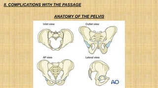
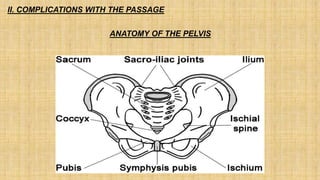
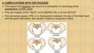
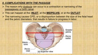
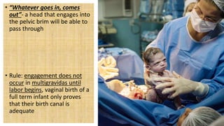
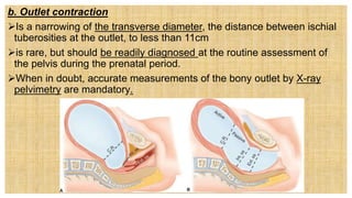
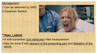
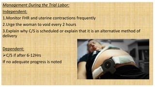
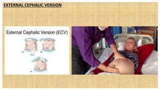
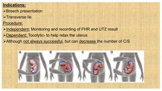
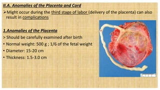
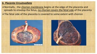
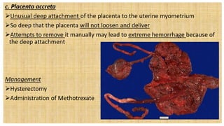
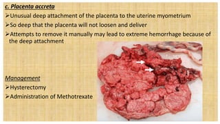
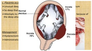
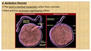
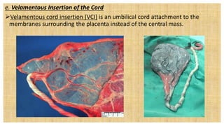
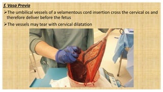
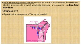
NCM 109- Care of Mother
and Child at Risk or with
Problems
(Acute and Chronic)-LECTURE
Wesleyan University –Philippines
Cabanatuan City
CONAMS
Jhonee Balmeo
Instructor
II. COMPLICATIONS WITH THE PASSAGE
ANATOMY OF THE PELVIS
II. COMPLICATIONS WITH THE PASSAGE
ANATOMY OF THE PELVIS
II. COMPLICATIONS WITH THE PASSAGE
ANATOMY OF THE PELVIS
II. COMPLICATIONS WITH THE PASSAGE
 The reason why dystocia can occur is a contraction or narrowing of the
passageway or birth canal.
 This can happen at the: INLET, at the MIDPELVIS, or at the OUTLET
 The narrowing causes CPD, or a disproportion between the size of the fetal head
and the pelvic diameters, that results in failure to progress in labor.
a. Inlet Contraction
Is narrowing of the anteroposterior diameter of the pelvis to less than 11 cm
or of the transverse diameter to 12 cm or less.
Usually caused by rickets in early life or by an inherited small pelvis
Rickets- caused by lack of calcium
Primigravidas – fetal head engages between weeks 36 and 38 of pregnancy
• If occurs anytime before labor begins, the pelvis is of adequate size
• Literally means, the fetal head sunk below the inlet
• “Whatever goes in, comes
out”- a head that engages into
the pelvic brim will be able to
pass through
• Rule: engagement does not
occur in multigravidas until
labor begins, vaginal birth of a
full term infant only proves
that their birth canal is
adequate
• Every primigravida should have pelvic
measurements taken and recorder before
week 24 of pregnancy.
• If CPD occurs, fetus may not engage but
remains “floating”, consider the possibility of
cord prolapse
b. Outlet contraction
Is a narrowing of the transverse diameter, the distance between ischial
tuberosities at the outlet, to less than 11cm
is rare, but should be readily diagnosed at the routine assessment of
the pelvis during the prenatal period.
When in doubt, accurate measurements of the bony outlet by X-ray
pelvimetry are mandatory.
Management:
1.Can be delivered by NSD
2.Cesarean Section
*TRIAL LABOR
If with borderline (just adequate) inlet measurement
May be done if with descent of the presenting part and dilatation of the
cervix
Management During the Trial Labor:
Independent:
1.Monitor FHR and uterine contractions frequently
2.Urge the woman to void every 2 hours
3.Explain why C/S is scheduled or explain that it is an alternative method of
delivery
Dependent:
C/S if after 6-12Hrs
If no adequate progress is noted
• EXTERNAL CEPHALIC VERSION
Is the turning of a fetus from a breech to a cephalic positions before birth
Done 34-45 weeks of gestation
EXTERNAL CEPHALIC VERSION
Indications:
Breech presentation
Transverse lie
Procedure:
Independent: Monitoring and recording of FHR and UTZ result
Dependent: Tocolytic- to help relax the uterus
Although not always successful, but can decrease the number of C/S
Contraindications:
Multiple gestation
Severe oligohydramnios
Small pelvic diameters
Nuchal cord
Unexpected third trimester bleeding
Contraindications:
Multiple gestation
Severe oligohydramnios
Small pelvic diameters
Nuchal cord
Unexpected third trimester bleeding
It is a condition where the baby’s head or body is
too large to fit through the mother’s pelvis.
• A. CPD
• B. Nuchal Cord
• C. Outlet contraction
• D. Inversion
A pregnant woman who’s having her trial labor has
already exceeded more than 6 hours can now be a
candidate for CS?
•Yesh
•Nooh!
The following are possible causes of
cephalopelvic disproportion (CPD), but one:
A. Fetal positions
B. Small pelvis
C. Abnormally shaped pelvis
D. Diabetes
E. Eclampsia
II.A. Anomalies of the Placenta and Cord
Might occur during the third stage of labor (delivery of the placenta) can also
result in complications
1.Anomalies of the Placenta
> Should be carefully examined after birth
• Normal weight: 500 g ; 1/6 of the fetal weight
• Diameter: 15-20 cm
• Thickness: 1.5-3.0 cm
a. Placenta Succenturiata
A placenta that have one or more accessory lobes connected to the main
placenta by blood vessels
an accessory lobe of the placenta may be found outside of the main disc.
appears torn at the edge, or torn blood vessels extend beyond the edge of the
placenta
a. Placenta Succenturiata
A placenta that have one or more accessory lobes connected to the main
placenta by blood vessels
an accessory lobe of the placenta may be found outside of the main disc.
appears torn at the edge, or torn blood vessels extend beyond the edge of the
placenta
a. Placenta Succenturiata
Should be carefully recognized, the small lobes may be retained in the uterus
after birth and can cause hemorrhage
b. Placenta Circumvallata
Normally , the chorion membrane begins at the edge of the placenta and
spreads to envelop the fetus, no chorion covers the fetal side of the placenta
The fetal side of the placenta is covered to some extent with chorion
b. Placenta Circumvallata
Placenta circumvallate refers to a placental abnormality in which the
membranous chorion transitions to a villous chorion in from the placental
edges.
c. Placenta accreta
Placenta accreta is a serious pregnancy condition that occurs when the
placenta grows too deeply into the uterine wall.
Typically, the placenta detaches from the uterine wall after childbirth. With
placenta accreta, part or all of the placenta remains attached. This can cause
severe blood loss after delivery.
c. Placenta accreta
Unusual deep attachment of the placenta to the uterine myometrium
So deep that the placenta will not loosen and deliver
Attempts to remove it manually may lead to extreme hemorrhage because of
the deep attachment
Management
Hysterectomy Dependent Intervention
Administration of Methotrexate
d. Battledore Placenta
The cord is inserted marginally rather than centrally
Rare and has no known significance either
d. Battledore Placenta
The cord is inserted marginally rather than centrally
Rare and has no known significance either
e. Velamentous Insertion of the Cord
The cord, instead of entering the placenta directly, separates into small vessels
that reach the placenta by spreading across a fold of amnion
Usually found with multiple gestation
Infant born with this anomalies should be examined carefully after birth
e. Velamentous Insertion of the Cord
Velamentous cord insertion (VCI) is an umbilical cord attachment to the
membranes surrounding the placenta instead of the central mass.
f. Vasa Previa
Vasa previa is a complication that can occur during pregnancy. Some of the
blood vessels that connect the umbilical cord to the placenta lie over or near
the entrance to the birth canal.
f. Vasa Previa
The umbilical vessels of a velamentous cord insertion cross the cervical os and
therefore deliver before the fetus
The vessels may tear with cervical dilatation
Before inserting any instrument such as an internal fetal monitor, be certain to
identify structures to prevent accidental tearing of a vasa previa – sudden fetal
blood loss
Diagnosis: UTZ
If positive for vasa previa, C/S may be needed
2.Anomalies of the Cord
1.two-vessel cord
The umbilical cord is responsible for transporting oxygen-rich blood to a baby
and taking away oxygen-poor blood and waste products from a baby.
2.Anomalies of the Cord
1.two-vessel cord
Normal cord: one vein and two arteries
Absence of one artery suggests congenital heart and kidney anomalies because
the insult that caused the loss of the vessel may have also affected other
mesoderm germ layer structures
Management:
Independent:
1.Perform physical assessment among newborn: inspection of the presence of
one vein and two arteries
2.Document the findings
3.Referral to attending physician in case of anomalies
2.Unusual Cord length
Unusual short cord – premature separation of the placenta or an abnormal
fetal lie
Unusual long cord- tendency to twist or knot/nuchal cord
2.Unusual Cord length
Unusual short cord – premature separation of the placenta or an abnormal
fetal lie
Unusual long cord- tendency to twist or knot/nuchal cord
As a nurse in the NICU, you just observed that
a newborn has only one umbilical artery. What
condition do you think is associated with this
condition?
A. Battledore Placenta
B. Congenital Heart Disease.
C. Rheumatic Heart Disease
D. Liver Disease
During “araw ng mga buntis” you noticed the
UTZ of your client that the umbilical cord of
the baby seems short. This condition may lead
to?
A. Cephalopelvic disproportion
B. Preterm Labor
C. Premature separation of placentA
D. Macrosomia
You were assigned in the post delivery setting,
you found out that there are/is mall lobes that
were retained in the uterus after birth in
placenta succenturiata, this may lead to:
A. Severe dehydration
B. Coma
C. Hypotension
D. Severe hemorrhage
Pssst… ano ang mga senyales ng vasa previa?
Huh?
A. A single umbilical artery
B. Sudden painless bleeding.
C. A short umbilical cord
D. The fetal side of the placenta is covered by chorion
Chill na dude, ala na sasagutan. That concludes
the discussion for today. May natutunan ka
ba?
A. Yes po!
B. Uhm, opo!
C. Yeiz, meron!
D. Naman!
End of morning
session
prepare for your
quiz/assignment!

NCM 109 WEEK 8

  • 1.
    NCM 109- Careof Mother and Child at Risk or with Problems (Acute and Chronic)-LECTURE Wesleyan University –Philippines Cabanatuan City CONAMS Jhonee Balmeo Instructor
  • 2.
    II. COMPLICATIONS WITHTHE PASSAGE ANATOMY OF THE PELVIS
  • 3.
    II. COMPLICATIONS WITHTHE PASSAGE ANATOMY OF THE PELVIS
  • 4.
    II. COMPLICATIONS WITHTHE PASSAGE ANATOMY OF THE PELVIS
  • 5.
    II. COMPLICATIONS WITHTHE PASSAGE  The reason why dystocia can occur is a contraction or narrowing of the passageway or birth canal.  This can happen at the: INLET, at the MIDPELVIS, or at the OUTLET  The narrowing causes CPD, or a disproportion between the size of the fetal head and the pelvic diameters, that results in failure to progress in labor.
  • 6.
    II. COMPLICATIONS WITHTHE PASSAGE  The reason why dystocia can occur is a contraction or narrowing of the passageway or birth canal.  This can happen at the: INLET, at the MIDPELVIS, or at the OUTLET  The narrowing causes CPD, or a disproportion between the size of the fetal head and the pelvic diameters, that results in failure to progress in labor.
  • 7.
    a. Inlet Contraction Isnarrowing of the anteroposterior diameter of the pelvis to less than 11 cm or of the transverse diameter to 12 cm or less.
  • 8.
    Usually caused byrickets in early life or by an inherited small pelvis Rickets- caused by lack of calcium Primigravidas – fetal head engages between weeks 36 and 38 of pregnancy • If occurs anytime before labor begins, the pelvis is of adequate size • Literally means, the fetal head sunk below the inlet
  • 9.
    • “Whatever goesin, comes out”- a head that engages into the pelvic brim will be able to pass through • Rule: engagement does not occur in multigravidas until labor begins, vaginal birth of a full term infant only proves that their birth canal is adequate
  • 10.
    • Every primigravidashould have pelvic measurements taken and recorder before week 24 of pregnancy. • If CPD occurs, fetus may not engage but remains “floating”, consider the possibility of cord prolapse
  • 11.
    b. Outlet contraction Isa narrowing of the transverse diameter, the distance between ischial tuberosities at the outlet, to less than 11cm is rare, but should be readily diagnosed at the routine assessment of the pelvis during the prenatal period. When in doubt, accurate measurements of the bony outlet by X-ray pelvimetry are mandatory.
  • 12.
    Management: 1.Can be deliveredby NSD 2.Cesarean Section *TRIAL LABOR If with borderline (just adequate) inlet measurement May be done if with descent of the presenting part and dilatation of the cervix
  • 13.
    Management During theTrial Labor: Independent: 1.Monitor FHR and uterine contractions frequently 2.Urge the woman to void every 2 hours 3.Explain why C/S is scheduled or explain that it is an alternative method of delivery Dependent: C/S if after 6-12Hrs If no adequate progress is noted
  • 14.
    • EXTERNAL CEPHALICVERSION Is the turning of a fetus from a breech to a cephalic positions before birth Done 34-45 weeks of gestation
  • 15.
  • 16.
    Indications: Breech presentation Transverse lie Procedure: Independent:Monitoring and recording of FHR and UTZ result Dependent: Tocolytic- to help relax the uterus Although not always successful, but can decrease the number of C/S
  • 17.
    Contraindications: Multiple gestation Severe oligohydramnios Smallpelvic diameters Nuchal cord Unexpected third trimester bleeding
  • 18.
    Contraindications: Multiple gestation Severe oligohydramnios Smallpelvic diameters Nuchal cord Unexpected third trimester bleeding
  • 19.
    It is acondition where the baby’s head or body is too large to fit through the mother’s pelvis. • A. CPD • B. Nuchal Cord • C. Outlet contraction • D. Inversion
  • 20.
    A pregnant womanwho’s having her trial labor has already exceeded more than 6 hours can now be a candidate for CS? •Yesh •Nooh!
  • 21.
    The following arepossible causes of cephalopelvic disproportion (CPD), but one: A. Fetal positions B. Small pelvis C. Abnormally shaped pelvis D. Diabetes E. Eclampsia
  • 22.
    II.A. Anomalies ofthe Placenta and Cord Might occur during the third stage of labor (delivery of the placenta) can also result in complications 1.Anomalies of the Placenta > Should be carefully examined after birth • Normal weight: 500 g ; 1/6 of the fetal weight • Diameter: 15-20 cm • Thickness: 1.5-3.0 cm
  • 23.
    a. Placenta Succenturiata Aplacenta that have one or more accessory lobes connected to the main placenta by blood vessels an accessory lobe of the placenta may be found outside of the main disc. appears torn at the edge, or torn blood vessels extend beyond the edge of the placenta
  • 24.
    a. Placenta Succenturiata Aplacenta that have one or more accessory lobes connected to the main placenta by blood vessels an accessory lobe of the placenta may be found outside of the main disc. appears torn at the edge, or torn blood vessels extend beyond the edge of the placenta
  • 25.
    a. Placenta Succenturiata Shouldbe carefully recognized, the small lobes may be retained in the uterus after birth and can cause hemorrhage
  • 26.
    b. Placenta Circumvallata Normally, the chorion membrane begins at the edge of the placenta and spreads to envelop the fetus, no chorion covers the fetal side of the placenta The fetal side of the placenta is covered to some extent with chorion
  • 27.
    b. Placenta Circumvallata Placentacircumvallate refers to a placental abnormality in which the membranous chorion transitions to a villous chorion in from the placental edges.
  • 28.
    c. Placenta accreta Placentaaccreta is a serious pregnancy condition that occurs when the placenta grows too deeply into the uterine wall. Typically, the placenta detaches from the uterine wall after childbirth. With placenta accreta, part or all of the placenta remains attached. This can cause severe blood loss after delivery.
  • 29.
    c. Placenta accreta Unusualdeep attachment of the placenta to the uterine myometrium So deep that the placenta will not loosen and deliver Attempts to remove it manually may lead to extreme hemorrhage because of the deep attachment Management Hysterectomy Dependent Intervention Administration of Methotrexate
  • 30.
    c. Placenta accreta Unusualdeep attachment of the placenta to the uterine myometrium So deep that the placenta will not loosen and deliver Attempts to remove it manually may lead to extreme hemorrhage because of the deep attachment Management Hysterectomy Dependent Intervention Administration of Methotrexate
  • 31.
    c. Placenta accreta Unusualdeep attachment of the placenta to the uterine myometrium So deep that the placenta will not loosen and deliver Attempts to remove it manually may lead to extreme hemorrhage because of the deep attachment Management Hysterectomy Dependent Intervention Administration of Methotrexate
  • 32.
    d. Battledore Placenta Thecord is inserted marginally rather than centrally Rare and has no known significance either
  • 33.
    d. Battledore Placenta Thecord is inserted marginally rather than centrally Rare and has no known significance either
  • 34.
    e. Velamentous Insertionof the Cord The cord, instead of entering the placenta directly, separates into small vessels that reach the placenta by spreading across a fold of amnion Usually found with multiple gestation Infant born with this anomalies should be examined carefully after birth
  • 35.
    e. Velamentous Insertionof the Cord Velamentous cord insertion (VCI) is an umbilical cord attachment to the membranes surrounding the placenta instead of the central mass.
  • 36.
    f. Vasa Previa Vasaprevia is a complication that can occur during pregnancy. Some of the blood vessels that connect the umbilical cord to the placenta lie over or near the entrance to the birth canal.
  • 37.
    f. Vasa Previa Theumbilical vessels of a velamentous cord insertion cross the cervical os and therefore deliver before the fetus The vessels may tear with cervical dilatation
  • 38.
    Before inserting anyinstrument such as an internal fetal monitor, be certain to identify structures to prevent accidental tearing of a vasa previa – sudden fetal blood loss Diagnosis: UTZ If positive for vasa previa, C/S may be needed
  • 39.
    2.Anomalies of theCord 1.two-vessel cord The umbilical cord is responsible for transporting oxygen-rich blood to a baby and taking away oxygen-poor blood and waste products from a baby.
  • 40.
    2.Anomalies of theCord 1.two-vessel cord Normal cord: one vein and two arteries Absence of one artery suggests congenital heart and kidney anomalies because the insult that caused the loss of the vessel may have also affected other mesoderm germ layer structures
  • 41.
    Management: Independent: 1.Perform physical assessmentamong newborn: inspection of the presence of one vein and two arteries 2.Document the findings 3.Referral to attending physician in case of anomalies
  • 42.
    2.Unusual Cord length Unusualshort cord – premature separation of the placenta or an abnormal fetal lie Unusual long cord- tendency to twist or knot/nuchal cord
  • 43.
    2.Unusual Cord length Unusualshort cord – premature separation of the placenta or an abnormal fetal lie Unusual long cord- tendency to twist or knot/nuchal cord
  • 44.
    As a nursein the NICU, you just observed that a newborn has only one umbilical artery. What condition do you think is associated with this condition? A. Battledore Placenta B. Congenital Heart Disease. C. Rheumatic Heart Disease D. Liver Disease
  • 45.
    During “araw ngmga buntis” you noticed the UTZ of your client that the umbilical cord of the baby seems short. This condition may lead to? A. Cephalopelvic disproportion B. Preterm Labor C. Premature separation of placentA D. Macrosomia
  • 46.
    You were assignedin the post delivery setting, you found out that there are/is mall lobes that were retained in the uterus after birth in placenta succenturiata, this may lead to: A. Severe dehydration B. Coma C. Hypotension D. Severe hemorrhage
  • 47.
    Pssst… ano angmga senyales ng vasa previa? Huh? A. A single umbilical artery B. Sudden painless bleeding. C. A short umbilical cord D. The fetal side of the placenta is covered by chorion
  • 48.
    Chill na dude,ala na sasagutan. That concludes the discussion for today. May natutunan ka ba? A. Yes po! B. Uhm, opo! C. Yeiz, meron! D. Naman!
  • 49.
    End of morning session preparefor your quiz/assignment!

Editor's Notes

  • #12 Contraction ring
  • #13 . A trial labor continues as long as descent of the presenting part and dilatation of the cervix continue to occur.
  • #18 Nuchal cord is the term used by medical professionals when your baby has their umbilical cord wrapped around their neck.
  • #19 Nuchal cord is the term used by medical professionals when your baby has their umbilical cord wrapped around their neck.
  • #21 6-12hrs
  • #22 6-12hrs
  • #36 VCI should be deemed a high-risk pregnancy 
  • #43 N: 30-70cm average is 55 (some 40-70) S: less than L: more than 70
  • #44 N: 30-70cm average is 55 (some 40-70) S: less than L: more than 70
  • #45 If the newborn is observed to have one umbilical artery, congenital heart anomaly should be suspected.
  • #46 A short umbilical cord may predispose to premature separation of placenta.