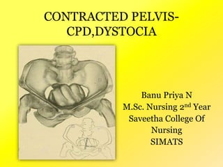
3. • Anatomical - It is a pelvis in which one or more of
its diameters is reduced below the normal by one or
more centimeters.
• Obstetric - It is a pelvis in which one or more of its
diameters is reduced so that it interferes with the
normal mechanism of labor.
4. • Common causes of contracted pelvis are:-
Nutritional and environmental defects:-
minor variation;- common
major :- rachitic and osteomalacic –rare
or injury affecting the bone
fracture ,tumors, tubercular artheritis.
Disease
pelvis:-
spine:- kyphosis, scoliosis, coccygeal deformity
lower limbs:- poliomyeitis, hip joint disease
Developmental defects:- naegele’s pelvis, Robert's
pelvis
5. Classified by:-
• A) type of distortion of pelvic architecture
• B) degree of contraction
6. A) Classification by Pelvic Architecture
1. Pelvis aequabiliter justo minor
• Characterized by general reduction of all diameters;
equally shortened usually by 1-2cm
• Occurs in short. Also occurs in women with massive
skeletal bones and developed muscles, the pelvis has
masculine features such as narrow sacrum, narrow
pubic outlet {funnel-shaped)
7. 2. Flat Pelvis
• Reduced anteroposterior diameters with
normal transverse and oblique diameters
• Has 2 types of contracture
a)Simple flat (or platypellic) pelvis
Entire sacral platform is dislocated toward
the symphysis hence all the anteroposterior
diameters of all pelvic planes are reduced
8.
9.
10. b) Flat rachitic
Anteroposterior diameter of the pelvic inlet
only is reduced
3. Generally Contracted Pelvis
• All diameters reduced, but the anteroposterior
diameters are shortened greater then the
others
• Usually connected with rickets of the childhood
11.
12. Rare forms of contracted pelvis
• Otto’s pelvis – develop as result of
inflammatory process in the hip or knee
• Beaked (rostrate) pelvis – under development
of both sacral wings
• Spondylolithetic pelvis – formed due to
partial dislocation of last lumbar vertebra in
front of 1st sacral vertebra
• Osteomalacic pelvis
• Scoliotic pelvis – only the lumber region
cause deformity of the pelvis. The acetabulum
is pushed inwards on the weight bearing side.
13.
14. B) Classification by degree of contracture
4 degrees
i. First degree: true conjugate <11cm but not
<9cm, spontaneous delivery is possible
ii. Second degree: true conjugate = 9-7.5cm
spontaneous delivery possible but complications
may arise
iii. Third degree: true conjugate 7.5-6cm
spontaneous delivery impossible, use C-section
iv. Fourth degree: true conjugate <6cm, impossible
delivery, only way is C-section ; also known as
absolutely contracted pelvis
15. Diagnosis
History
• Rickets: is expected if there is a history of delayed
walking and dentition.
• Trauma or diseases: of the pelvis, spines or lower
limbs.
• Infantilism
• Previous tuberculosis of bones and joints
16. • Bad obstetric history: e.g. prolonged labour
ended by;
Difficult forceps
Caesarean section or
Still birth.
Weight of the baby
Evidence of maternal injuries such as
complete perineal tear, vesico vaginal
istula, recto vaginal fistula
17. B. Generalexamination:
Abnormal gait :-
Assess woman for stockily built with
bull neck.
Broad shoulder and short thigh
Obese and male distribution of hair
Stature :women < 150 cm or 5 feet
18. Abdomen examination
Pendulous abdomen in primigravida
Fetal head fails to enter a contracted
pelvis at the end of pregnancy and
floats high above inlet, failed growth
of uterus deviates upward and
anteriorly
Non engagement in last 3-4 wks. in
primigravida
19. 2 shapes of abdomen
• Acuminate (pointed)abdomen in primigravida
with a resilient abdominal wall
• Pendulous abdomen in multiparous women
20. • Patient is placed in dorsal position with thigh
flexes and separated.
• The head is grasped by the left hand.
• 2 fingers (index and middle) of theright hand
are placed above the symphysis pubis to note
the degree of overlapping. If when the head is
pushed downward and backward.
21. • The head can be pushed down in the pelvis
without overlapping of the parietal bone on
the symphysis pubis:- No disproportion
• Head can be pushed down a little but ther is
slightly overlapping of the parietal bone
evidence by touch on the under surface of
finger overlapping by 0.5cm:- Moderate
disproportion
22. • Head can not be pushed down and instead the
partial bone overhangs the symphysis pubis
displacing the finger – sever disproportion
Some times the degree of disproportion is
difficult to found by this method because of:-
• Deflexed head
• Thick abdominal wall
• Irritable uterus
• High floating head
23. • It is also called as MULLER – MUNRO KERR
• It is bimanual method.
24. Results :-
• The head can be pushed down up to the level
of ischia spines and there is no overlapping
of the parietal bone over the symphysis
pubis:- no disproportion
• The head can be pushed down a little but not
up to the level of ischia spine and there is
slight overlapping of the parietal bone:-
slight or moderate disproportion
• The head can not be pushed down and
instead the parietal bone overhangs the
symphysis pubis displacing the thumb:-
sever disproportion.
25. D. Pelvimetry
It is assessment of the pelvic diameters and
capacity done at 38-39 weeks. It includes:
Clinical pelvimetry
Internal pelvimetry for
Inlet,
Cavity, and
Outlet.
External pelvimetry for:
Inlet and
Outlet.
Imaging pelvimetry:
•X-ray.
•Computerized tomography (CT).
•Magnetic resonance imaging (MRI) .
30. Elective cesarean section at term is indicated
in:-
Major degree of contraction
Major disproportion
Absolute contraction
Dead fetus
Patient not fit for trial labor
The operation is done in planned way any
time during last week of pregnancy.
Emergency
When trial labor is failed
31. Careful fetal and maternal monitoring by
electronic fetal monitoring and non stress test
Oral feeding remain suspended and hydration is
maintained by intravenous drip
Adequate analgesic is administered
Augmentation of labor by pitocin
32. The progress of labor is mapped with
partograph:-
i) progressive descent of the head
ii) progressive dilatation of the cervix
After the membrane rupture, pelvic examination
is to be done:-
i) to exclude cord prolapse
ii) to note the color of liquor
iii) to assess the pelvis once or more
iv) to note the condition of the cervix
including pressure of the presenting part of the
cervix
33. in favorable cases, end spontaneously, low forcep and low
ventose.
In unfavorable cases, do caesarean section.
Successful trial:-
A trial is called successful, if a healthy baby is born
vaginally, spontaneous or by forcep or ventose with the
mother in good condition
Failure of trial labor:-
Delivery is by cesarean section or delivery of a dead
baby spontaneously or by craniotomy is called failure of trial
labor
34. • Lower incidence of cesarean section.
• A successful trial ensures the women a good
future obstetrics.
35. • May end before full cervix dilatation
• Increased fetal mortality and morbidity
• In failed trial operative risk increases.
36. • Check vitals every 4 hourly
• Monitor both contraction and fetus continuously
• Report immediately the sign of fetal distress
• Position the mother in ways to increase the pelvic
diameter such as sitting or squatting which increase
the outlet diameter and also aid in fetal descent
• Assess the fetus for hypoxia
• Provide support to the client and the family
members in coping with stress of a complicated
labor
41. •Cephalo pelvic disproportion is the disparity in
relation between the head of baby and the mother’s
pelvis.
• It is a pelvis in which one or more of its diameter is
reduced below the normal by one or more
centimeter
42. It is based on clinical findings and pelvimetry:-
Severe disproportion:- when the obstetric
conjugate is less than 7.5 cm (3”) then it is said
to be severe disproportion.
Borderline disproportion:- when the obstetric
conjugate is between 9.5 and 10 cm. In inlet the
anterior posterior diameter is less than 10 cm
and transverse diameter is less than 12 cm.
43. According to American College of Nursing Midwives,
occur 20 out of 250 pregnancy.
“It has been seen through studies that 65% of
women who have been diagnosed with CPD in
previous pregnancies, deliver vaginally in
subsequent pregnancies.”
44. • Nutritional deficiency
• Disease / injury to pelvic bones
• Developmental defects
• A large size baby
• Abnormal fetal position
• Problem with genital tract
45. Absolute causes:- it is a true mechanical obstruction
due to:-
Permanent maternal cause such as contracted
pelvis, anterior sacrococcygeal tumor.
Temporary fetal causes such as hydrocephalus,
large baby etc.
Relative cause:- the relative cause includes brow
presentation, face presentation, mento posterior,
occipito posterior position, deflexed head in vertex
presentation
46. MANAGEMENT:
The treatment for CPD is to continue with labour or move on to a
caesarean section. The goal of treatment is to have a safe delivery, so the
doctors will decide how to treat the condition based on how the delivery is
going.
TRIAL OF LABOR:
When there is a possibility of CPD, the doctors may decide to let you
try to labour. If your labor is moving along well, it may continue along
with:
Close monitoring of your contractions, dilation, and the baby's
progression down the birth canal.
Close monitoring of the baby's movements and heart rate.
Confirmation of the baby's position with a vaginal exam.
Other tests such as X-ray, ultrasound, or MRI to visualize the baby's
head and your pelvis.
47. • During the trial of labour, you can help to open your pelvis and
move the labour along by changing positions with the help of
your nurse, doula, or partner. You can try:
• Sitting
• Squatting
• Changing sides
• Going on your hands and knees
• If labour continues, forceps or a vacuum may be needed to help
deliver the baby. But, if problems arise such as ineffective
contractions, slow dilation and effacement, no descent, or fetal
distress, the doctors will end the trial, and a C-section will be
48. Caesarean Section
When the labour is very long, not progressing as it should,
or causing complications for you or the baby, the next step is a C-
section.
You may need a C-section if:
• You have had a previous C-section.
• You are an older first-time mom.
• The baby is not in a good position for delivery.
• The baby is overdue by a week or more.
• You are having complications such as pre-eclampsia.
• You or the baby are having other medical issues.
50. Definition
When fetal head is delivered,but
shoulders are stuck and cannot
be delivered it is known as
shoulder dystocia
51. Shoulder dystocia
The anterior shoulder becomes trapped
behind on the symphysis pubis, whilst
the posterior shoulder may be in the
hollow of the sacrum or high above the
sacral promontory.
54. Warning signs and
diagnosis
The delivery may have been uncomplicated
initially, but the head may have advanced
slowly and the chin may have had difficulty in
sweeping over the perineum.
Once the head is delivered it may look as if it
is trying to return into the vagina, which is
caused by reverse traction.
Diagnosed when maneouvers normally used
by the midwife fail to accomplish delivery.
56. Management
Principles
DONTs’:
– Do not be panicky
– Do not give traction over baby’s head
– Do not apply fundal pressure
Dos’
– Call for extra help
– Clear the infant’s mouth and nose
– Involve the anaesthesist and the paediatrician
– Perform episiotomy if not performed earlier
61. Rubin’s Maneuver
3. If the shoulder is still not delivered:
insert a hand into the vagina and apply
pressure to the anterior shoulder in the
direction of the baby’s sternum to rotate
the shoulder and decrease the shoulder
diameter.
• If the needed, apply pressure to the
posterior shoulder in the direction of
the baby’s sternum
62. Wood’s maneuver
• 4. If the shoulder is still not delivered
despite the above measures:
• Insert a hand into the vagina
• Grasp the humerus of the posterior arm and
keeping the arm flexed at the elbow, sweep
the arm across the chest, grasp the hand
and deliver the entire arm.
• With one hand on each side of the fetal
head, apply firm, continuous traction
downward to move the anterior shoulder
under the symphysis pubis
64. Cockscrew maneyver
If the posterior arm cannot be
extracted, perform the cockscrew
maneuver.
65. Cleidotomy
If all of the measures fail to deliver the
anterior shoulder;
Another option is to fracture the baby’s
anterior clavicle to decrease the width of
the shoulder. This is done by pressing
the anterior clavicle against the
symphysis pubis.
After birth, facilitate urgent and
immediate newborn care or transfer of
the newborn.
69. Post Procedure care
Repair the episiotomy
If needed, provide emotional
support to the woman and family
following a traumatic birth and
possible death of the newborn or
injury to the baby.
70. JOURNAL PRESENTATION:
Topic: “Fetal pelvic index to predict
cephalopelvic disproportion – a retrospective
clinical cohort study”
Author: Pekka Taipale et al..,
First published: 12 February 2019
Published At: Acta obstetrician and gynaecological
scandinavica
71. ABSTRACT
OBJECTIVE:
To investigate the diagnostic accuracy of the fetal pelvic
index to predict cephalopelvic disproportion.
DESIGN: Retrospective observational cohort study.
SETTING: Pregnant women who had been examined by
X‐ray or magnetic resonance imaging pelvimetry
because of an increased risk of fetal–pelvic disproportion
during 2000–2008 in North Karelia Central Hospital.
72. POPULATION: A total of 274 pregnant women.
METHODS: Univariable and multivariable regression
analyses were carried out to identify risk factors for
caesarean section.
Diagnostic accuracy was tested with a receiver
operating characteristic curve, and the optimal cut‐off
value for fetal pelvic index was calculated.
73. RESULTS
A total of 242 women delivered vaginally, and 32 delivered
with caesarean section caused by labour arrest. In multivariable
modelling, the fetal pelvic index, maternal pelvic inlet size, fetal
head circumference and maternal age were significantly associated
with a risk of caesarean section. In the receiver operating
characteristic analysis, the area under curve was 0.686 with
a p‐value of 0.001 and a 95% confidence interval of 0.595–0.778.
The optimal fetal pelvic index cut‐off value according to the
receiver operating characteristic was −0.65.
The caesarean section rate was 8% below the fetal pelvic
index value of −0.65 and 20% above the fetal pelvic index value of
−0.65.
CONCLUSIONS:
The fetal pelvic index was not a clinically useful tool to
predict the mode of delivery for patients at high risk of
cephalopelvic disproportion. The pooled analysis of the current
and previous studies strengthened this conclusion.