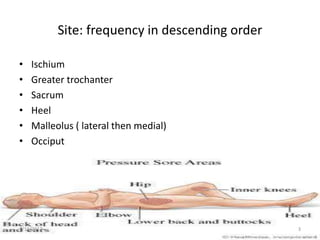
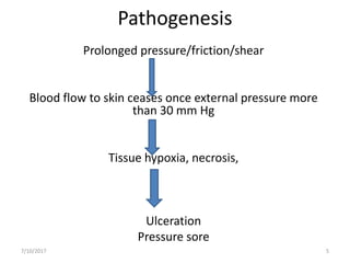
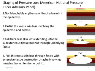
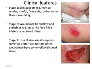
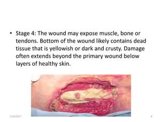
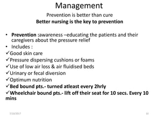
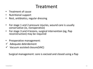
Pressure Sores
By
Vivek Ghosh
Intern
Dept. of General Surgery, GMCTH
7/10/2017 1
Introduction
• Defined as tissue necrosis with ulceration due
to prolonged pressure
• Aka Bedsores/ pressure ulcers/ decubitus
ulcers/ trophic ulcers
• Incidence: 5% of all hospitalized pts.
80% of paraplegic pts.
7/10/2017 2
Site: frequency in descending order
• Ischium
• Greater trochanter
• Sacrum
• Heel
• Malleolus ( lateral then medial)
• Occiput
7/10/2017 3
• Risk factors:
Malnutrition
 Diabetes mellitus
 Peripheral vascular disease
Cerebrovascular accidents
 Peripheral nerve injury
 Old age
 Smoking
 Altered mental status
 Urinary and fecal incontinence
7/10/2017 4
Pathogenesis
Prolonged pressure/friction/shear
Blood flow to skin ceases once external pressure more
than 30 mm Hg
Tissue hypoxia, necrosis,
Ulceration
Pressure sore
7/10/2017 5
Staging of Pressure sore (American National Pressure
Ulcer Advisory Panel)
1.Nonblanchable erythema without a breach in
the epidermis
2.Partial thickness skin loss involving the
epidermis and dermis
3.Full thickness skin loss extending into the
subcutaneous tissue but not through underlying
fascia
4. Full thickness skin loss through fascia with
extensive tissue destruction ,maybe involving
muscles ,bone , tendon or joint .
7/10/2017 6
Clinical features
• Stage 1: Skin appears red, may be
tender, painful, firm, soft, cool or warm
than surrounding
• Stage 2: Wound may be shallow and
pinkish or red, looks like fluid filled
blisters or ruptured blister
• Stage 3: Loss of skin, usually exposes
some fat, crater-like. Bottom of the
wound may have some yellowish dead
tissue
7/10/2017 7
• Stage 4: The wound may expose muscle, bone or
tendons. Bottom of the wound likely contains dead
tissue that is yellowish or dark and crusty. Damage
often extends beyond the primary wound below
layers of healthy skin.
7/10/2017 8
Investigations
• Study of discharge
• Blood sugar
• Biopsy from the edge
• X-ray of part ,X-ray of spine
7/10/2017 9
Management
Prevention is better than cure
Better nursing is the key to prevention
• Prevention :awareness –educating the patients and their
caregivers about the pressure relief
• Includes :
Good skin care
Pressure dispersing cushions or foams
Use of low air loss & air fluidised beds
Urinary or fecal diversion
Optimum nutrition
Bed bound pts.- turned atleast every 2hrly
Wheelchair bound pts.- lift off their seat for 10 secs. Every 10
mins
7/10/2017 10
Treatment
• Treatment of cause
• Nutritional support
• Rest, antibiotics, regular dressing
• For stage 1 and 2 pressure injuries, wound care is usually
conservative (ie, nonoperative)
• For stage 3 and 4 lesions, surgical intervention (eg, flap
reconstruction) may be required
• Preoperative management:
 Adequate debridement
 Vacuum assisted closure(VAC)
Surgical management: sore is excised and closed using a flap
7/10/2017 11
• Treatment options currently being studied:
Hyperbaric oxygen therapy
Electrotherapy
Growth factors
Negative-pressure wound therapy (NPWT)
7/10/2017 12
7/10/2017 13
Complications:
• Sepsis
• Cellulitis
• Osteomyelitis
• Malignant transformation
7/10/2017 14
References
1. Bailey & Love’s Short Practice of Surgery, 26th
Edition
2. Manipal Manual of Surgery, K.R. Shenoy et al,
4th Edition
3. SRB’s Manual of Surgery, S. Bhat M, 4th
Edition
4. www.medscape.com
7/10/2017 15
7/10/2017 16

Pressure sores

  • 1.
    Pressure Sores By Vivek Ghosh Intern Dept.of General Surgery, GMCTH 7/10/2017 1
  • 2.
    Introduction • Defined astissue necrosis with ulceration due to prolonged pressure • Aka Bedsores/ pressure ulcers/ decubitus ulcers/ trophic ulcers • Incidence: 5% of all hospitalized pts. 80% of paraplegic pts. 7/10/2017 2
  • 3.
    Site: frequency indescending order • Ischium • Greater trochanter • Sacrum • Heel • Malleolus ( lateral then medial) • Occiput 7/10/2017 3
  • 4.
    • Risk factors: Malnutrition Diabetes mellitus  Peripheral vascular disease Cerebrovascular accidents  Peripheral nerve injury  Old age  Smoking  Altered mental status  Urinary and fecal incontinence 7/10/2017 4
  • 5.
    Pathogenesis Prolonged pressure/friction/shear Blood flowto skin ceases once external pressure more than 30 mm Hg Tissue hypoxia, necrosis, Ulceration Pressure sore 7/10/2017 5
  • 6.
    Staging of Pressuresore (American National Pressure Ulcer Advisory Panel) 1.Nonblanchable erythema without a breach in the epidermis 2.Partial thickness skin loss involving the epidermis and dermis 3.Full thickness skin loss extending into the subcutaneous tissue but not through underlying fascia 4. Full thickness skin loss through fascia with extensive tissue destruction ,maybe involving muscles ,bone , tendon or joint . 7/10/2017 6
  • 7.
    Clinical features • Stage1: Skin appears red, may be tender, painful, firm, soft, cool or warm than surrounding • Stage 2: Wound may be shallow and pinkish or red, looks like fluid filled blisters or ruptured blister • Stage 3: Loss of skin, usually exposes some fat, crater-like. Bottom of the wound may have some yellowish dead tissue 7/10/2017 7
  • 8.
    • Stage 4:The wound may expose muscle, bone or tendons. Bottom of the wound likely contains dead tissue that is yellowish or dark and crusty. Damage often extends beyond the primary wound below layers of healthy skin. 7/10/2017 8
  • 9.
    Investigations • Study ofdischarge • Blood sugar • Biopsy from the edge • X-ray of part ,X-ray of spine 7/10/2017 9
  • 10.
    Management Prevention is betterthan cure Better nursing is the key to prevention • Prevention :awareness –educating the patients and their caregivers about the pressure relief • Includes : Good skin care Pressure dispersing cushions or foams Use of low air loss & air fluidised beds Urinary or fecal diversion Optimum nutrition Bed bound pts.- turned atleast every 2hrly Wheelchair bound pts.- lift off their seat for 10 secs. Every 10 mins 7/10/2017 10
  • 11.
    Treatment • Treatment ofcause • Nutritional support • Rest, antibiotics, regular dressing • For stage 1 and 2 pressure injuries, wound care is usually conservative (ie, nonoperative) • For stage 3 and 4 lesions, surgical intervention (eg, flap reconstruction) may be required • Preoperative management:  Adequate debridement  Vacuum assisted closure(VAC) Surgical management: sore is excised and closed using a flap 7/10/2017 11
  • 12.
    • Treatment optionscurrently being studied: Hyperbaric oxygen therapy Electrotherapy Growth factors Negative-pressure wound therapy (NPWT) 7/10/2017 12
  • 13.
  • 14.
    Complications: • Sepsis • Cellulitis •Osteomyelitis • Malignant transformation 7/10/2017 14
  • 15.
    References 1. Bailey &Love’s Short Practice of Surgery, 26th Edition 2. Manipal Manual of Surgery, K.R. Shenoy et al, 4th Edition 3. SRB’s Manual of Surgery, S. Bhat M, 4th Edition 4. www.medscape.com 7/10/2017 15
  • 16.

Editor's Notes

  • #3 B&L 26th edition