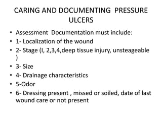
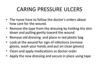
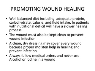
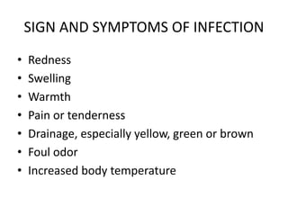
SKIN AND WOUND CARE
Author: Raisa Metauten . Doctorate in Medicine, RN, BSN
KEY TERMS
• Abrasion: A rubbing on the surface of skin
• Blister: (ampolla in spanish) : A swelling on the skin filled with
watery matter.
• Crater: A cup-shaped opening.
• Diabetes: A chronic disease caused due not enough production or
use of insuline by the body.
• Pressure ulcer: Inflammation or sore that develops over areas when
the skin and underneath tissues are injured as result of constant
pressure
• Skin tear: A break in the skin that occurs when the top layer of the
skin is separated from the underlying layers.
• Wound: Any physical injury involving a break in the skin
• Venous: Related to the veins, the blood vessels that carry blood
toward the heart
HOW & WHY
• Pressure ulcers are sometimes
called bedsores, pressure
sores, decubitus ulcer and
decubiti
OOLDER ADULTS
• The skin of an older person is thinner and drier.
• The older person has less subcutaneous or fat tissue
under the skin.
• Older people are more likely to develop skin tears
than younger people
• Skin breakdown can occur after only 90 minutes of
continuous pressure
• Notify any rash that you observe to the doctor
• Keep patient clean and dry
• Check the skin and notify rash or redness
CHILDREN
• Infants and toddlers are more likely to have to
skin problems in the diaper area
• Keep patient clean and dry
• Check the skin and notify rash or redness
PATIENTS AT RISK FOR PRESSURE
SORES ARE THOSE WHO:
• Are unable to move because of paralysis, weakness or coma
• Are unwilling to move because of severe pain, depression or
confusion
• Are unable to control bowel and bladder function
• Have poor food or fluid intake, are dehydrated.
• Have poor circulation, especially patients with diabetes
• Are elderly, obese or very thin
• Have cast, braces or splints
BRADEN SCALE
• Is a standardized way to predicting pressure sore risk, giving
points to measure some topic:
• Sensory perception :ability to respond meaningfully to
pressure related discomfort
• Grade of moisture: Degree to which skin is exposed to
moisture
• Activity: Grade of physical activity
• Mobility: Ability to change and control body position
• Nutrition: Usual food intake pattern
• Friction and shear: Assistance to moving on bed needed
PREVENTING SKIN BREAKDOWN
• Most pressure ulcers are preventable
• Is most difficult and costly to treat a pressure ulcer that to
prevent one.
• Turn and reposition patient at least each 2 hours
• Use pressure-relief devices such as pillows, cushions, special
mattresses
• Keep the skin clean and dry
• Apply lotion to intact skin to prevent dry skin
• Be gentle when lifting or moving
• Pay special attention to the skin during routine assessment
CULTURE
• It may be harder to detect the early signs
of a pressure ulcer in patient with dark
skin tones. A stage I pressure ulcer may
appear blue or purple in color rather
than red. Dark skin that is damaged is
more likely to appear shiny than skin that
is lighter in color
CARING AND DOCUMENTING PRESSURE
ULCERS
• Assessment Documentation must include:
• 1- Localization of the wound
• 2- Stage (I, 2,3,4,deep tissue injury, unsteageable
)
• 3- Size
• 4- Drainage characteristics
• 5-Odor
• 6- Dressing present , missed or soiled, date of last
wound care or not present
CARING PRESSURE ULCERS
• The nurse have to follow the doctor’s orders about
how care for the wound.
• Remove the tape from the dressing by holding the skin
down and pulling gently toward the wound
• Remove old dressing and place in red plastic bag
• Look at the wound for sign of infections (remove
gloves, wash your hands and put on clean gloves)
• Clean and apply medications as doctor order
• Apply the new dressing and secure in place using tape
PROMOTING WOUND HEALING
• Well balanced diet including adequate protein,
carbohydrate, calorie, and fluid intake. In patients
with nutritional deficit will have a slower healing
process.
• The wound must also be kept clean to prevent
wound infection
• A clean, dry dressing may cover every wound
because proper moisten help in healing and
prevent infection
• Always follow medical orders and never use
Alcohol or Iodine in a wound
SIGN AND SYMPTOMS OF INFECTION
• Redness
• Swelling
• Warmth
• Pain or tenderness
• Drainage, especially yellow, green or brown
• Foul odor
• Increased body temperature
COMMON SITES OF PRESSURE ULCERS
STAGE 1
• Stage 1 Pressure Injury: Non-
blanchable erythema of intact
skin
Intact skin with a localized area
of non-blanchable erythema,
which may appear differently in
darkly pigmented skin. Presence
of blanchable erythema or
changes in sensation,
temperature, or firmness may
precede visual changes. Color
changes do not include purple or
maroon discoloration; these may
indicate deep tissue pressure
injury.
BETWEEN US
-SKIN INTACT
-NO BLANCHEABLE
REDNESS
STAGE 2
• Stage 2 Pressure Injury: Partial-thickness
skin loss with exposed dermis
Partial-thickness loss of skin with exposed
dermis. The wound bed is viable, pink or
red, moist, and may also present as an
intact or ruptured serum-filled blister.
Adipose (fat) is not visible and deeper
tissues are not visible. Granulation tissue,
slough and eschar are not present. These
injuries commonly result from adverse
microclimate and shear in the skin over the
pelvis and shear in the heel. This stage
should not be used to describe moisture
associated skin damage (MASD) including
incontinence associated dermatitis (IAD),
intertriginous dermatitis (ITD), medical
adhesive related skin injury (MARSI), or
traumatic wounds (skin tears, burns,
abrasions).
BETWEEN US
-INTACT OR RUPTURED
SERUM- FILLED BLISTER
STAGE 3
Stage 3 Pressure Injury: Full-thickness skin loss
Full-thickness loss of skin, in which adipose
(fat) is visible in the ulcer and granulation
tissue and epibole (rolled wound edges) are
often present. Slough and/or eschar may be
visible. The depth of tissue damage varies
by anatomical location; areas of
significant adiposity can develop deep
wounds. Undermining and tunneling may
occur. Fascia, muscle, tendon, ligament,
cartilage and/or bone are not exposed. If
slough or eschar obscures the extent of
tissue loss this is an Unstageable Pressure
Injury.
BETWEEN US
-LOSS OF SKIN
-FAT IS VISIBLE
-SLOUGH OR ESCHAR
VISIBLE
-TUNNELING AND
UNDERMINING MAY
OCCUR
STAGE 4
• Stage 4 Pressure Injury: Full-
thickness skin and tissue loss
Full-thickness skin and tissue
loss with exposed or directly
palpable fascia, muscle, tendon,
ligament, cartilage or bone in
the ulcer. Slough and/or eschar
may be visible. Epibole (rolled
edges), undermining and/or
tunneling often occur. Depth
varies by anatomical location. If
slough or eschar obscures the
extent of tissue loss this is an
Unstageable Pressure Injury.
BETWEEN US
-LOSS OF SKIN
-FAT + MUSCLE,
TENDON,LIGAMENT,
CARTILAGE OR BONE IS
VISIBLE
-SLOUGH OR ESCHAR
VISIBLE
-TUNNELING AND
UNDERMINING MAY
OCCUR
UNSTAGEABLE
• Unstageable Pressure Injury:
Obscured full-thickness skin
and tissue loss
Full-thickness skin and tissue
loss in which the extent of tissue
damage within the ulcer cannot
be confirmed because it is
obscured by slough or eschar. If
slough or eschar is removed, a
Stage 3 or Stage 4 pressure
injury will be revealed. Stable
eschar (i.e. dry, adherent, intact
without erythema or fluctuance)
on the heel or ischemic limb
should not be softened or
removed.
BETWEEN US
-THE TISSUE CAN NOT
BE
CONFIRMED BECAUSE
IT IS
OBSCURED BY
SLOUGH OR
ESCHAR
DEEP TISSUE
INJURY
• Deep Tissue Pressure Injury: Persistent
non-blanchable deep red, maroon or
purple discoloration
Intact or non-intact skin with localized area
of persistent non-blanchable deep red,
maroon, purple discoloration or epidermal
separation revealing a dark wound bed or
blood filled blister. Pain and temperature
change often precede skin color changes.
Discoloration may appear differently in
darkly pigmented skin. This injury results
from intense and/or prolonged pressure
and shear forces at the bone-muscle
interface. The wound may evolve rapidly to
reveal the actual extent of tissue injury, or
may resolve without tissue loss. If necrotic
tissue, subcutaneous tissue, granulation
tissue, fascia, muscle or other underlying
structures are visible, this indicates a full
thickness pressure injury (Unstageable,
Stage 3 or Stage 4). Do not use DTPI to
describe vascular, traumatic, neuropathic,
or dermatologic conditions.
BETWEEN US
-PERSISTENT
NON-BLANCHEABLE
DEEP
RED,MAROON OR
PURPLE
DECOLORATION
REFERENCES
• Wound Care Resources. (2016, December 27).
Retrieved March 29, 2017, from
http://woundeducators.com/resources/
• Become. (n.d.). Retrieved March 29, 2017, from
http://www.wcei.net/
• Understanding the Healing Stages of Wounds.
(2014, August 26). Retrieved March 29, 2017,
from
https://www.advancedtissue.com/understandin
g-healing-stages-wounds/

Pp skin and wound care

  • 1.
    SKIN AND WOUNDCARE Author: Raisa Metauten . Doctorate in Medicine, RN, BSN
  • 2.
    KEY TERMS • Abrasion:A rubbing on the surface of skin • Blister: (ampolla in spanish) : A swelling on the skin filled with watery matter. • Crater: A cup-shaped opening. • Diabetes: A chronic disease caused due not enough production or use of insuline by the body. • Pressure ulcer: Inflammation or sore that develops over areas when the skin and underneath tissues are injured as result of constant pressure • Skin tear: A break in the skin that occurs when the top layer of the skin is separated from the underlying layers. • Wound: Any physical injury involving a break in the skin • Venous: Related to the veins, the blood vessels that carry blood toward the heart
  • 3.
    HOW & WHY •Pressure ulcers are sometimes called bedsores, pressure sores, decubitus ulcer and decubiti
  • 4.
    OOLDER ADULTS • Theskin of an older person is thinner and drier. • The older person has less subcutaneous or fat tissue under the skin. • Older people are more likely to develop skin tears than younger people • Skin breakdown can occur after only 90 minutes of continuous pressure • Notify any rash that you observe to the doctor • Keep patient clean and dry • Check the skin and notify rash or redness
  • 5.
    CHILDREN • Infants andtoddlers are more likely to have to skin problems in the diaper area • Keep patient clean and dry • Check the skin and notify rash or redness
  • 6.
    PATIENTS AT RISKFOR PRESSURE SORES ARE THOSE WHO: • Are unable to move because of paralysis, weakness or coma • Are unwilling to move because of severe pain, depression or confusion • Are unable to control bowel and bladder function • Have poor food or fluid intake, are dehydrated. • Have poor circulation, especially patients with diabetes • Are elderly, obese or very thin • Have cast, braces or splints
  • 7.
    BRADEN SCALE • Isa standardized way to predicting pressure sore risk, giving points to measure some topic: • Sensory perception :ability to respond meaningfully to pressure related discomfort • Grade of moisture: Degree to which skin is exposed to moisture • Activity: Grade of physical activity • Mobility: Ability to change and control body position • Nutrition: Usual food intake pattern • Friction and shear: Assistance to moving on bed needed
  • 8.
    PREVENTING SKIN BREAKDOWN •Most pressure ulcers are preventable • Is most difficult and costly to treat a pressure ulcer that to prevent one. • Turn and reposition patient at least each 2 hours • Use pressure-relief devices such as pillows, cushions, special mattresses • Keep the skin clean and dry • Apply lotion to intact skin to prevent dry skin • Be gentle when lifting or moving • Pay special attention to the skin during routine assessment
  • 9.
    CULTURE • It maybe harder to detect the early signs of a pressure ulcer in patient with dark skin tones. A stage I pressure ulcer may appear blue or purple in color rather than red. Dark skin that is damaged is more likely to appear shiny than skin that is lighter in color
  • 10.
    CARING AND DOCUMENTINGPRESSURE ULCERS • Assessment Documentation must include: • 1- Localization of the wound • 2- Stage (I, 2,3,4,deep tissue injury, unsteageable ) • 3- Size • 4- Drainage characteristics • 5-Odor • 6- Dressing present , missed or soiled, date of last wound care or not present
  • 11.
    CARING PRESSURE ULCERS •The nurse have to follow the doctor’s orders about how care for the wound. • Remove the tape from the dressing by holding the skin down and pulling gently toward the wound • Remove old dressing and place in red plastic bag • Look at the wound for sign of infections (remove gloves, wash your hands and put on clean gloves) • Clean and apply medications as doctor order • Apply the new dressing and secure in place using tape
  • 12.
    PROMOTING WOUND HEALING •Well balanced diet including adequate protein, carbohydrate, calorie, and fluid intake. In patients with nutritional deficit will have a slower healing process. • The wound must also be kept clean to prevent wound infection • A clean, dry dressing may cover every wound because proper moisten help in healing and prevent infection • Always follow medical orders and never use Alcohol or Iodine in a wound
  • 13.
    SIGN AND SYMPTOMSOF INFECTION • Redness • Swelling • Warmth • Pain or tenderness • Drainage, especially yellow, green or brown • Foul odor • Increased body temperature
  • 14.
    COMMON SITES OFPRESSURE ULCERS
  • 17.
    STAGE 1 • Stage1 Pressure Injury: Non- blanchable erythema of intact skin Intact skin with a localized area of non-blanchable erythema, which may appear differently in darkly pigmented skin. Presence of blanchable erythema or changes in sensation, temperature, or firmness may precede visual changes. Color changes do not include purple or maroon discoloration; these may indicate deep tissue pressure injury. BETWEEN US -SKIN INTACT -NO BLANCHEABLE REDNESS
  • 18.
    STAGE 2 • Stage2 Pressure Injury: Partial-thickness skin loss with exposed dermis Partial-thickness loss of skin with exposed dermis. The wound bed is viable, pink or red, moist, and may also present as an intact or ruptured serum-filled blister. Adipose (fat) is not visible and deeper tissues are not visible. Granulation tissue, slough and eschar are not present. These injuries commonly result from adverse microclimate and shear in the skin over the pelvis and shear in the heel. This stage should not be used to describe moisture associated skin damage (MASD) including incontinence associated dermatitis (IAD), intertriginous dermatitis (ITD), medical adhesive related skin injury (MARSI), or traumatic wounds (skin tears, burns, abrasions). BETWEEN US -INTACT OR RUPTURED SERUM- FILLED BLISTER
  • 19.
    STAGE 3 Stage 3Pressure Injury: Full-thickness skin loss Full-thickness loss of skin, in which adipose (fat) is visible in the ulcer and granulation tissue and epibole (rolled wound edges) are often present. Slough and/or eschar may be visible. The depth of tissue damage varies by anatomical location; areas of significant adiposity can develop deep wounds. Undermining and tunneling may occur. Fascia, muscle, tendon, ligament, cartilage and/or bone are not exposed. If slough or eschar obscures the extent of tissue loss this is an Unstageable Pressure Injury. BETWEEN US -LOSS OF SKIN -FAT IS VISIBLE -SLOUGH OR ESCHAR VISIBLE -TUNNELING AND UNDERMINING MAY OCCUR
  • 20.
    STAGE 4 • Stage4 Pressure Injury: Full- thickness skin and tissue loss Full-thickness skin and tissue loss with exposed or directly palpable fascia, muscle, tendon, ligament, cartilage or bone in the ulcer. Slough and/or eschar may be visible. Epibole (rolled edges), undermining and/or tunneling often occur. Depth varies by anatomical location. If slough or eschar obscures the extent of tissue loss this is an Unstageable Pressure Injury. BETWEEN US -LOSS OF SKIN -FAT + MUSCLE, TENDON,LIGAMENT, CARTILAGE OR BONE IS VISIBLE -SLOUGH OR ESCHAR VISIBLE -TUNNELING AND UNDERMINING MAY OCCUR
  • 21.
    UNSTAGEABLE • Unstageable PressureInjury: Obscured full-thickness skin and tissue loss Full-thickness skin and tissue loss in which the extent of tissue damage within the ulcer cannot be confirmed because it is obscured by slough or eschar. If slough or eschar is removed, a Stage 3 or Stage 4 pressure injury will be revealed. Stable eschar (i.e. dry, adherent, intact without erythema or fluctuance) on the heel or ischemic limb should not be softened or removed. BETWEEN US -THE TISSUE CAN NOT BE CONFIRMED BECAUSE IT IS OBSCURED BY SLOUGH OR ESCHAR
  • 22.
    DEEP TISSUE INJURY • DeepTissue Pressure Injury: Persistent non-blanchable deep red, maroon or purple discoloration Intact or non-intact skin with localized area of persistent non-blanchable deep red, maroon, purple discoloration or epidermal separation revealing a dark wound bed or blood filled blister. Pain and temperature change often precede skin color changes. Discoloration may appear differently in darkly pigmented skin. This injury results from intense and/or prolonged pressure and shear forces at the bone-muscle interface. The wound may evolve rapidly to reveal the actual extent of tissue injury, or may resolve without tissue loss. If necrotic tissue, subcutaneous tissue, granulation tissue, fascia, muscle or other underlying structures are visible, this indicates a full thickness pressure injury (Unstageable, Stage 3 or Stage 4). Do not use DTPI to describe vascular, traumatic, neuropathic, or dermatologic conditions. BETWEEN US -PERSISTENT NON-BLANCHEABLE DEEP RED,MAROON OR PURPLE DECOLORATION
  • 23.
    REFERENCES • Wound CareResources. (2016, December 27). Retrieved March 29, 2017, from http://woundeducators.com/resources/ • Become. (n.d.). Retrieved March 29, 2017, from http://www.wcei.net/ • Understanding the Healing Stages of Wounds. (2014, August 26). Retrieved March 29, 2017, from https://www.advancedtissue.com/understandin g-healing-stages-wounds/

Editor's Notes

  • #11 Remember to follow Standard Precautions and the Blood borne Pathogen Standard when caring a patient who has an open area on the skin
  • #13 Wound is any physical injury involving a break in the skin (E.g. Cutting in the kin when using a kitchen knife, wound for surgical procedure causes a wound closed with staples.)
  • #14 SYMPTOM: The patient complain SIGN: Clinical findings in assessment