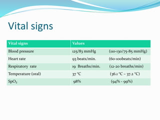
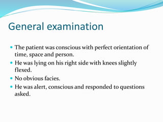
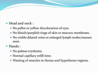
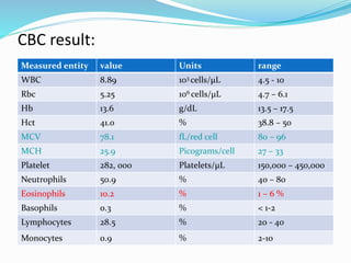
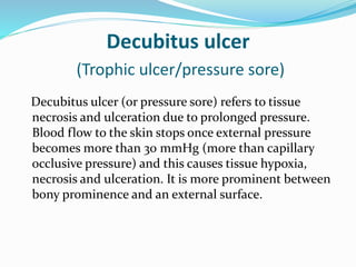
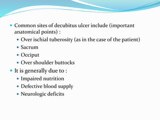
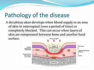
By:
Namratha Raj
MBBS YR - 3
Patient details
 Name : Rashed
 Age : 52 years
 Gender : Male
 Occupation : NIL
 Nationality : Emirati
 Marital status : unmarried
 MRN – MSQ – 00123***
 Date of admission: 18th October, 20**
 Chief complaint : hip pain ( buttocks
region).
( patient also mentioned that he has “inflammation” in the mentioned
region in Arabic )
History of present illness
 Site : Buttocks region
 Onset : 4 years ago
 Character : mild burning like
 Radiation of pain : absent
 Associated symptoms: Numbness in the surrounding
area
 Time of pain : present constantly throughout the day
 Exacerbating / relieving factors : None
 Severity : 2-3
Past medical history
 Patient met with a major accident on 30th March, 2000 which
resulted in cervical spine ( c₂ - c₇) injury, he lost all sensory and
motor functions of hip and lower limbs (paraplegia) and partial
loss of motor function in upper limbs. He also had right clavicle
fracture.
 He did not undergo any surgery for management of the damages
caused due to accident. Instead he went to U.S.A and underwent
physiotherapy for about 2 years.
 From 2003 – 2013, he was in Kerala (India) undergoing ayurvedic
treatment which included oil massages and oral ayurvedic
drugs. This helped him regain partial sensory function in his
lower limbs and partial motor function in upper limbs.
 He is diabetic (since about 16 years).
 On urine drainage bag since the accident.
 Underwent surgery for hemorrhoids 9 years ago.
 Negative history of asthma , hypertension or heart
diseases.
 General weakness present.
Surgical history
 Patient had developed abscess in the same area where
he experiences pain 4 years ago for which he
underwent incision and drainage. NO local anesthesia
was given for this procedure.
 The patient believes that the wound had not healed
and developed into an ulcer soon.
History of ulcer
 Mode of onset: ulcer developed in the buttocks region
after I & D of an abscess.
 Duration : 4 years
 Pain : mild pain.
 Change in size : No known change in size.
 Discharge : absent
Family history
 Patient’s mother had hypertension.
 Negative history of Diabetes/ hypertension/
CVD/asthma in father.
 He has 3 sisters and 2 brother, the eldest brother has
diabetes which he developed during later stages of life.
Drug history
 Patient is on anti diabetic drugs since 16 years.
(drug name and dosage unknown).
 He had been on oral ayurvedic tablets for a period of
ten years (2003 – 2013).
 He has also been taking venlafaxine (anti-depressant),
BID , since two months.
 Negative history of vitamin or mineral supplements.
Personal & social history
 Non-smoker.
 Does not consume alcohol.
 Consumes healthy food (mostly Arab cuisine).
 Consumes about 2 L of water a day.
 No known addictions.
 Accommodation : lives in an independent house with
family.
 Support : the patient has appointed a man to help him
in his day-to-day basic tasks (like shifting positions
while he is on bed )
 Animal contact : None. The patient doesn’t own any
pets
 Travel history : He has been to U.S.A for about 4 1/2
months after the accident and then was in India from
2003 – 2013. After which he went to Jordan for about 3
months for physiotherapy.
History of allergy and
immunizations.
 Allergic to egg.
 No known allergy to drugs.
 History of immunizations unknown.
Review of systems
 Respiratory system: NO cough/ haemoptysis/
dyspnoea/ wheezing.
 Cardio-vascular system : NO palpitations, no history
of chest pain.
 Gastro-intestinal : mild indigestion, NO abdominal
pain/ nausea/vomiting/diarrhoea/ change in bowel
habits.
 Nervous system : NO headache/dizziness. Paraplegic.
Negative history of seizures.
 Urinary system : Patient is on urine drainage bag.
Data analysis and generation of
1st hypothesis
The patient has had cervical spine injury that has
impaired the functions of his upper limbs as well as
resulted in paraplegia, this indicates that the patient
has been bed-ridden or wheel chair bound for a very
large period of time. In addition to it, he is also
diabetic.
Due to this reason, he has probably developed a
pressure sore.
Differential diagnosis
 Moisture associated dermatitis.
 Osteomyelitis.
 Diabetic ulcer.
Examination
Vital signs
Vital signs Values
Blood pressure 125/83 mmHg (110-130/75-85 mmHg)
Heart rate 93 beats/min. (60-100beats/min)
Respiratory rate 19 Breaths/min. (12-20 breaths/min)
Temperature (oral) 37 °C (36.1 °C – 37.2 °C)
SpO₂ 98% (94% - 99%)
General examination
 The patient was conscious with perfect orientation of
time, space and person.
 He was lying on his right side with knees slightly
flexed.
 No obvious facies.
 He was alert, conscious and responded to questions
asked.
 Head and neck :
 No pallor or yellow discoloration of eyes.
 No bluish/purplish tinge of skin or mucous membrane.
 No visible dilated veins or enlarged lymph nodes/masses
seen.
 Hands :
 No palmar erythema.
 Normal capillary refill time.
 Wasting of muscles in thenar and hypothenar regions.
 Legs :
 Slight bilateral pedal edema present.
 Skin appears dry.
 Normal heart sounds : S₁ and S₂ heard clearly, no
murmur.
Local examination
(note : local examination could not be performed as
permission was provided only to remove the bandage
and see)
 Inspection :
 Shape and size: about 3-5 cm in diameter, almost round.
 Number : 1
 Position : inferio-medial part of the right gluteal region
(near ischial tuberosity ).
 Margin: regular, non edematous, pinkish in colour with
peripheral pigmentation.
 Edge : other part whitish in colour (sloping edge as
stated by the doctor) .
 floor of ulcer not seen due to dressing.
 Surrounding area of skin appears normal.
 Palpation :
 Mild tenderness on pressure.
 Outer part of edge does not bleed on touch or when
mildly scratched.
 Base :
 Consistency (hard / firm) : unknown due to dressing.
 Fixed/mobile : unknown due to dressing.
Clinical Provisional Diagnosis
Decubitus ulcer
(pressure sore)
Investigations
 CBC - A complete blood count (CBC) with differential
may show an elevated white blood cell (WBC) count
indicative of inflammation or infection.
CBC result:
Measured entity value Units range
WBC 8.89 103 cells/μL 4.5 - 10
Rbc 5.25 106 cells/μL 4.7 – 6.1
Hb 13.6 g/dL 13.5 – 17.5
Hct 41.0 % 38.8 – 50
MCV 78.1 fL/red cell 80 – 96
MCH 25.9 Picograms/cell 27 – 33
Platelet 282, 000 Platelets/μL 150,000 – 450,000
Neutrophils 50.9 % 40 – 80
Eosinophils 10.2 % 1 – 6 %
Basophils 0.3 % < 1-2
Lymphocytes 28.5 % 20 - 40
Monocytes 0.9 % 2-10
 HIV test – to see if the patient is HIV+ (therefore
immunocompromised) or not and if positive, to
ensure necessary steps are taken to prevent its spread
among the staff , students, visitors etc.
Result : Negative.
 Fasting blood glucose – to monitor the status of his
diabetes.
Result : 125 mg/dL (normal:<100mg/dL)
 Electrolyte panel - to check for electrolyte balance
since it is critically important for things like hydration,
nerve impulses, muscle function, and pH level.
Measured
entity
value units range
Sodium 136 mEq/L 135 - 145
Potassium 3.8 mEq/L 3.5 – 5.0
Chloride 99 mEq/L 96 - 106
CO2 29 mEq/L 23-29
Anion gap 7 mEq/L 8-16
 Wound culture – to check for bacterial/fungal
infection of the ulcer and to know which anti-biotic
/anti-fungal can inhibit the growth or kill the
bacteria/fungus.
Result : Not updated in system.
 Chest X-ray – is important in case of tuberculous
ulcers to detect any primary focus in the lung and also
for pre-operative care.
Result :Not updated in system.
 X-ray of pelvis – to look for radiologic appearances in
x-ray that linked directly or indirectly to pressure ulcer
development (like free air, fistula development) and
represent pathological changes in skeletal system.
Result :Not updated in system.
Missing investigations
 LFT – for albumin levels.
 Hemoglobin A1C test.
 Urine test.
Management Map
 Medical treatment :
 Treatment of cause – patient asked to lay on his sides
(periodically alternating positions , once in 2 hours).
 Rest, anti-biotics, slough excision (if present), regular
dressings.
 Vacuum-assisted closure (VAC) : It is the creation of
intermittent negative pressure of -125 mmHg to promote
formation of healthy granulation tissue. Negative
pressure reduces tissue oedema, clears interstitial fluid
and improves the perfusion thereby promoting wound
healing.
 Surgical treatment:
 Surgical intervention is considered once there is
complete loss of epidermis and dermis and the ulcer
starts extending into the subcutaneous tissue.
 It includes :
 Flap cover or skin grafting once ulcer granulates well.
or
 Excision of ulcer and skin grafting .
 Actual treatment given :
 Patient is put on a special diet because he is diabetic.
advised to take oral anti-biotic (clarithromycin).
 Silvercel dressing done every 8 hours. (silvercel dressing
is a non-woven pad composed of alginate,
carboxymethylcellulose (CMC) and silver coated nylon
fibers, with a non-adherent. wound contact layer.)
 Patient is kept under observation.
Pharmacologic data of each drug
Scientific name
(group)
Mode of action Side effects Antagonism /
synergism
Clarithromycin
(macrolide)
Clarithromycin
prevents bacterial
growth/
multiplication by
acting as a protein
synthesis
inhibitor. It binds
to 23S rRNA, a
component of the
50S subunit of the
bacterial
ribosome , thus
inhibiting
the translation
of peptides.
•stomach pain,
indigestion, gas.
•vomiting, mild
diarrhea.
•unusual or
unpleasant taste in
your mouth.
•headache, sleep
problems
(insomnia).
•Antagonism with
cefuroxime (and
other bactericidal
drugs )
•Synergism with
anti- tuberculous
drugs
Ulcer
 Defined as a break in the continuity of the covering
epithelium , either skin or mucous membrane due to
molecular death.
Anatomy of skin
 Skin has three layers:
 The epidermis, the
outermost layer of skin,
provides a waterproof barrier
and creates our skin tone.
 The dermis, beneath the
epidermis, contains tough
connective tissue, hair
follicles, and sweat glands.
 The deeper subcutaneous
tissue (hypodermis) is made
of fat and connective tissue.
 Function of organ affected , the skin :
 Protection: covers the body and provides a physical
barrier that protects underlying tissues from physical
abrasion, bacterial invasion, dehydration and UV
radiation.
 Regulation: regulates temperature by the use of
sweating and changes in the blood flow when exposed
to extremes of temperature
 Sensation: skin contains abundant nerve endings and
receptors to detect stimuli related to temperature,
touch, pressure and pain
(The above functions are impaired in an ulcerated area)
Decubitus ulcer
(Trophic ulcer/pressure sore)
Decubitus ulcer (or pressure sore) refers to tissue
necrosis and ulceration due to prolonged pressure.
Blood flow to the skin stops once external pressure
becomes more than 30 mmHg (more than capillary
occlusive pressure) and this causes tissue hypoxia,
necrosis and ulceration. It is more prominent between
bony prominence and an external surface.
 Common sites of decubitus ulcer include (important
anatomical points) :
 Over ischial tuberosity (as in the case of the patient)
 Sacrum
 Occiput
 Over shoulder buttocks
 It is generally due to :
 Impaired nutrition
 Defective blood supply
 Neurologic deficits
Pathology of the disease
A decubitus ulcer develops when blood supply to an area
of skin in interrupted (over a period of time) or
completely blocked. This can occur when layers of
skin are compressed between bone and another hard
surface.
 Due to the presence of neurologic deficit (spinal cord
injury , in case of the patient), it begins as a callosity
due to repeated trauma and pressure, under which
suppuration takes place, the pus comes out and the
central hole forms the ulcer which gradually burrows
through the muscles and the tendons to the bone. The
skin surrounding the ulcer would have no sensation ,
the cause being spinal injury (as in case of the
patient), diabetic neuropathy, peripheral nerve injury
etc.
Complications of the disease
 Sepsis – It occurs when bacteria enter the
bloodstream through broken skin and spread
throughout the body. It's a rapidly progressing, life-
threatening condition that can cause organ failure.
 Cellulitis: It is an infection of the skin and connected
soft tissues. It can cause severe pain, redness and
swelling. (People with nerve damage often do not feel
pain with this condition). Cellulitis can lead to life-
threatening complications.
 Bone and joint infections. An infection from a
pressure sore can burrow into joints and bones. Joint
infections (septic arthritis) can damage cartilage and
tissue. Bone infections (osteomyelitis) may reduce the
function of joints and limbs. Such infections can lead
to life-threatening complications.
 Cancer. Another complication is the development of a
type of squamous cell carcinoma that develops in
chronic, non-healing wounds. This type of cancer is
aggressive and usually requires surgery
Management of complications:
 Management of sepsis :
 Start adequate antibiotic therapy as early as possible.
 Resuscitate the patient, using supportive measures to
correct hypoxia, hypotension, and impaired tissue
oxygenation (hypoperfusion).
 Identify the source of infection (ulcer), and treat with
antimicrobial therapy/ surgery, or both.
 Maintain adequate organ system function, guided by
cardiovascular monitoring, and interrupt the
progression to multiple organ dysfunction syndrome.
 Management of cellulitis : Cause is often
polymicrobial, empiric coverage is recommended,
(which include broad coverage of gram-positive, gram-
negative and anaerobic organisms, coverage of MRSA
is also recommended until sensitivity culture proves
otherwise.)
 Management of bone and joint infections : IV
antibiotics and/or surgery.
 Management of cancer : surgery.
Psychological impact of disease
 On Patient: Anxiety regarding the progress,
management and recurrence of ulcer, depression, self-
pity.
 On Family: Anxiety about the outcome of the surgery
and about complications.
 On Community: General anxiety whether the disease
could spread among individuals
References
 Manual on Clinical surgery by S. Das
 SRB’s manual of surgery
 www.mayoclinic.org
 www.icid.salisbury.nhs.uk
 emedicine.medscape.com

case presentation - decubitus ulcer

  • 1.
  • 2.
    Patient details  Name: Rashed  Age : 52 years  Gender : Male  Occupation : NIL  Nationality : Emirati  Marital status : unmarried  MRN – MSQ – 00123***  Date of admission: 18th October, 20**
  • 3.
     Chief complaint: hip pain ( buttocks region). ( patient also mentioned that he has “inflammation” in the mentioned region in Arabic )
  • 4.
    History of presentillness  Site : Buttocks region  Onset : 4 years ago  Character : mild burning like  Radiation of pain : absent  Associated symptoms: Numbness in the surrounding area  Time of pain : present constantly throughout the day  Exacerbating / relieving factors : None  Severity : 2-3
  • 5.
    Past medical history Patient met with a major accident on 30th March, 2000 which resulted in cervical spine ( c₂ - c₇) injury, he lost all sensory and motor functions of hip and lower limbs (paraplegia) and partial loss of motor function in upper limbs. He also had right clavicle fracture.  He did not undergo any surgery for management of the damages caused due to accident. Instead he went to U.S.A and underwent physiotherapy for about 2 years.  From 2003 – 2013, he was in Kerala (India) undergoing ayurvedic treatment which included oil massages and oral ayurvedic drugs. This helped him regain partial sensory function in his lower limbs and partial motor function in upper limbs.  He is diabetic (since about 16 years).  On urine drainage bag since the accident.
  • 6.
     Underwent surgeryfor hemorrhoids 9 years ago.  Negative history of asthma , hypertension or heart diseases.  General weakness present.
  • 7.
    Surgical history  Patienthad developed abscess in the same area where he experiences pain 4 years ago for which he underwent incision and drainage. NO local anesthesia was given for this procedure.  The patient believes that the wound had not healed and developed into an ulcer soon.
  • 8.
    History of ulcer Mode of onset: ulcer developed in the buttocks region after I & D of an abscess.  Duration : 4 years  Pain : mild pain.  Change in size : No known change in size.  Discharge : absent
  • 9.
    Family history  Patient’smother had hypertension.  Negative history of Diabetes/ hypertension/ CVD/asthma in father.  He has 3 sisters and 2 brother, the eldest brother has diabetes which he developed during later stages of life.
  • 10.
    Drug history  Patientis on anti diabetic drugs since 16 years. (drug name and dosage unknown).  He had been on oral ayurvedic tablets for a period of ten years (2003 – 2013).  He has also been taking venlafaxine (anti-depressant), BID , since two months.  Negative history of vitamin or mineral supplements.
  • 11.
    Personal & socialhistory  Non-smoker.  Does not consume alcohol.  Consumes healthy food (mostly Arab cuisine).  Consumes about 2 L of water a day.  No known addictions.  Accommodation : lives in an independent house with family.
  • 12.
     Support :the patient has appointed a man to help him in his day-to-day basic tasks (like shifting positions while he is on bed )  Animal contact : None. The patient doesn’t own any pets  Travel history : He has been to U.S.A for about 4 1/2 months after the accident and then was in India from 2003 – 2013. After which he went to Jordan for about 3 months for physiotherapy.
  • 13.
    History of allergyand immunizations.  Allergic to egg.  No known allergy to drugs.  History of immunizations unknown.
  • 14.
    Review of systems Respiratory system: NO cough/ haemoptysis/ dyspnoea/ wheezing.  Cardio-vascular system : NO palpitations, no history of chest pain.  Gastro-intestinal : mild indigestion, NO abdominal pain/ nausea/vomiting/diarrhoea/ change in bowel habits.  Nervous system : NO headache/dizziness. Paraplegic. Negative history of seizures.  Urinary system : Patient is on urine drainage bag.
  • 15.
    Data analysis andgeneration of 1st hypothesis The patient has had cervical spine injury that has impaired the functions of his upper limbs as well as resulted in paraplegia, this indicates that the patient has been bed-ridden or wheel chair bound for a very large period of time. In addition to it, he is also diabetic. Due to this reason, he has probably developed a pressure sore.
  • 16.
    Differential diagnosis  Moistureassociated dermatitis.  Osteomyelitis.  Diabetic ulcer.
  • 17.
  • 18.
    Vital signs Vital signsValues Blood pressure 125/83 mmHg (110-130/75-85 mmHg) Heart rate 93 beats/min. (60-100beats/min) Respiratory rate 19 Breaths/min. (12-20 breaths/min) Temperature (oral) 37 °C (36.1 °C – 37.2 °C) SpO₂ 98% (94% - 99%)
  • 19.
    General examination  Thepatient was conscious with perfect orientation of time, space and person.  He was lying on his right side with knees slightly flexed.  No obvious facies.  He was alert, conscious and responded to questions asked.
  • 20.
     Head andneck :  No pallor or yellow discoloration of eyes.  No bluish/purplish tinge of skin or mucous membrane.  No visible dilated veins or enlarged lymph nodes/masses seen.  Hands :  No palmar erythema.  Normal capillary refill time.  Wasting of muscles in thenar and hypothenar regions.
  • 21.
     Legs : Slight bilateral pedal edema present.  Skin appears dry.  Normal heart sounds : S₁ and S₂ heard clearly, no murmur.
  • 22.
    Local examination (note :local examination could not be performed as permission was provided only to remove the bandage and see)  Inspection :  Shape and size: about 3-5 cm in diameter, almost round.  Number : 1  Position : inferio-medial part of the right gluteal region (near ischial tuberosity ).  Margin: regular, non edematous, pinkish in colour with peripheral pigmentation.
  • 23.
     Edge :other part whitish in colour (sloping edge as stated by the doctor) .  floor of ulcer not seen due to dressing.  Surrounding area of skin appears normal.  Palpation :  Mild tenderness on pressure.  Outer part of edge does not bleed on touch or when mildly scratched.  Base :  Consistency (hard / firm) : unknown due to dressing.  Fixed/mobile : unknown due to dressing.
  • 24.
  • 25.
    Investigations  CBC -A complete blood count (CBC) with differential may show an elevated white blood cell (WBC) count indicative of inflammation or infection.
  • 26.
    CBC result: Measured entityvalue Units range WBC 8.89 103 cells/μL 4.5 - 10 Rbc 5.25 106 cells/μL 4.7 – 6.1 Hb 13.6 g/dL 13.5 – 17.5 Hct 41.0 % 38.8 – 50 MCV 78.1 fL/red cell 80 – 96 MCH 25.9 Picograms/cell 27 – 33 Platelet 282, 000 Platelets/μL 150,000 – 450,000 Neutrophils 50.9 % 40 – 80 Eosinophils 10.2 % 1 – 6 % Basophils 0.3 % < 1-2 Lymphocytes 28.5 % 20 - 40 Monocytes 0.9 % 2-10
  • 27.
     HIV test– to see if the patient is HIV+ (therefore immunocompromised) or not and if positive, to ensure necessary steps are taken to prevent its spread among the staff , students, visitors etc. Result : Negative.  Fasting blood glucose – to monitor the status of his diabetes. Result : 125 mg/dL (normal:<100mg/dL)
  • 28.
     Electrolyte panel- to check for electrolyte balance since it is critically important for things like hydration, nerve impulses, muscle function, and pH level. Measured entity value units range Sodium 136 mEq/L 135 - 145 Potassium 3.8 mEq/L 3.5 – 5.0 Chloride 99 mEq/L 96 - 106 CO2 29 mEq/L 23-29 Anion gap 7 mEq/L 8-16
  • 29.
     Wound culture– to check for bacterial/fungal infection of the ulcer and to know which anti-biotic /anti-fungal can inhibit the growth or kill the bacteria/fungus. Result : Not updated in system.
  • 30.
     Chest X-ray– is important in case of tuberculous ulcers to detect any primary focus in the lung and also for pre-operative care. Result :Not updated in system.  X-ray of pelvis – to look for radiologic appearances in x-ray that linked directly or indirectly to pressure ulcer development (like free air, fistula development) and represent pathological changes in skeletal system. Result :Not updated in system.
  • 31.
    Missing investigations  LFT– for albumin levels.  Hemoglobin A1C test.  Urine test.
  • 32.
    Management Map  Medicaltreatment :  Treatment of cause – patient asked to lay on his sides (periodically alternating positions , once in 2 hours).  Rest, anti-biotics, slough excision (if present), regular dressings.  Vacuum-assisted closure (VAC) : It is the creation of intermittent negative pressure of -125 mmHg to promote formation of healthy granulation tissue. Negative pressure reduces tissue oedema, clears interstitial fluid and improves the perfusion thereby promoting wound healing.
  • 33.
     Surgical treatment: Surgical intervention is considered once there is complete loss of epidermis and dermis and the ulcer starts extending into the subcutaneous tissue.  It includes :  Flap cover or skin grafting once ulcer granulates well. or  Excision of ulcer and skin grafting .
  • 34.
     Actual treatmentgiven :  Patient is put on a special diet because he is diabetic. advised to take oral anti-biotic (clarithromycin).  Silvercel dressing done every 8 hours. (silvercel dressing is a non-woven pad composed of alginate, carboxymethylcellulose (CMC) and silver coated nylon fibers, with a non-adherent. wound contact layer.)  Patient is kept under observation.
  • 35.
    Pharmacologic data ofeach drug Scientific name (group) Mode of action Side effects Antagonism / synergism Clarithromycin (macrolide) Clarithromycin prevents bacterial growth/ multiplication by acting as a protein synthesis inhibitor. It binds to 23S rRNA, a component of the 50S subunit of the bacterial ribosome , thus inhibiting the translation of peptides. •stomach pain, indigestion, gas. •vomiting, mild diarrhea. •unusual or unpleasant taste in your mouth. •headache, sleep problems (insomnia). •Antagonism with cefuroxime (and other bactericidal drugs ) •Synergism with anti- tuberculous drugs
  • 36.
    Ulcer  Defined asa break in the continuity of the covering epithelium , either skin or mucous membrane due to molecular death.
  • 37.
    Anatomy of skin Skin has three layers:  The epidermis, the outermost layer of skin, provides a waterproof barrier and creates our skin tone.  The dermis, beneath the epidermis, contains tough connective tissue, hair follicles, and sweat glands.  The deeper subcutaneous tissue (hypodermis) is made of fat and connective tissue.
  • 38.
     Function oforgan affected , the skin :  Protection: covers the body and provides a physical barrier that protects underlying tissues from physical abrasion, bacterial invasion, dehydration and UV radiation.  Regulation: regulates temperature by the use of sweating and changes in the blood flow when exposed to extremes of temperature  Sensation: skin contains abundant nerve endings and receptors to detect stimuli related to temperature, touch, pressure and pain (The above functions are impaired in an ulcerated area)
  • 39.
    Decubitus ulcer (Trophic ulcer/pressuresore) Decubitus ulcer (or pressure sore) refers to tissue necrosis and ulceration due to prolonged pressure. Blood flow to the skin stops once external pressure becomes more than 30 mmHg (more than capillary occlusive pressure) and this causes tissue hypoxia, necrosis and ulceration. It is more prominent between bony prominence and an external surface.
  • 40.
     Common sitesof decubitus ulcer include (important anatomical points) :  Over ischial tuberosity (as in the case of the patient)  Sacrum  Occiput  Over shoulder buttocks  It is generally due to :  Impaired nutrition  Defective blood supply  Neurologic deficits
  • 41.
    Pathology of thedisease A decubitus ulcer develops when blood supply to an area of skin in interrupted (over a period of time) or completely blocked. This can occur when layers of skin are compressed between bone and another hard surface.
  • 42.
     Due tothe presence of neurologic deficit (spinal cord injury , in case of the patient), it begins as a callosity due to repeated trauma and pressure, under which suppuration takes place, the pus comes out and the central hole forms the ulcer which gradually burrows through the muscles and the tendons to the bone. The skin surrounding the ulcer would have no sensation , the cause being spinal injury (as in case of the patient), diabetic neuropathy, peripheral nerve injury etc.
  • 43.
    Complications of thedisease  Sepsis – It occurs when bacteria enter the bloodstream through broken skin and spread throughout the body. It's a rapidly progressing, life- threatening condition that can cause organ failure.  Cellulitis: It is an infection of the skin and connected soft tissues. It can cause severe pain, redness and swelling. (People with nerve damage often do not feel pain with this condition). Cellulitis can lead to life- threatening complications.
  • 44.
     Bone andjoint infections. An infection from a pressure sore can burrow into joints and bones. Joint infections (septic arthritis) can damage cartilage and tissue. Bone infections (osteomyelitis) may reduce the function of joints and limbs. Such infections can lead to life-threatening complications.  Cancer. Another complication is the development of a type of squamous cell carcinoma that develops in chronic, non-healing wounds. This type of cancer is aggressive and usually requires surgery
  • 45.
    Management of complications: Management of sepsis :  Start adequate antibiotic therapy as early as possible.  Resuscitate the patient, using supportive measures to correct hypoxia, hypotension, and impaired tissue oxygenation (hypoperfusion).  Identify the source of infection (ulcer), and treat with antimicrobial therapy/ surgery, or both.  Maintain adequate organ system function, guided by cardiovascular monitoring, and interrupt the progression to multiple organ dysfunction syndrome.
  • 46.
     Management ofcellulitis : Cause is often polymicrobial, empiric coverage is recommended, (which include broad coverage of gram-positive, gram- negative and anaerobic organisms, coverage of MRSA is also recommended until sensitivity culture proves otherwise.)  Management of bone and joint infections : IV antibiotics and/or surgery.  Management of cancer : surgery.
  • 47.
    Psychological impact ofdisease  On Patient: Anxiety regarding the progress, management and recurrence of ulcer, depression, self- pity.  On Family: Anxiety about the outcome of the surgery and about complications.  On Community: General anxiety whether the disease could spread among individuals
  • 48.
    References  Manual onClinical surgery by S. Das  SRB’s manual of surgery  www.mayoclinic.org  www.icid.salisbury.nhs.uk  emedicine.medscape.com