This document discusses tissue engineering of the nasal septum using a chitosan-based material. Chitosan is derived from crustacean shells and has advantages like biodegradability and biocompatibility. The author creates a hybrid polymer by mixing a chitosan solution with hyaluronic acid and collagen. Human inferior turbinate tissue is treated and seeded onto the collagen-coated hybrid polymer. Morphological examinations are conducted to analyze pore size, cell proliferation, ciliary beat, histology, and immuno-histochemistry. The goal is to develop a tissue-engineered nasal septum with sufficient control over shape and improved biocompatibility for cell seeding.
The defense mechanism of gingiva includes GCF, Saliva, epithelial barrier and connective tissue cells. All these protect the periodontium from bacterial invasion.
Introduction
Artificial skin
Invention
Structure of human skin
Importance of skin
Key development
Biomaterials
Methods to produce artificial skin
Application
Problems
Future development
Conclusions
references
Wound management has made rapid advances over the last 25 years. New innovations in dressing technology could have a huge impact on the greater wound care industry.
Proposal for Protein-DNA Mapping using AFM for Lab on a ChipAnthony Salvagno
This was a report written for a class I am taking in conjunction with Ken Seal, John Montoya, and Laura Pawlikowski. We discuss functionalizing an AFM tip with antibodies for protein detection. Conceptually, proteins bound to DNA can be flowed down a nanochannel and can be detected by an AFM tip in the channel. The detection will be based on the interaction between antibodies and their antigens (the proteins). This proposal details everything we imagined. We presented on this topic as well.
Imaging, spectroscopy and manipulation of C60 molecule on semiconductor surf...Cristina Chiutu
Scanning probe microscopy techniques were employed to investigate C60 molecules adsorbed on Si(111)-(7x7) and Ag-Si(111)-(√3x√3)R30o using imaging, spectroscopy, and manipulation methods. First, dynamic scanning tunnelling microscopy revealed the lowest unoccupied molecular orbital features of C60 molecules adsorbed on Si(111)-(7x7) with extremely high resolution at 77 K. Experimental data were compared with Hückel molecular orbital theory simulations to determine the orientation of the molecules on these surfaces. Second, C60 molecules were imaged with a qPlus atomic force microscope, in the attractive force regime and appeared as bright spherical protrusions. The potential energy of interaction between the AFM tip and C60 molecules adsorbed on Si(111)-(7x7) was quantified by force spectroscopy.
Furthermore, a C60 molecule was transferred to the scanning probe microscope tip and used as molecular probe to image the Si(111)-(7x7) surface and other C60 molecules. The on-tip C60 molecule was imaged with high precision. Hückel molecular orbital theory calculations accurately predicted the shape and characteristics of molecular orbitals observed with dynamic scanning tunnelling microscopy, which were strongly dependent on molecular symmetry, orientation, and adsorption angle. Using qPlus atomic force microscopy, chemical reactivity was probed close to or at the carbon atom positions in the C60 cage. Density functional theory simulations showed that an (iono)covalent bond formed between a carbon atom and the underlying Si adatom was responsible for contrast formation.
The pair potential for two C60 molecules was also determined experimentally and found to be in very good agreement with the Girifalco potential (Girifalco, L.A., J. Phys. Chem., 1992. 96(2): p. 858). Using Hückel molecular orbital theory, the mutual orientation of a C60 molecule adsorbed on the STM/AFM tip and a C60 molecule adsorbed on the Si(111)-(7x7) surface was determined via comparison of simulated images to the experimental data. Individual C60 molecules were also manipulated with qPlus atomic force microscopy.
Manipulation of single C60 molecules was performed on the Ag-Si(111)-(√3x√3)R30o surface using scanning tunnelling microscopy at room temperature and at 100 K. The interaction was predominantly attractive. Due to weak molecule-substrate interaction, a short-range chemical force between the C60 molecule and the tip was considered to be responsible for the manipulation process.
This is the presentation based on our propsal of the same name. The presenters were Laura Pawlikowski, John Montoya, Ken Seal, and myself. We discuss functionalizing an AFM tip with antibodies for protein detection and discuss several possible uses for such a device. Conceptually, proteins bound to DNA can be flowed down a nanochannel and can be detected by an AFM tip in the channel. The detection will be based on the interaction between antibodies and their antigens (the proteins).
Defense mechanism of gingiva / /certified fixed orthodontic courses by Indi...Indian dental academy
Welcome to Indian Dental Academy
The Indian Dental Academy is the Leader in continuing dental education , training dentists in all aspects of dentistry and offering a wide range of dental certified courses in different formats.
Indian dental academy has a unique training program & curriculum that provides students with exceptional clinical skills and enabling them to return to their office with high level confidence and start treating patients
State of the art comprehensive training-Faculty of world wide repute &Very affordable.
Genotoxicity Evaluation of Polystyrene Membrane with Collagen and Norbixin by...inventionjournals
The biocompatible membranes are widely applied in the medical field in order to stimulate tissue repair. The biological principle of this type of treatment is the repair and guided regeneration. In the literature, there are few reports of studies evaluating the effects and biological properties of norbixin in animal tissues. Thus, the present study was to evaluate the effect of polystyrene membrane with collagen and norbixin, through the micronucleus test and comet assay in rats, as part of the recommended test battery to evaluate the mutagenic potential. The research project was approved by CEP / FACID Protocol 069/2014. For this study, 15 rats were divided into 3 groups were used: A - the membrane was introduced into the peritoneum of the animals through a laparotomy; B - received cyclophosphamide at a dose of 50mg / kg intraperitoneally; C - were performed only one laparotomy. A peripheral blood sample was collected from the animals for conducting Comet assay and 72 hours after the start of the experiment were euthanized. It was collected bone marrow material of each rat to perform the micronucleus test. In conclusion, through the tests, the membrane is not genotoxic
Genotoxicity Evaluation of Polystyrene Membrane with Collagen and Norbixin by...inventionjournals
The biocompatible membranes are widely applied in the medical field in order to stimulate tissue repair. The biological principle of this type of treatment is the repair and guided regeneration. In the literature, there are few reports of studies evaluating the effects and biological properties of norbixin in animal tissues. Thus, the present study was to evaluate the effect of polystyrene membrane with collagen and norbixin, through the micronucleus test and comet assay in rats, as part of the recommended test battery to evaluate the mutagenic potential. The research project was approved by CEP / FACID Protocol 069/2014. For this study, 15 rats were divided into 3 groups were used: A - the membrane was introduced into the peritoneum of the animals through a laparotomy; B - received cyclophosphamide at a dose of 50mg / kg intraperitoneally; C - were performed only one laparotomy. A peripheral blood sample was collected from the animals for conducting Comet assay and 72 hours after the start of the experiment were euthanized. It was collected bone marrow material of each rat to perform the micronucleus test. In conclusion, through the tests, the membrane is not genotoxic.
Genotoxicity Evaluation of Polystyrene Membrane with Collagen and Norbixin by...inventionjournals
The biocompatible membranes are widely applied in the medical field in order to stimulate tissue repair. The biological principle of this type of treatment is the repair and guided regeneration. In the literature, there are few reports of studies evaluating the effects and biological properties of norbixin in animal tissues. Thus, the present study was to evaluate the effect of polystyrene membrane with collagen and norbixin, through the micronucleus test and comet assay in rats, as part of the recommended test battery to evaluate the mutagenic potential. The research project was approved by CEP / FACID Protocol 069/2014. For this study, 15 rats were divided into 3 groups were used: A - the membrane was introduced into the peritoneum of the animals through a laparotomy; B - received cyclophosphamide at a dose of 50mg / kg intraperitoneally; C - were performed only one laparotomy. A peripheral blood sample was collected from the animals for conducting Comet assay and 72 hours after the start of the experiment were euthanized. It was collected bone marrow material of each rat to perform the micronucleus test. In conclusion, through the tests, the membrane is not genotoxic
Comparative evaluation of antimicrobial efficacy of q mix™/ dental implant co...Indian dental academy
The Indian Dental Academy is the Leader in continuing dental education , training dentists in all aspects of dentistry and
offering a wide range of dental certified courses in different formats.
Engineering of a Microscale Niche for Pancreatic Tumor Cells Using Bioactive ...Arun kumar
Two-photon polymerization has recently emerged as a promising technique to fabricate scaffolds for three-dimensional (3D) cell culture and tissue engineering. Here, we combined 3D-printed microscale scaffolds fabricated using two-photon polymerization with a bioactive layer-by-layer film coating. This bioactive coating consists of hyaluronic acid and poly(l-lysine) of controlled stiffness, loaded with fibronectin and bone morphogenic proteins 2 and 4 (BMP2 and BMP4) as matrix-bound proteins. Planar films were prepared using a liquid handling robot directly in 96-well plates to perform high-content studies of cellular processes, especially cell adhesion, proliferation, and BMP-induced signaling. The behaviors of two human pancreatic cell lines PANC1 (immortalized) and PAN092 (patient-derived cell line) were systematically compared and revealed important context-specific cell responses, notably in response to film stiffness and matrix-bound BMPs (bBMPs). Fibronectin significantly increased cell adhesion, spreading, and proliferation for both cell types on soft and stiff films; BMP2 increased cell adhesion and inhibited proliferation of PANC1 cells and PAN092 on soft films. BMP4 enhanced cell adhesion and proliferation of PANC1 and showed a bipolar effect on PAN092. Importantly, PANC1 exhibited a strong dose-dependent BMP response, notably for bBMP2, while PAN092 was insensitive to BMPs. Finally, we proved that it is possible to combine a microscale 3D Ormocomp scaffold fabricated using the two-photon polymerization technique with the bioactive film coating to form a microscale tumor tissue and mimic the early stages of metastatic cancer.
wo-photon polymerization has recently emerged as a promising technique to fabricate scaffolds for three-dimensional (3D) cell culture and tissue engineering. Here, we combined 3D-printed microscale scaffolds fabricated using two-photon polymerization with a bioactive layer-by-layer film coating. This bioactive coating consists of hyaluronic acid and poly(l-lysine) of controlled stiffness, loaded with fibronectin and bone morphogenic proteins 2 and 4 (BMP2 and BMP4) as matrix-bound proteins. Planar films were prepared using a liquid handling robot directly in 96-well plates to perform high-content studies of cellular processes, especially cell adhesion, proliferation, and BMP-induced signaling. The behaviors of two human pancreatic cell lines PANC1 (immortalized) and PAN092 (patient-derived cell line) were systematically compared and revealed important context-specific cell responses, notably in response to film stiffness and matrix-bound BMPs (bBMPs). Fibronectin significantly increased cell adhesion, spreading, and proliferation for both cell types on soft and stiff films; BMP2 increased cell adhesion and inhibited proliferation of PANC1 cells and PAN092 on soft films. BMP4 enhanced cell adhesion and proliferation of PANC1 and showed a bipolar effect on PAN092. Importantly, PANC1 exhibited a strong dose-dependent BMP response, notably for bBMP2, while PAN092 was insensitive to BMPs. Finally, we proved that it is possible to combine a microscale 3D Ormocomp scaffold fabricated using the two-photon polymerization technique with the bioactive film coating to form a microscale tumor tissue and mimic the early stages of metastatic cancer.
3D collagen fibrillar microstructure guides pancreatic cancer cell phenotype ...Arun kumar
Pancreatic cancer, one of the deadliest cancers, is characterized by high rates of metastasis
and intense desmoplasia, both of which are associated with changes in fibrillar type I collagen
composition and microstructure. Epithelial to mesenchymal transition (EMT), a critical
step of metastasis, also involves a change in extracellular matrix (ECM) context as cells
detach from basement membrane (BM) and engage interstitial matrix (IM). The objective of
this work was to develop and apply an in-vitro three-dimensional (3D) tumor-ECM model to
define how ECM composition and biophysical properties modulate pancreatic cancer EMT.
Three established pancreatic ductal adenocarcinoma (PDAC) lines were embedded within
3D matrices prepared with type I collagen Oligomer (IM) at various fibril densities to control
matrix stiffness or Oligomer and Matrigel combined at various ratios while maintaining constant
matrix stiffness
A protamine-conjugated gold decorated graphene oxide composite as an electroc...Arun kumar
In this study, an effective electrochemical sensor was developed for heparin detection using a protamineconjugated
graphene oxide/gold (GO/Au) composite. Protamine is an antidote that can act as an affinity ligand
for heparin. The GO was used as support for signal amplification, and Au nanoparticles (NPs) were employed
to immobilize the protamine. This Au NPs also increasing the electron transfer rate and enhancing the signal response
during protamine-heparin integration. The proposed affinity sensor had a simple fabrication process, a
low detection limit (0.9 nM), a wide linear range (1.9 × 10⁻⁷ M to 1.5 × 10⁻⁹ M), high stability, and high selectivity
in the detection of heparin.
3D tumor spheroid models for in vitro therapeutic screening: a systematic app...Arun kumar
The potential of a spheroid tumor model composed of cells in different proliferative and metabolic
states for the development of new anticancer strategies has been amply demonstrated. However, there
is little or no information in the literature on the problems of reproducibility of data originating from
experiments using 3D models. Our analyses, carried out using a novel open source software capable of
performing an automatic image analysis of 3D tumor colonies, showed that a number of morphology
parameters affect the response of large spheroids to treatment. In particular, we found that both
spheroid volume and shape may be a source of variability. We also compared some commercially
available viability assays specifically designed for 3D models. In conclusion, our data indicate the need
for a pre-selection of tumor spheroids of homogeneous volume and shape to reduce data variability to
a minimum before use in a cytotoxicity test. In addition, we identified and validated a cytotoxicity test
capable of providing meaningful data on the damage induced in large tumor spheroids of up to diameter
in 650 μm by different kinds of treatments.
Covalent Triazine Polymer–Fe3O4 Nanocomposite for Strontium Ion Removal from ...Arun kumar
A microporous covalent triazine polymer (CTP) is synthesized via a Friedel–Crafts reaction and used as a solid support to immobilize magnetite Fe3O4 nanoparticles. Thermogravimetric analysis shows that approximately 60 wt % Fe3O4 is loaded onto the composite, and transmission electron microscopy analysis illustrates that the Fe3O4 nanoparticles are uniformly impregnated into the CTP surface. The CTP–Fe3O4 nanocomposite is an efficient adsorbent for the removal of strontium ion (Sr2+) from seawater. Response surface methodology, employed to optimize the removal of Sr2+, confirms that the optimal conditions for this removal are 0.55 mg, pH 7, 40 °C, and 250 min. The experimental results illustrate that the adsorption process fits well with the Freundlich isotherm, with a correlation coefficient of 0.976 and a maximum adsorption capacity of 128 mg g–1. The kinetic study demonstrates that the adsorption behavior follows pseudo-second-order kinetics. The adsorbent is easily recovered from seawater using an external magnetic field, thereby offering facile and economic separation of the adsorbent.
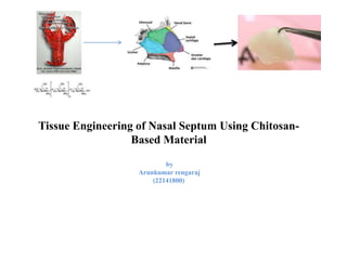
1. Tissue Engineering of Nasal Septum Using Chitosan-
Based Material
by
Arunkumar rengaraj
(22141800)
2. Nasal septum
Nasal septum divides the nasal chamber into two cavities.
The turbinates are made of bone and soft tissue.
Conventional repair of nasal septum involves autologous rib or septum cartilage grafts and prosthetic devices.
Its having limitations like tissue availability, and prosthesis related infection and extrusion.
Major reason leading to the failure
Lack of sufficient control over shape is the lack of appropriate scaffolds
Less biocompatibility for cell seeding
3. Chitosan
• Chitosan, a linear polysaccharide derived from crustacean shells and from many natural sources.
• It having advantages like polycationic nature, biodegradability, biocompatibility, mucoadhesiveness.
• In addition, it enhances epithelial permeability through the opening of tight junctions between epithelial cells.
4. Glycosaminoglycans
It is long unbranched polysaccharides.
GAGs in enhancing chondrogenesis in vitro.
One of the chief components of the extracellular matrix .
It contributes significantly to cell proliferation and migration.
Hyaluronan recognizing receptor
• CD44
• Receptor for HA-mediated motility (RHAMM)
• Intercellular adhesion molecule-1 (ICAM-1)
It may also be used postoperatively to induce tissue healing.
5. Materials and methods
2 % solution of chitosan + 1 M acetic acid
Add Hyaluronic acid/ MeOH 1:1
Mix 40 μg type I collagen
Hybrid polymer
6. Tissues (Human inferior turbinates ) were treated with 0.5% pronase+1:1 mixture (DMEM) +
Ham’s nutrient F12
centrifugation
suspended in DMEM/F12
1.5-mL cell suspension was seeded on collagen coated
Morphological Examination
• Calculate the pore size of the mess
• The DNA content of the cell have to study for calculating the proliferation.
• The ciliary beat was observed under inverted microscope (Leica, DMI 6000, Solms, Germany) with 400 magnification.
• Imaging of cilia movement was recorded at 240 frames per second with a high-speed complementary metal oxide
semiconductor (CMOS) camera (CMC1300; VDS Vossku¨ hler GmbH, Osnabru¨ ck, Germany).
• Histological and immuno-histochemical apperance of the tissue.
• Finally want to perform animal study.
7. Reference
• Dong-Won Lee, Shawna A Shirley, Richard F Lockey and Shyam Mohapatra. Thiolated chitosan nanoparticles enhance anti-inflammatory
effects of intranasally delivered theophylline. Respiratory Research 2006; 7:112.
• Tsung-Wei Huang, Yi-Ho Young, Po-Wen Cheng, Yen-Hui Chan, Tai-Horng Young. Culture of Nasal Epithelial Cells Using
Chitosan-Based Membranes. The Laryngoscope 2009; 119:2066-2070.