Download to read offline

![Reference
1. Titus A, Marappa-Ganeshan R. Physiology, Endothelin. [Updated 2022 May 8].
2. Dagamajalu S, A network map of endothelin mediated signaling pathway. J Cell Commun Signal.
2021 Jun;15(2):277-282.](https://image.slidesharecdn.com/endothelin-220825142517-eda68cf2/85/Endothelin-pptx-16-320.jpg)

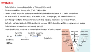
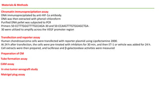
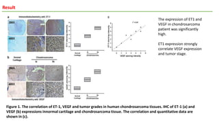
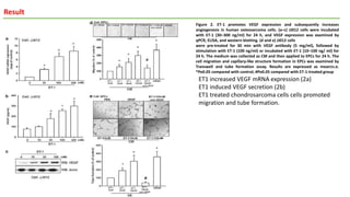
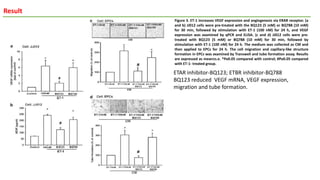
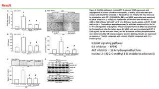
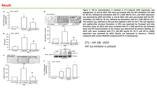
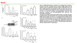
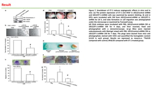
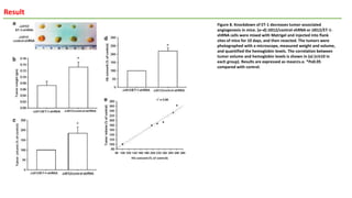
1) Endothelin-1 (ET-1) promotes vascular endothelial growth factor (VEGF) expression and angiogenesis in human chondrosarcoma cells. ET-1 increases VEGF levels through the ETAR receptor, integrin-linked kinase, Akt, and hypoxia-inducible factor-1α signaling pathways. 2) Knockdown of ET-1 decreases VEGF expression and inhibits angiogenesis both in vitro and in vivo. ET-1 promotes migration and tube formation of endothelial progenitor cells through increased VEGF. 3) ET-1 activates HIF-1α, which binds to the VEGF promoter region to increase VEGF expression. This ET-1/ETAR/ILK/Akt/


![CTEV [ clubfoot] DR ARUN LAL ,DR MOHAMED ASHRAF travancore medical college k...](https://cdn.slidesharecdn.com/ss_thumbnails/ctevclubfootdrarunlaldrmohamedashraftravancoremedicalcollegekollamkeralaindia-260208063247-18fc466c-thumbnail.jpg?width=640&height=640&fit=bounds)


