This document provides instructions for preparing biological specimens for light microscopy. It discusses the key steps of sample collection, killing and fixation, dehydration, clearing, paraffin embedding, microtomy, staining, and observation. Specific fixation fluids, dehydration reagents, clearing agents, and staining methods are described. The goal is to preserve specimens while modifying properties like refractive index to allow examination under a light microscope.
The "Telome theory" of Walter Zimmermann (1930, 1952) is the most accepted theory that is based on fossil record and synthesizes the major steps in the evolution of vascular plants.
It describes how the primitive type of vascular plants developed from Rhynia like plants.
Alternation of generation in archegoniatesSumit Sangwan
Altrenation of generations:
All plants undergo a life cycle that takes them through both haploid and diploid generations. The multicellular diploid plant structure is called the sporophyte, which produces spores through meiotic (asexual) division. The multicellular haploid plant structure is called the gametophyte, which is formed from the spore and give rise to the haploid gametes. The fluctuation between these diploid and haploid stages that occurs in plants is called the alternation of generations.
Bryophyte generations
Bryophytes are nonvascularized plants that are still dependent on a moist environment for survival (see Plant Classification, Bryophytes . Like all plants, the bryophyte life cycle goes through both haploid (gametophyte) and diploid (sporophyte) stages. The gametophyte comprises the main plant (the green moss or liverwort), while the diploid sporophyte is much smaller and is attached to the gametophyte. The haploid stage, in which a multicellular haploid gametophyte develops from a spore and produces haploid gametes, is the dominant stage in the bryophyte life cycle. The mature gametophyte produces both male and female gametes, which join to form a diploid zygote. The zygote develops into the diploid sporophyte, which extends from the gametophyte and produces haploid spores through meiosis. Once the spores germinate, they produce new gametophyte plants and the cycle continues.
Tracheophyte Generations
Tracheophytes are plants that contain vascular tissue; two of the major classes of tracheophytes are gymnosperms (conifers) and angiosperms (flowering plants). Tracheophytes, unlike bryophytes, have developed seeds that encase and protect their embryos. The dominant phase in the tracheophyte life cycle is the diploid (sporophyte) stage. The gametophytes are very small and cannot exist independent of the parent plant. The reproductive structures of the sporophyte (cones in gymnosperms and flowers in angiosperms), produce two different kinds of haploid spores: microspores (male) and megaspores (female). This phenomenon of sexually differentiated spores is called heterospory. These spores give rise to similarly sexually differentiated gametophytes, which in turn produce gametes. Fertilization occurs when a male and female gamete join to form a zygote. The resulting embryo, encased in a seed coating, will eventually become a new sporophyte.
Structure, Development & Function of PeridermFatima Ramay
A group of secondary tissues forming a protective layer which replaces the epidermis of many plant stems, roots, and other parts.
Although periderm may develop in leaves and fruits, its main function is to protects stems and roots.
The periderm consists of three different layers:
Phelloderm
Phellogen (cork cambium)
Phellem (cork)
Its main function is to protect the underlying tissues from:
Desiccation
Freezing
Heat injury
Mechanical destruction
Disease
Loss of epidermis.
Bounding tissue restricting the pathogen & insects.
Allowing gaseous exchange through lenticels.
This is a detailed presentation on Morphology, anatomy and reproduction of Marchantia spp. with high quality pics and eye capturing transitions and animations
This ppterrestrial habitt explains about the archegoniate plants, their adaptations, development of different support systems in transition from aquatic to terrestrial habit, about their alternation of generations, etc.
Pteridophytes are vascular plants and have leaves (known as fronds), roots and sometimes true stems, and tree ferns have full trunks. Examples include ferns, horsetails and club-mosses. Fronds in the largest species of ferns can reach some six metres in length!
Many ferns from tropical rain forests are epiphytes, which means they only grow on other plant species; their water comes from the damp air or from rainfall running down branches and tree trunks. There are also some purely aquatic ferns such as water fern or water velvet (Salvinia molesta) and mosquito ferns (Azolla species).
Pteridophytes do not have seeds or flowers either, instead they also reproduce via spores.
There are around 13,000 species of Pteridophytes.
Gymnosperm is from the Greek “gymnos” naked, and “sperma” seeds. They are groups of vascular plants that reproduce by means of an exposed seeds or ovules. They are phanerogams according to A. W. Eichler.
Chemotaxonomy is a little bit difficult task for the students to learn and understand. This slide helps the teachers and students to take class and understood it in a liable way
techniques used for preparing serial sections using microtomes include dehydrating agents and clearing agents ,this slide includes some details on dehydrating and clearing agents
Demonstration of different fixatives used in Histopathology
Demonstration of different Microtome used in Histopathology
To demonstrate the activity of enzyme
Demonstration of following enzymes activity in a Tissue
Demonstration of Laboratory method that uses antibodies
Demonstrate the FIC and FITC techniques
Demonstration of the technique used to separate DN
Demonstration of technique for rapidly producing
Demonstrate the Flow cytometer technique
I. OBJECTIVES OF PRESERVATION
In this guideline, we are mainly concerned with the taxonomic reasons for preservation. The scientific description of an animal species requires the detailed examination and description of a representative type specimen and a series of specimens which are subsequently deposited, catalogued and maintained in a museum or zoological collection. This remains a reference for other workers to consult in future.
Specimens from any field collection should be deposited in a reference collection in an institutional for the long-term maintenance and access for the future. The animals should therefore be preserved in the best possible condition and where possible, ensure that the natural colour is retained, their external appendages (e.g. fins) are erected and stomach contents intact.
Care should be taken to ensure that specimens are undamaged. Features important in the taxonomic study of fish, for example, are easily damaged with contact even after preservation. Live crabs before preservation should be kept individually as some species will damage each other and other animals, especially fish even when they are being directly preserved.
Acetabularia Information For Class 9 .docxvaibhavrinwa19
Acetabularia acetabulum is a single-celled green alga that in its vegetative state is morphologically differentiated into a basal rhizoid and an axially elongated stalk, which bears whorls of branching hairs. The single diploid nucleus resides in the rhizoid.
Embracing GenAI - A Strategic ImperativePeter Windle
Artificial Intelligence (AI) technologies such as Generative AI, Image Generators and Large Language Models have had a dramatic impact on teaching, learning and assessment over the past 18 months. The most immediate threat AI posed was to Academic Integrity with Higher Education Institutes (HEIs) focusing their efforts on combating the use of GenAI in assessment. Guidelines were developed for staff and students, policies put in place too. Innovative educators have forged paths in the use of Generative AI for teaching, learning and assessments leading to pockets of transformation springing up across HEIs, often with little or no top-down guidance, support or direction.
This Gasta posits a strategic approach to integrating AI into HEIs to prepare staff, students and the curriculum for an evolving world and workplace. We will highlight the advantages of working with these technologies beyond the realm of teaching, learning and assessment by considering prompt engineering skills, industry impact, curriculum changes, and the need for staff upskilling. In contrast, not engaging strategically with Generative AI poses risks, including falling behind peers, missed opportunities and failing to ensure our graduates remain employable. The rapid evolution of AI technologies necessitates a proactive and strategic approach if we are to remain relevant.
Welcome to TechSoup New Member Orientation and Q&A (May 2024).pdfTechSoup
In this webinar you will learn how your organization can access TechSoup's wide variety of product discount and donation programs. From hardware to software, we'll give you a tour of the tools available to help your nonprofit with productivity, collaboration, financial management, donor tracking, security, and more.
The Roman Empire A Historical Colossus.pdfkaushalkr1407
The Roman Empire, a vast and enduring power, stands as one of history's most remarkable civilizations, leaving an indelible imprint on the world. It emerged from the Roman Republic, transitioning into an imperial powerhouse under the leadership of Augustus Caesar in 27 BCE. This transformation marked the beginning of an era defined by unprecedented territorial expansion, architectural marvels, and profound cultural influence.
The empire's roots lie in the city of Rome, founded, according to legend, by Romulus in 753 BCE. Over centuries, Rome evolved from a small settlement to a formidable republic, characterized by a complex political system with elected officials and checks on power. However, internal strife, class conflicts, and military ambitions paved the way for the end of the Republic. Julius Caesar’s dictatorship and subsequent assassination in 44 BCE created a power vacuum, leading to a civil war. Octavian, later Augustus, emerged victorious, heralding the Roman Empire’s birth.
Under Augustus, the empire experienced the Pax Romana, a 200-year period of relative peace and stability. Augustus reformed the military, established efficient administrative systems, and initiated grand construction projects. The empire's borders expanded, encompassing territories from Britain to Egypt and from Spain to the Euphrates. Roman legions, renowned for their discipline and engineering prowess, secured and maintained these vast territories, building roads, fortifications, and cities that facilitated control and integration.
The Roman Empire’s society was hierarchical, with a rigid class system. At the top were the patricians, wealthy elites who held significant political power. Below them were the plebeians, free citizens with limited political influence, and the vast numbers of slaves who formed the backbone of the economy. The family unit was central, governed by the paterfamilias, the male head who held absolute authority.
Culturally, the Romans were eclectic, absorbing and adapting elements from the civilizations they encountered, particularly the Greeks. Roman art, literature, and philosophy reflected this synthesis, creating a rich cultural tapestry. Latin, the Roman language, became the lingua franca of the Western world, influencing numerous modern languages.
Roman architecture and engineering achievements were monumental. They perfected the arch, vault, and dome, constructing enduring structures like the Colosseum, Pantheon, and aqueducts. These engineering marvels not only showcased Roman ingenuity but also served practical purposes, from public entertainment to water supply.
The French Revolution, which began in 1789, was a period of radical social and political upheaval in France. It marked the decline of absolute monarchies, the rise of secular and democratic republics, and the eventual rise of Napoleon Bonaparte. This revolutionary period is crucial in understanding the transition from feudalism to modernity in Europe.
For more information, visit-www.vavaclasses.com
Palestine last event orientationfvgnh .pptxRaedMohamed3
An EFL lesson about the current events in Palestine. It is intended to be for intermediate students who wish to increase their listening skills through a short lesson in power point.
How to Make a Field invisible in Odoo 17Celine George
It is possible to hide or invisible some fields in odoo. Commonly using “invisible” attribute in the field definition to invisible the fields. This slide will show how to make a field invisible in odoo 17.
Instructions for Submissions thorugh G- Classroom.pptxJheel Barad
This presentation provides a briefing on how to upload submissions and documents in Google Classroom. It was prepared as part of an orientation for new Sainik School in-service teacher trainees. As a training officer, my goal is to ensure that you are comfortable and proficient with this essential tool for managing assignments and fostering student engagement.
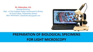
1. PREPARATION OF BIOLOGICAL SPECIMENS
FOR LIGHT MICROSCOPY
Dr. Abdussalam, A.K.
Assistant Professor,
Dept. of Post Graduate Studies and Research in Botany
Sir Syed College, Taliparamba, Kannur
Mob: 9847654285, salamkoduvally@gmail.com
3. 1. Killing and Fixation
Essential requirement
Performed by fixative
Killing – Sudden stoppage of life processes
Fixation - Preservation of a “life-like” state
Purposes –
Preservation of natural form
Modifying RI
Making material resistant and hard
Preparing material for improving staining
4. Reagents of Fixatives
No single reagent for the purpose
Combinations of reagents
Principle – Keep balance between properties
1. Ethyl alcohol
Water soluble
Reducing agent
Rapid Penetrability
Shrink tissues
Hardening effect
Makes tissues difficult to stain
5. 2. Formalin
Aqueous Formaldehyde
Reducing agent
Water miscible
Slow Penetration
Causes shrinkage
Great Hardening effect
Makes staining difficult
Reagents of Fixatives
6. 3. Acetic Acid
Water miscible
Rapid penetration
No hardening effect
Makes tissues soft
4. Chromic acid
Water miscible
Oxidiser
Slow Penetration
Reagents of Fixatives
7. Killing and Fixing Fluids (Fixatives)
Many groups based on their ingredients
Selection depends on specific requirement
Some stable
Some unstable
Some known by their ingredients
Some by their investigators
8. 1. Farmer’ s Formula
Glacial Acetic acid- 5m ml
Absolute Alcohol - 15 ml
Ideal for cytological preparations – Root tips, Anther
Fixation time – Root tips – 15 m, Anthers – 1 h
Washing and storage in 70% alcohol
2. Carnoy’s Formula
Absolute alcohol - 10 ml
Chloroform - 15 ml
Glacial Acetic acid- 5 ml
Ideal for cytological preparations
Fixation time – 10- 15 m.
Washing and storage in 85% alcohol
Killing and Fixing Fluids (Fixatives)
Acetic
Acid
Alcohol
Mixtures
9. Killing and Fixing Fluids (Fixatives)
1. Rawlin’ Formula
95% Ethyl alcohol - 50 ml
Glacial Acetic acid - 5 ml
Formalin - 10 ml
Water - 35 ml
For delicate materials
Good hardening action and
Materials may be stored in this for years even
For hard woody materials decrease acid and increased formalin
Fixation time: 18 hrs
Wash in alcohol and store in same
Formalin
Acetic
acid
Alcohol
(FAA)
Mixtures
10. Killing and Fixing Fluids (Fixatives)
1. Chromo acetic acid ( Weak)
Chromic acid 1% - 50ml
Acetic acid 1% - 50 ml
2. Chromo acetic (Medium)
Chromic acid 1% - 70ml
Acetic acid 1% - 20 ml
Water - 10 ml
3. Chromoic acetic : Strong
Chromic acid 1% - 97 ml
Acetic acid 1% - 3 ml
ChromoChromo
AceticAcetic
AcidAcid
MixturesMixtures
11. Killing and Fixing Fluids (Fixatives)
Recommended for delicate objects like
filamentous and thalloid plants, root tips, Floral
organs and small sections of leaves or stems
Fixation time: Few minutes for algae, 12 hours
for small leaf and root tips
24 hours for larger pieces of tissue
Wash well in running water for 24 hours and
then in distilled water for 12 hours
ChromoChromo
AceticAcetic
AcidAcid
MixturesMixtures
12. 1. Navaschin’s Formula
Sol. A: Chromic acid (1%) - 15 ml
Glacial acetic acid - 10 ml
Distilled water - 90 ml
Sol. B: Formalin - 40 ml
Distilled water - 60 ml
Mix equal quantities of A and B just before use
Fixation time : 12 hours
Washing in water not required
Navashin’s original formula has been modified by
many investigators and the name CRAF has been
coined for such types
ChromoChromo
AceticAcetic
AcidAcid
FormalinFormalin
MixturesMixtures
Killing and Fixing Fluids (Fixatives)
13. Craf I
Chromic acid 1% 20 ml
Acetic acid 1% 75 ml
Formalin 5 ml
Craf II
Chromic acid 1 % 20 ml
Acetic acid 10% 10 ml
Formalin 5 ml
Distilled water 65 ml
ChromoChromo
AceticAcetic
AcidAcid
FormalinFormalin
MixturesMixtures
Killing and Fixing Fluids (Fixatives)
14. ZIRKLE-ERLIKI FORMULA
Potassium bichromate 1.25 gm
Ammonium bichromate 1.25 gm
Cupric sulphate 1.00 gm
Distilled water 200 ml
Recommended for studies of mitochondria, nucleoplasm, nucleoli and vacuoles
Dissolves chromatin spindles
Fixation time 24-28 hours
Wash in water
Potassium
Chromate
Mixtures
Killing and Fixing Fluids (Fixatives)
15. Materials should be fixed as soon as possible after collection – if possible on
the spot
Bearing in mind the properties of the reagent used, decide upon the proper
fixative
Materials for anatomical studies should be cut into pieces of 1x1x0.5 cm
without injuring tissues
Place them in flat bottomed tubes with cork and use the fixative and material
in the proportion by volume of 100 to 1 respectively
If pieces of materials do not sink in the fluid at once, air should be removed by
using an aspirator in repeatedly until the pieces sink at lest under the surface of
the liquid
Wash thoroughly after fixing for the required time
TECHNIQUES
TECHNIQUES OF FIXING
16. Dehydration
Chemical removal of water and fixative from the specimen
Replace them with dehydrating fluid - dehydrant
Many dehydrants are alcohols. Several are hydrophilic so attract water from tissue.
Practiced in graded series
Progressively decreasing concentration of water
Progressively increasing concentration of dehydrant
17. Dehydrants – Reagents in dehydration
Some merely removes water
Some acts also as solvents of mounting media
Common dehydrants are ethyl alcohol, acetone, normal butyl alcohol , tertiary
butyl alcohol Glycerine, Dioxan etc.
Ethyl Alcohol/Isopropyl alcohol
Most common
Progressively increasing concentrations – 10%, 20%, 30%, 40% …… 100%
Begin with a grade same as the water content in the tissue
Time required – soft tissues ~30 minutes – Hard/ large tissue- ~6-12 hrs.
18. Normal Butyl Alcohol
Advantage – solvents of paraffin – directly followed to impregnation
Grades are prepared in combination with ethyl alcohol
Series No. 95 % Ethyl
Alcohol (ml)
Normal butyl
alcohol (ml)
Distilled water
(ml)
1.
2.
3.
4.
5.
6.
7.
8.
20
25
30
30
25
20
15
0
10
15
25
40
55
70
85
100
70
60
45
30
20
10
0
0
1 hour
2 hour
19. Tertiary Butyl Alcohol (TBA)
Series No. Absolute Alcohol (ml) 95% Ethyl
Alcohol (ml)
TBA
(ml)
Dist. Water
(ml)
1.
2.
3.
4.
5.
0
0
0
0
25
50
50
50
50
0
10
20
35
50
75
100
40
30
15
0
0
Dehydrate first in ethyl alcohol upto 50%
Three changes in absolute TBA
20. 3. Clearing (Dealcoholization)
Removal of alcohol from the tissues
Replacing the dehydrating fluid with a fluid that is totally miscible with
both the dehydrating fluid and the embedding medium- Paraffin
Transition step between dehydration and infiltration
Only needed when the dehydrants are not solvents of wax
Clearing agents- Xylene, Toluene, Chloroform, Benzene, Petrol etc.
21. Reagents in Clearing - Xylene
Xylene- Conventional reagent in dealcoholization
Practiced in graded series (30 min 1hr in each)
Series No. Ethyl alcohol (ml) Xylene (ml)
1
2
3
4
5
6
7
8
9
10
90
80
70
60
50
40
30
20
10
0
10
20
30
40
50
60
70
80
90
100
22.
23. Clearing
Clearing is transition step between dehydration and infiltration with the embedding medium
Many dehydrants are immiscible with paraffin wax
a solvent imiscible with both the dehydrant and the embedding medium us used to facilitate the
transition between dehydratin and infiltration step
Replacing the dehydrating fluid with a fluid that is totally miscible with both the dehydrating fluid and the
embedding medium.
Choice of a clearing agent depends upon the following:
- The type of tissues to be processed, and the type of processing to be undertaken.
- The processor system to be used.
- Intended processing conditions such as temperature, vacuum and pressure.
- Safety factors.
- Cost and convenience.
- Speedy removal of dehydrating agent .
- Ease of removal by molten paraffin wax .
- Minimal tissue damage .
25. Mounting
The final stage in the preparation of tissues for microscopy is
mounting
For stained preparations, the mounting medium or mount ant should
have the same refractive index as the section or nearer to that
To be effective, a mountant should possess certain characteristics.
These include the following
26. It should be colourless and transparent
It should be able to completely permeate and fill tissue spaces
It should have no adverse effect on tissue components
It should be resistant to contamination particularly by
microorganisms
It should be completely miscible with dehydrant or clearing agent
The mountant may be hydrophobic or hydrophilic
27. Hydrophobic mountant
Canada balsam
This is an oleorosin obtained from the bark of the fir Abis balsamea of the
family Pinaceae
The dried resin is freely soluble in xylene and other organic solvents DPX
(Distrene, Polystyrene Xylene)
DPX is one of the most commonly used mountants
It is a colouless, neutral medium in which most standard stains are well
preserved
This fast drying mounting medium prevents moisture from developing
under the cover glass and the consequent clouding of the specimen
28. `
Hydrophilinc mountant
Water
Glycerol
Used as temporary mountant
Having higher refractive index (1.460)
Having longer drying time that water
Phosphate buffered glycerol is commonly used
29. 4. Paraffin infiltration (Embedding)
Most commonly used waxes for infiltration are
the commercial paraffin waxes
It us solid at room temperature but melts at
temperatures up to about 65°C or 70°C.
Available in melting points at different
temperatures
Dehydrated material is gradually infiltrated
with wax
Liquid wax is recommended for the initial
infiltration
31. Paraffin Embedding
Three changes in 100 % wax
Paraffin block-material preparation
Attachment of the block into the holder of the
microtome
Sectioning with microtome
32. Steps involved
1. Killing and fixation
2. Dehydration
3. Clearing
4. Paraffin infiltration
5. Casting of wax impregnated material into blocks
6. Attachment of the block into the holder of the microtome
7. Microtomy
8. Affixing paraffin ribbon on glass slides
9. Removal of wax
10.Staining and mounting
33. Sections
Sectioning allows light pass through the material
FREE HAND SECTIONS
SERIAL SECTIONS
FREE HAND SECTIONS
Can be done if the material is hard
Thin sections - 10 µM can be taken
Sectioning with razor
34. Serial sections
Serial sections are produced by paraffin method
Paraffin infiltrated material are affixed on
wooden blocks
Objects are cut into a series of sections
Serial sections are placed on adhesive smeared
glass slides
Serial sections enables the reconstruction of
structure of organ
Orientation of vasculature, cellular organization
etc. can be studied
36. Stains and Staining
Staining - Use of dyes to provide color to various tissue constituents
Different tissue constituents react differently to dyes – contrast
Chromogen
Chromophore
Auxochrome – acid/ alkali radicals. Responsible for solubility
37. Stains - classification
Principle Chemical Nature
Chemical Nature Basic : Colored organic base+ uncolored acetate, chloride or sulphate radical
(safranin, methylene blue, crystal violet)
Acidic : Metallic base (Na, K) + Colored organic radical (Aniline Blue, Eosin,
Orange G )
Neutral : Combinations of acidic and basic dyes (Giesma stain, Sudan black B)
Affinity to different
plant parts
Nuclear : Nucleus
Cytoplasmic: Cytoplasm
Microtechnical
purposes
Histological: defines tissues (xylem, phloem etc.)
Cytological : Define cell components (nucleus, chromosomes etc.)
38. Stains
Natural Dyes – dyes obtained from plant/ animal (Brazilin, Hematoxylin, Carmine)
Synthetic dyes – made from Coal tar – (Orange G, Safranine, Fast Green)
Brazilin (Timber of Caesalpinia crista, C. echinata)
Hematoxylin Hematoxylon campechianum
Carmine Insect Dactylopius coccus
Staining Methods
1. Progressive staining
2. Regressive (Retrogressive staining)
3. Counter staining
4. Double, triple and quadruple staining
39. Methods of Staining
Progressive Staining
Useful for beginners
Tissues are understained first
Gradually more stain is added until the desired intensity attained
Staining interval required is determined by trial
Regressive (Retrogressive) Staining
Overstained first
Then destained until the desired intensity is attained
Destaining agent – 70% alcohol with 1% acetic acid
Proper washing after differentiation
40. Counterstaining
Staining certain part of cells/ tissues with one stain
Other parts with a contrasting color
Double/ Triple/ Quadruple staining
Use of 2, 3, 4 colors on same section
Double staining - Safranin O and Fast Green
Triple staining - Safranin O, Gentian Violet and Orange G
Quadruple Staining - Safranin O, Methyl violet, Fast Green and Orange G
Methods of Staining
41. Whole Mounts
Used to preserve and retain natural color, form and shape of
whole plants/ plant parts
Microscopic museum materials preserved in ethyl alcohol,
formalin
Water – 72 ml
Formaldehyde – 5 ml
Glacial acetic acid – 3 ml
Glycerine - 20 ml
Temporary whole mounts – small filamentous algae- in 10%
glycerine/ coverslip
42. Whole Mounts
Permanent whole mounts – Microscopic Material
Constant handling requires preparation of permanent nature
1. Killing and fixation
2. Washing in water
3. Staining with hematoxylin for 30 min -1 hour
4. Destaining in 0.1% HCl
5. Transfer to glass slide
6. Covering with DPX and cover slip
43. Cytological Methods
Used to study the minute details of the cell structure – nucleus
Smear and squash methods are the most common.
Smear – Smearing material on glass slide (Acetocarmine
method, Feulgen method)
Squashes - component parts separate and are not studied
intact.
MACERATION
Separation of cells of fixed plant or animal material through
hydrolysis
Useful to visualize the 3d nature of structural elements
Reagents used depends – nature of middle lamella
44. Maceration
Middle lamella
Herbaceous – Pectin (boiling in water)
Woody - Lignin (alkali/ acid/ enzyme treatment)
3 common methods in practice
1. Schultze’ s Method
Treatment with con. H2SO4+KClO3 and warming
After thorough bleaching washing in water
2. Jeffrey’ s Method
Treatment in equal vol 10% HNO3+K2CrO4 at 30° C - 40° C for 1 -2 days
Thorough washing
45. 3. Harlow’ s Method
Treatment in chorine water –2 hours
Washing in running water
Boiling in 3% Na2SO3 – 15 min
Washing
Staining in Safranin
Washing in water
Dehydration with hygrobutol
Infiltration with Canada balsum
After placing the material on glass slides tease with needles
Mount with cover slip
Maceration