Pregnancy Diagnose through Ultrasound + X-Ray in Veterinary Field
•
3 likes•2,258 views

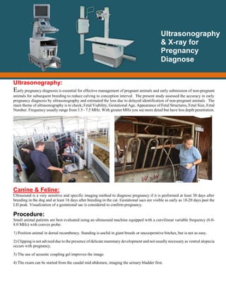
Early pregnancy diagnosis is essential for effective management of pregnant animals and early submission of non-pregnant animals for subsequent breeding to reduce calving to conception interval. The present study assessed the accuracy in early pregnancy diagnosis by ultrasonography and estimated the loss due to delayed identification of non-pregnant animals. The main theme of ultrasonography is to check; Fetal Viability, Gestational Age, Appearance of Fetal Structures, Fetal Size, Fetal Number. Frequency usually range from 3.5 - 7.5 MHz. With greater MHz you see more detail but have less depth penetration. Presented By: Dr. Fakhar-e-Alam Kulyar DVM, M.Phil CMS University of Agriculture Faislalabad

Recommended
Recommended
More Related Content
What's hot
What's hot (20)
Similar to Pregnancy Diagnose through Ultrasound + X-Ray in Veterinary Field
Similar to Pregnancy Diagnose through Ultrasound + X-Ray in Veterinary Field (20)
More from Dr. Fakhar
Recently uploaded
Recently uploaded (20)
Pregnancy Diagnose through Ultrasound + X-Ray in Veterinary Field
- 1. Ultrasonography: Early pregnancy diagnosis is essential for effective management of pregnant animals and early submission of non-pregnant animals for subsequent breeding to reduce calving to conception interval. The present study assessed the accuracy in early pregnancy diagnosis by ultrasonography and estimated the loss due to delayed identification of non-pregnant animals. The main theme of ultrasonography is to check; Fetal Viability, Gestational Age, Appearance of Fetal Structures, Fetal Size, Fetal Number. Frequency usually range from 3.5 - 7.5 MHz. With greater MHz you see more detail but have less depth penetration. Canine & Feline: Ultrasound is a very sensitive and specific imaging method to diagnose pregnancy if it is performed at least 30 days after breeding in the dog and at least 16 days after breeding in the cat. Gestational sacs are visible as early as 18-20 days past the LH peak. Visualization of a gestational sac is considered to confirm pregnancy. Procedure: Small animal patients are best evaluated using an ultrasound machine equipped with a curvilinear variable frequency (6.0- 8.0 MHz) with convex probe. 1) Position animal in dorsal recumbency. Standing is useful in giant breeds or uncooperative bitches, but is not as easy. 2) Clipping is not advised due to the presence of delicate mammary development and not usually necessary as ventral alopecia occurs with pregnancy. 3) The use of acoustic coupling gel improves the image. 4) The exam can be started from the caudal mid abdomen, imaging the urinary bladder first. Ultrasonography & X-ray for Pregnancy Diagnose
- 2. 5) The normal uterus is best located by scanning transversely between the urinary bladder and the colon. The cervix and uterine body are seen as a continuous hypoechoic round structure dorsal to the anechoic urinary bladder and ventral to the hyperechoic, crescent shaped colon 6) Examining dorsally for the uterine body. The left uterine horn is followed cranially and then the right uterine horn is followed caudally. Following are the pictures captured at different stages of gestation. Uterus Normal Ultrasound At 25 day Head Gestational sac Day 41 Stomach At 30-32 days’ gestation the vesicle is larger than transverse small bowel, making identification easy as seen in figure 1. Each fetus is contained in an oval, fluid filled gestational sac. The flicker of the heartbeat is regularly seen even without Doppler after 25-28 days’ gestation. Normal canine fetal heartrates should be >180-200 after LH Peak. To check the viability “Cardiac activity” and “fetal movement” are predictable signs. Litter size determination is best at 30 days’ gestation. In later gestation (>45-50 days) the fetuses are so large the uterine horns overlap making the correct count difficult.
- 3. Late Pregnancy The determination of gestational age can be of vital importance. Prolonged gestation is a form of dystocia. Prediction of gestational age can be accomplished by measuring the gestation sac (GSD), crown-rump length (CRL), and head diameter (HD) and body diameters (BD) of the fetus. Gestational age estimates of canine fetus: Age (days) C-R Length (mm) 20 8 30 20 35 35 38 55 40 60 45 80 50 118 53 140 57 160 57-63 160-185 Gestational age estimates of feline fetus: Age (days) C-R Length (mm) 18 8 20 10 24-26 18-20 27 25 30 35 37 60 43 80 46 95 50 110
- 4. 55 125 60 143 60-63 143-150 Formulas to predict gestational age and days before parturition: Bovines: The transrectal ultrasonography is done at 30 days’ post breeding and repeated after 45 days’ post breeding for confirmation. Ultrasound scanning of the uterus and ovaries were made using a 6.5 MHz rectal linear probe for diagnosis and confirmation of pregnancy. A technician needs to ensure that confusion between fluid accumulation in the chorioallantois during early pregnancy and uterine fluid within the uterus during proestrus and estrus are not confused when making the diagnosis. Gestational age estimates of bovine fetus through Ultrasound: Age Relative Size C-R Measurement 30 days 10 mm 35 days 15 mm 40 days 25 mm 45 days 35 mm 50 days 40 mm 55 days 50 mm 60 days 65 mm 2 months Mouse 6 to 8 cm 3 months Rat 13 to 17 cm 4 months Small Cat 22 to 32 cm 5 months Large Cat 30 to 45 cm 6 months Small Dog 40 to 60 cm 7 months Medium Dog 55 to 75 cm 8 months Large Dog 60 to 85 cm
- 5. Following are the pictures captured at different stages of gestation of bovines. Embryo at 25 days Embryo at 30 days Embryo at 37 days 40 days old 21 mm embryo Fetus on day 59 with the first view of the ribs Measuring the crown-rump length Three placentomes in the pregnant horn on day 74 of gestation of a 53-day old fetus
- 6. Placentomes in the pregnant horn on day 110 of gestation Placentomes in the pregnant horn on day 159 of gestation Fetal Sexing: At approximately day 50 of gestation, male and female fetuses can be differentiated by the relative location of the genital tubercle and development of the genital swellings into the scrotum in male fetuses. Caprine & Ovine: Early detection of pregnancy and determining fetal numbers have economical benefits to goats & sheep producers. Transabdominal B-mode Ultrasonography are able to accurately diagnose both pregnancy and fetal numbers. The optimum time for detecting pregnancy is from 45 to 90 days of gestation in sheep and goat both by using 3.8MHz convex probe. Method: Method 1 Method 2
- 7. Gestational age estimates of ovine and caprine fetus: Age (Weeks) C-R Measurement (cm) 3-4 0.3-2 5-6 2-9 7-9 9-15.5 10-13 15-35 14-18 35-40 19-21 40-48 Following are the pictures captured at different stages of gestation of Goats & Sheep. 28 Day Sheep Pregnancy 27 Day Sheep Pregnancy 45 Day Sheep Pregnancy Sheep Cranium - Thorax w heart - Front leg 75 day
- 8. Sheep Cranium & Cotyledons 75 days Non-echogenic area in goat on days 20 to 24 of gestation 28 days of gestation in goat 72 days of gestation in goat 103 days of gestation in goat 143 days of gestation in goat
- 9. References: Veterinary Obstetrics and Genital Diseases Theriogenology, 3rd Edition Roberts SJ. Veterinary Obstetrics and Genital Diseases Evans HE and Sack WO, Prenatal Development of Domestic and Laboratory Mammals Vinole-Gil C, Reproductive Ultrasound of Sheep and Goats Santos MHB, et al. Sexing of Boer goat fetuses using transrectal ultrasonography. Animal Reproduction Article “Using Portable Ultrasound for Early Pregnancy Detection in Sheep” submitted by: Gary Veserat MS, PAS* “American Registry of Professional Animal Scientists” Article “Determining Pregnancy in Cattle” by Bruce B. Carpenter and L. R. Sprott* with collaboration with http://AgriLifebookstore.org Article “Pregnancy diagnose by in goats by using two different probes” The Journal of Animal & Plant Sciences, 24(4): 2014, Page: 1026-1031 ISSN: 1018-7081 http://www.slideshare.net/GangaramChaudhary/current-approach-for-pregnancy-diagnosis-in-animals Radiography: Radiography is an imaging technique that uses electromagnetic radiation other than visible light, especially X-rays, to view the internal structure of a non-uniformly composed and opaque object A range of detectors including photographic film, scintillator and semiconductor diode arrays have been used to collect images. To a limited extent radiography has been used for pregnancy diagnosis in veterinary field. The technique is known to be good in evaluating fetal numbers in the bitch and cat, sheep, goat rarely use in large animals due to different reasons like restraining, but is poor in evaluating fetal viability. Moreover, the high cost and the hazards of exposure to growing fetuses. The bones of fetal kittens become mineralized (calcified) at around day 40-45 of cat pregnancy. Radiographs taken of pregnant cats after day 45 of gestation. Sheep and goat, fetuses are visible by day 70 of gestation.
- 10. Bitches fetal skeletons are visible with high accuracy only by the sixth week of pregnancy. Signs of fetal death as seen by radiography include the Spalding sign, (which is the overlapping of the cranial bones), gas shadows in the fetal heart and stomach and tightly flexed spine (seen in fetuses died for long time) Fetal skeletons begin to calcify only after the sixth week in sows and hence radiography should be performed only after this time for pregnancy diagnosis in sows. Advantages: Good in evaluating fetal numbers in the bitches. Fetus can be differentiated easily from abdominal contents due to mineralization of bones after 45 days. Position of the fetuses can be easily determined. Number of fetuses can be easily counted. Whelping date can be predicted Disadvantages: Poor in evaluating fetal viability. High cost of machine Hazards of exposure to X Rays to growing fetuses & operator Radiographic Films: Radiographic Pregnancy Diagnosis Radiography of Fetuses
- 11. Fetal dentation radiograph Dystocia radiograph lateral view Signs of Fetal Death: Spalding sign, (which is the overlapping of the cranial bones) Collapse of Skelton Gas shadows in the fetal heart, stomach, around the fetus Tightly flexed spine (seen in fetuses died for long time) Presented By: Dr. Fakhar-e-Alam Kulyar DVM, M.Phil CMS University of Agriculture Faislalabad