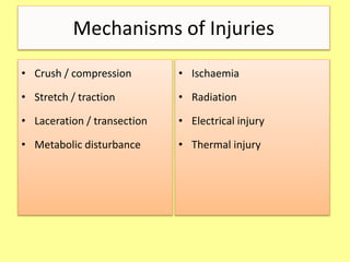
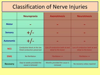
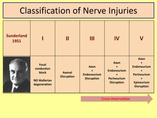
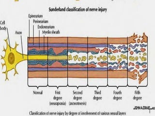
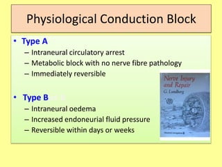
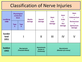
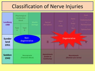
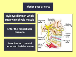
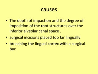
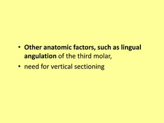
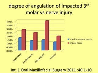
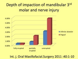
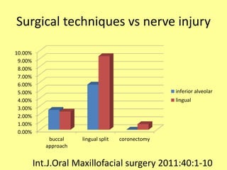
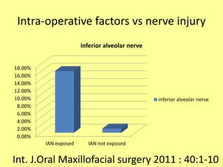
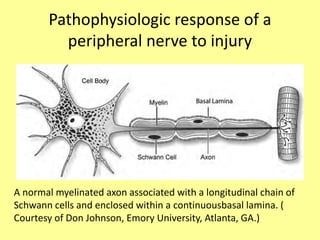
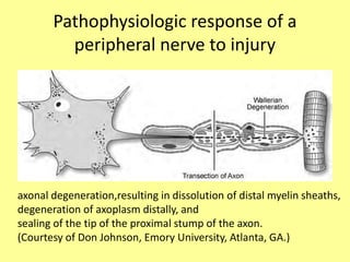
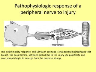
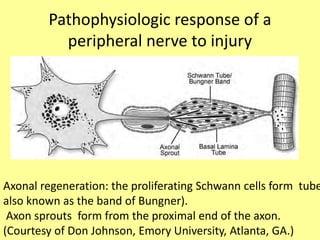
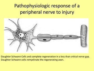
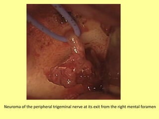
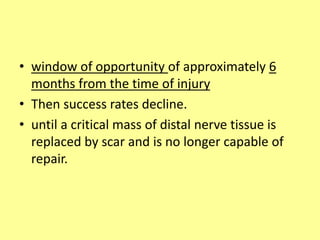
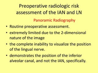
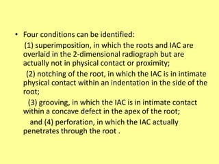
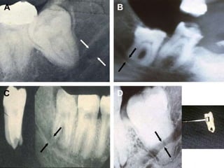
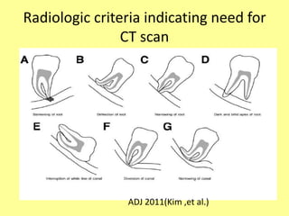
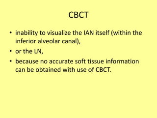
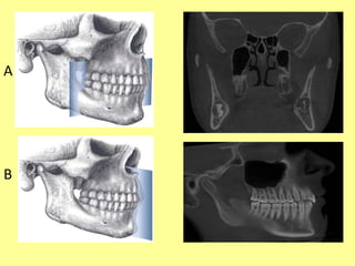
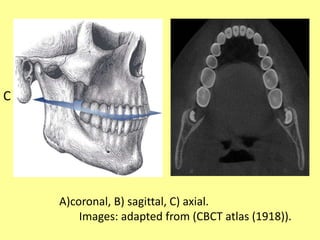
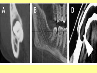
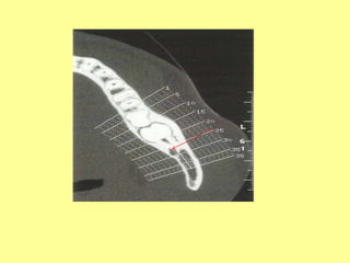
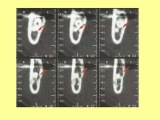
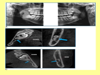
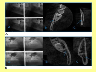
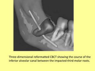
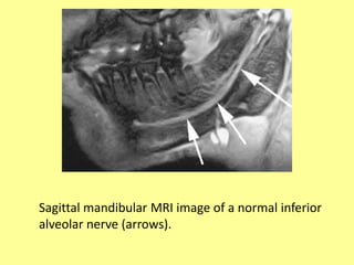
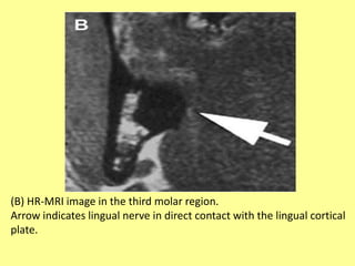
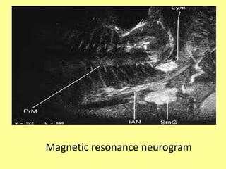
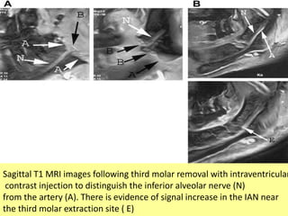
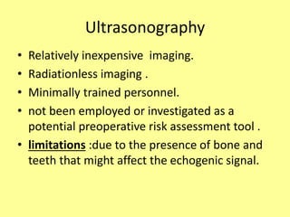
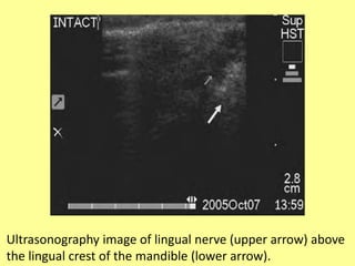
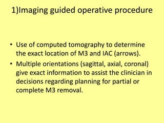
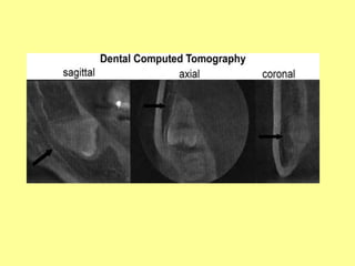
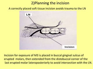
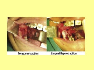
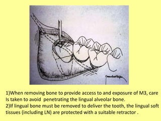
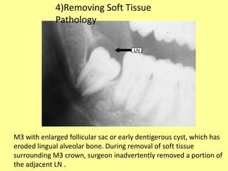
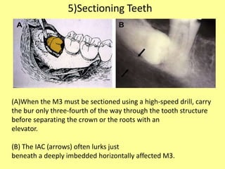
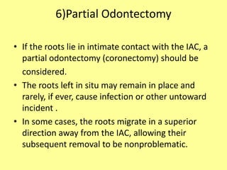
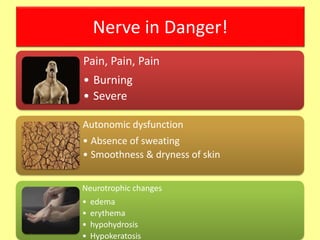
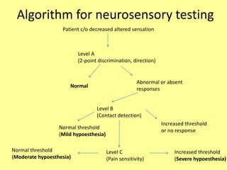
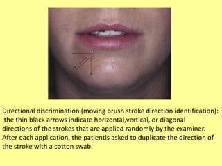
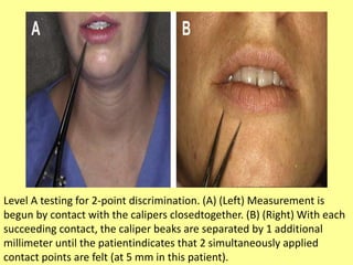
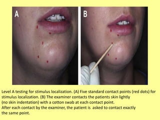
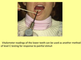
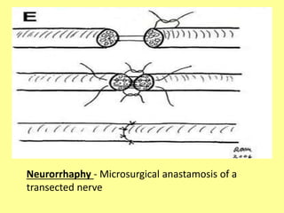
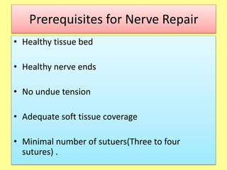
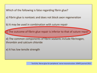
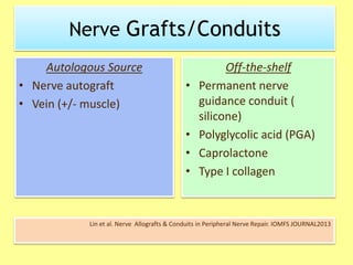
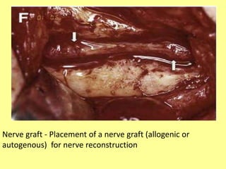
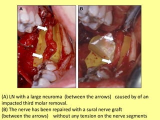
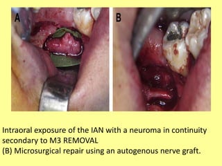
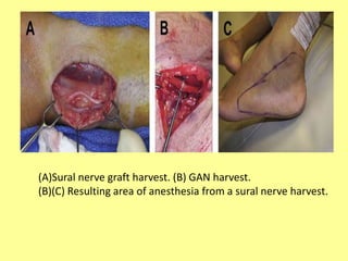
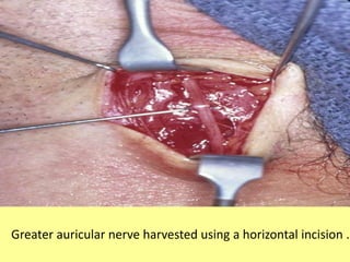
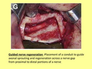
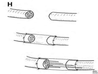
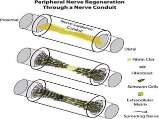
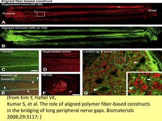
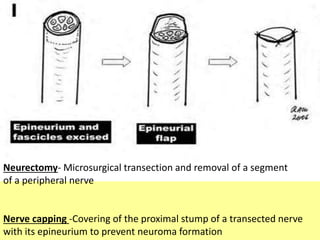
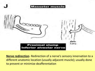
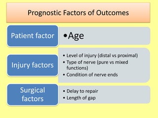
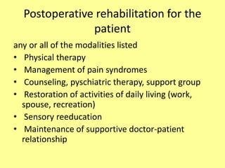
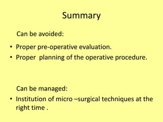
NERVE INJURIES DURING
MANDIBULAR IMPACTION
SURGERIES
By
A.Selva Arockiam,
CRI
Procedures associated with peripheral trigeminal nerve
injuries are listed in descending order of frequency, based
on a survey (1981 to 2010)
• Removal of lower third molar teeth
• Orthognathic surgery
• Maxillofacial trauma (fractures, soft tissue injury, gunshot
wound)
• Dental implants
• Cyst or tumor excision
• Preprosthetic surgery (vestibuloplasty, ridge augmentation)
• Root canal treatment (canal filling, apical surgery)
• Local anesthetic injection
• Salivary gland excision
• Biopsy DCNA MARCH
2011
Mechanisms of Injuries
• Crush / compression
• Stretch / traction
• Laceration / transection
• Metabolic disturbance
• Ischaemia
• Radiation
• Electrical injury
• Thermal injury
Classification of Nerve Injuries
Seddon
BMJ
1942
Neurapraxia
(Transient Block)
Axonotmesis
(Lesion in Continuity)
Neurotmesis
(Division of a nerve)
• Localised
degeneration of
the myelin sheaths
• Complete
interruption of
axons
• Preservation of
supporting
structures
(Schwann tubes,
endoneurium,
perineurium)
• All essential parts
destroyed
• Interruption can
occur without
apparent loss of
continuity
Classification of Nerve Injuries
Neurapraxia Axonotmesis Neurotmesis
Motor - - -
Sensory +/- - -
Autonomic +/- - -
NCS
Conduction block at the site
Distal conduction preserved
Loss of conduction both at and
distal to the lesion
Loss of conduction both at and
distal to the lesion
EMG No fibrillation Fibrillation ++ Fibrillation ++
Recovery
Days to weeks provided the
cause is removed
Months provided the cause is
removed
No recovery unless repaired
Classification of Nerve Injuries
Sunderland
1951 I II III IV V
Focal
conduction
block
NO Wallerian
degeneration
Axonal
Disruption
Axon
+
Endoneurium
Disruption
Axon
+
Endoneurium
+
Perineurium
Disruption
Axon
+
Endoneurium
+
Perineurium
+
Epineurium
Disruption
Cross-innervation
Physiological Conduction Block
• Type A
– Intraneural circulatory arrest
– Metabolic block with no nerve fibre pathology
– Immediately reversible
• Type Bpe B
– Intraneural oedema
– Increased endoneurial fluid pressure
– Reversible within days or weeks
Classification of Nerve Injuries
Lundborg
1988
Physiological
conduction
block
Myelin
damage
Axonal
damage
Axon
+
Endo
damage
Axon
+
Endo
+
Peri
damage
Axon
+
Endoneurium
+
Perineurium
+
Epineurium
damage
Type
A
Type
B
Sunder
land
1951
I II III IV V
Seddon
1942
Neurapraxia
(Transient Block)
Axonotmesis
(Lesion in
Continuity)
Neurotmesis
(Division of a nerve)
Classification of Nerve Injuries
Lundborg
1988
Physiological
conduction
block
Myelin
damage
Axonal
disruption
Axon
+
Endo
Axon
+
Endo
+
Peri
Axon
+
Endoneurium
+
Perineurium
+
Epineurium
Type
A
Type
B
Sunder
land
1951
I II III IV V
Seddon
1942
Neurapraxia
(Transient Block)
Axonotmesis
(Lesion in
Continuity)
Neurotmesis
(Division of a nerve)
Non-
degenerative
Degenerative
Classification of Nerve Injuries
Lundborg
1988
Physiological
conduction
block
Myelin
damage
Axonal
disruption
Axon
+
Endo
Axon
+
Endo
+
Peri
Axon
+
Endoneurium
+
Perineurium
+
Epineurium
Type
A
Type
B
Sunder
land
1951
I II III IV V
Seddon
1942
Neurapraxia
(Transient Block)
Axonotmesis
(Lesion in
Continuity)
Neurotmesis
(Division of a nerve)
Anteromedial to inferior
alveolar nerve
causes
• The depth of impaction and the degree of
imposition of the root structures over the
inferior alveolar canal space .
• surgical incisions placed too far lingually
• breaching the lingual cortex with a surgical
bur
• Other anatomic factors, such as lingual
angulation of the third molar,
• need for vertical sectioning
degree of angulation of impacted 3rd
molar vs nerve injury
0.00%
0.50%
1.00%
1.50%
2.00%
2.50%
3.00%
3.50%
4.00%
inferior alveolar nerve
lingual nerve
Int. j. Oral Maxillofacial.Surgery 2011 :40:1-10
Depth of impaction of mandibular 3rd
molar and nerve injury
0.00%
1.00%
2.00%
3.00%
4.00%
5.00%
6.00%
fully erupted partially
erupted
unerupted
inferior alveolar
lingual
Int. j. Oral Maxillofacial.Surgery 2011 :40:1-10
Surgical techniques vs nerve injury
0.00%
1.00%
2.00%
3.00%
4.00%
5.00%
6.00%
7.00%
8.00%
9.00%
10.00%
buccal
approach
lingual split coronectomy
inferior alveolar
lingual
Int.J.Oral Maxillofacial surgery 2011:40:1-10
Intra-operative factors vs nerve injury
0.00%
2.00%
4.00%
6.00%
8.00%
10.00%
12.00%
14.00%
16.00%
18.00%
IAN exposed IAN not exposed
inferior alveolar nerve
Int. J.Oral Maxillofacial surgery 2011 : 40:1-10
Pathophysiologic response of a
peripheral nerve to injury
A normal myelinated axon associated with a longitudinal chain of
Schwann cells and enclosed within a continuousbasal lamina. (
Courtesy of Don Johnson, Emory University, Atlanta, GA.)
Pathophysiologic response of a
peripheral nerve to injury
axonal degeneration,resulting in dissolution of distal myelin sheaths,
degeneration of axoplasm distally, and
sealing of the tip of the proximal stump of the axon.
(Courtesy of Don Johnson, Emory University, Atlanta, GA.)
Pathophysiologic response of a
peripheral nerve to injury
The inflammatory response. The Schwann cell tube is invaded by macrophages that
breach the basal lamina. Schwann cells distal to the injury site proliferate and
axon sprouts begin to emerge from the proximal stump.
Pathophysiologic response of a
peripheral nerve to injury
Axonal regeneration: the proliferating Schwann cells form tube
also known as the band of Bungner).
Axon sprouts form from the proximal end of the axon.
(Courtesy of Don Johnson, Emory University, Atlanta, GA.)
Pathophysiologic response of a
peripheral nerve to injury
Daughter Schwann Cells and complete regeneration in a less than critical nerve gap.
Daughter Schwann cells remyelinate the regenerating axon.
Neuroma of the peripheral trigeminal nerve at its exit from the right mental foramen
• window of opportunity of approximately 6
months from the time of injury
• Then success rates decline.
• until a critical mass of distal nerve tissue is
replaced by scar and is no longer capable of
repair.
Preoperative radiologic risk
assessment of the IAN and LN
Panoramic Radiography
• Routine preoperative assessment.
• extremely limited due to the 2-dimensional
nature of the image
• the complete inability to visualize the position
of the lingual nerve.
• demonstrates the position of the inferior
alveolar canal, and not the IAN, specifically.
Imaging Studies
• Imaging studies are the basis for evaluation of
contemplated M3 removal.
• In addition to the depth of M3 within the
mandible (soft tissue, partial bone, and complete
bone impaction), and the angulation of the tooth
within the alveolar bone (vertical, horizontal,
mesioangular, distoangular), perhaps, the most
important information is the position of the tooth
roots in relation to the IAC.
• Four conditions can be identified:
(1) superimposition, in which the roots and IAC are
overlaid in the 2-dimensional radiograph but are
actually not in physical contact or proximity;
(2) notching of the root, in which the IAC is in intimate
physical contact within an indentation in the side of the
root;
(3) grooving, in which the IAC is in intimate contact
within a concave defect in the apex of the root;
and (4) perforation, in which the IAC actually
penetrates through the root .
• Conditions other than superimposition might
require further evaluation with a computed
tomographic scan .
Radiologic criteria indicating need for
CT scan
ADJ 2011(Kim ,et al.)
CBCT
• inability to visualize the IAN itself (within the
inferior alveolar canal),
• or the LN,
• because no accurate soft tissue information
can be obtained with use of CBCT.
A
B
C
A)coronal, B) sagittal, C) axial.
Images: adapted from (CBCT atlas (1918)).
Three-dimensional reformatted CBCT showing the course of the
inferior alveolar canal between the impacted third molar roots.
High-Resolution Magnetic Resonance
Imaging
• Although detailed information can be
obtained with the use MRI, routine presurgical
evaluation of the route and integrity of the LN
and IAN is not undertaken.
• SUSPECTED CASES
Sagittal mandibular MRI image of a normal inferior
alveolar nerve (arrows).
(B) HR-MRI image in the third molar region.
Arrow indicates lingual nerve in direct contact with the lingual cortical
plate.
Magnetic Resonance Neurography
• distinguish nerves from surrounding
structures such as blood vessels, lymph nodes,
ligaments,adipose tissue, and ducts.
• nerve continuity versus discontinuity at the
fascicular level, and localize extraneural nerve
compression prior to nerve exploration
Magnetic resonance neurogram
Sagittal T1 MRI images following third molar removal with intraventricular
contrast injection to distinguish the inferior alveolar nerve (N)
from the artery (A). There is evidence of signal increase in the IAN near
the third molar extraction site ( E)
Ultrasonography
• Relatively inexpensive imaging.
• Radiationless imaging .
• Minimally trained personnel.
• not been employed or investigated as a
potential preoperative risk assessment tool .
• limitations :due to the presence of bone and
teeth that might affect the echogenic signal.
Ultrasonography image of lingual nerve (upper arrow) above
the lingual crest of the mandible (lower arrow).
PREVENTION OF NERVE INJURIES
1)Imaging guided operative procedure
• Use of computed tomography to determine
the exact location of M3 and IAC (arrows).
• Multiple orientations (sagittal, axial, coronal)
give exact information to assist the clinician in
decisions regarding planning for partial or
complete M3 removal.
Incision for exposure of M3 is placed in buccal gingival sulcus of
erupted molars, then extended from the distobuccal corner of the
last erupted molar lateroposteriorly to avoid intersection with the LN.
A correctly placed soft tissue incision avoids trauma to the LN
2)Planning the incision
• Injury inevitable.
• But significant sensory dysfunction of the LBN is
extremely rare.
(Henry 1996&Merrill
1979
LBN Nerve Jeoparadised
3)Soft Tissue Flap Retraction
• Soft tissue flap retraction affords not only
access to and visualization of the surgical field
but also protection to important adjacent soft
tissue structures, including the LN .
1)When removing bone to provide access to and exposure of M3, care
Is taken to avoid penetrating the lingual alveolar bone.
2)If lingual bone must be removed to deliver the tooth, the lingual soft
tissues (including LN) are protected with a suitable retractor .
M3 with enlarged follicular sac or early dentigerous cyst, which has
eroded lingual alveolar bone. During removal of soft tissue
surrounding M3 crown, surgeon inadvertently removed a portion of
the adjacent LN .
4)Removing Soft Tissue
Pathology
(A)When the M3 must be sectioned using a high-speed drill, carry
the bur only three-fourth of the way through the tooth structure
before separating the crown or the roots with an
elevator.
(B) The IAC (arrows) often lurks just
beneath a deeply imbedded horizontally affected M3.
5)Sectioning Teeth
6)Partial Odontectomy
• If the roots lie in intimate contact with the IAC, a
partial odontectomy (coronectomy) should be
considered.
• The roots left in situ may remain in place and
rarely, if ever, cause infection or other untoward
incident .
• In some cases, the roots migrate in a superior
direction away from the IAC, allowing their
subsequent removal to be nonproblematic.
(A)Preoperative view of distoangular M3 lying adjacent to IAC.
(B)Immediate postoperative film showing removal of M3 crown;
roots left in situ to avoid injury to IAN.
(C) After 5 years, regeneration
of bone and no reaction around retained M3 roots are noticed.
When closing the incision after M3 removal, look for LN
before placing needle through lingual soft tissue flap.
7)Placing Sutures
Neurosensory testing
• to grade the level of impairment of sensory
function as
• normal, or mild, moderate, or severe
hypoesthesia, or complete (ie, anesthesia).
Nerve in Danger!
Pain, Pain, Pain
• Burning
• Severe
Autonomic dysfunction
• Absence of sweating
• Smoothness & dryness of skin
Neurotrophic changes
• edema
• erythema
• hypohydrosis
• Hypokeratosis
Algorithm for neurosensory testing
Patient c/o decreased altered sensation
Level A
(2-point discrimination, direction)
Abnormal or absent
responses
Normal
Level B
(Contact detection)
Increased threshold
or no response
Normal threshold
(Mild hypoesthesia)
Level C
(Pain sensitivity)
Normal threshold
(Moderate hypoesthesia)
Increased threshold
(Severe hypoesthesia)
Directional discrimination (moving brush stroke direction identification):
the thin black arrows indicate horizontal,vertical, or diagonal
directions of the strokes that are applied randomly by the examiner.
After each application, the patientis asked to duplicate the direction of
the stroke with a cotton swab.
Level A testing for 2-point discrimination. (A) (Left) Measurement is
begun by contact with the calipers closedtogether. (B) (Right) With each
succeeding contact, the caliper beaks are separated by 1 additional
millimeter until the patientindicates that 2 simultaneously applied
contact points are felt (at 5 mm in this patient).
Level A testing for stimulus localization. (A) Five standard contact points (red dots) for
stimulus localization. (B) The examiner contacts the patients skin lightly
(no skin indentation) with a cotton swab at each contact point.
After each contact by the examiner, the patient is asked to contact exactly
the same point.
Semmes-Weinstein monofilament is an alternative method of evaluating
static light touch (level B testing
Vitalometer readings of the lower teeth can be used as another method
of level C testing for response to painful stimuli
Management of nerve injuries
George Bonney 1986
External decompression -Removal of surrounding bony, soft tissue
structures and/or foreign material around the nerve
Internal neurolysis -Opening of the epineurium to inspect and
decompress the nerve fascicles
Excision of neuroma - Removal of a neuroma associated with a nerve
Neurorrhaphy - Microsurgical anastamosis of a
transected nerve
Prerequisites for Nerve Repair
• Healthy tissue bed
• Healthy nerve ends
• No undue tension
• Adequate soft tissue coverage
• Minimal number of sutuers(Three to four
sutures) .
Which of the following is false regarding fibrin glue?
a) Fibrin glue is nontoxic and does not block axon regeneration
b) It may be used in combination with suture repair
c) The outcome of fibrin glue repair is inferior to that of suture repair
d) The common components of fibrin sealants include fibrinogen,
thrombin and calcium chloride
e) It has low tensile strength
Tse & Ko. Nerve glue for peripheral nerve reconstruction. IOMFS journal 2012
Nerve Grafts/Conduits
Autologous Source
• Nerve autograft
• Vein (+/- muscle)
Off-the-shelf
• Permanent nerve
guidance conduit (
silicone)
• Polyglycolic acid (PGA)
• Caprolactone
• Type I collagen
Lin et al. Nerve Allografts & Conduits in Peripheral Nerve Repair. IOMFS JOURNAL2013
Nerve graft - Placement of a nerve graft (allogenic or
autogenous) for nerve reconstruction
Principles of Nerve graft Transfers
• Donor nerve near target motor end plates
• Expendable donor nerve
• Donor-recipient size match
• Donor function synergy with recipient
function
• Sensory re-education improves function
Mackinnon SE, Novak CB. BDJ 1999
(A) LN with a large neuroma (between the arrows) caused by of an
impacted third molar removal.
(B) The nerve has been repaired with a sural nerve graft
(between the arrows) without any tension on the nerve segments
Intraoral exposure of the IAN with a neuroma in continuity
secondary to M3 REMOVAL
(B) Microsurgical repair using an autogenous nerve graft.
(A)Sural nerve graft harvest. (B) GAN harvest.
(B)(C) Resulting area of anesthesia from a sural nerve harvest.
Greater auricular nerve harvested using a horizontal incision .
Guided nerve regeneration -Placement of a conduit to guide
axonal sprouting and regeneration across a nerve gap
from proximal to distal portions of a nerve
Gore-Tex conduit used for nerve reconstruction,
demonstrating the distal repair
Neuro tube used as a nerve guide channel
(A) NeuraGen nerve guide can be used for nerve repair.
(B) The NeuroGen tube has been used to repair the IAN.
(from Kim Y, Haftel VK,
Kumar S, et al. The role of aligned polymer fiber-based constructs
in the bridging of long peripheral nerve gaps. Biomaterials
2008;29:3117; )
Neurectomy- Microsurgical transection and removal of a segment
of a peripheral nerve
Nerve capping -Covering of the proximal stump of a transected nerve
with its epineurium to prevent neuroma formation
Nerve redirection- Redirection of a nerve’s sensory innervation to a
different anatomic location (usually adjacent muscle); usually done
to prevent or minimize deafferentation
Prognostic Factors of Outcomes
•AgePatient factor
• Level of injury (distal vs proximal)
• Type of nerve (pure vs mixed
functions)
• Condition of nerve ends
Injury factors
• Delay to repair
• Length of gap
Surgical
factors
Postoperative rehabilitation for the
patient
any or all of the modalities listed
• Physical therapy
• Management of pain syndromes
• Counseling, pyschiatric therapy, support group
• Restoration of activities of daily living (work,
spouse, recreation)
• Sensory reeducation
• Maintenance of supportive doctor-patient
relationship
Summary
• Proper pre-operative evaluation.
• Proper planning of the operative procedure.
Can be managed:
• Institution of micro –surgical techniques at the
right time .
Can be avoided:

Nerve injuries during impaction surgeries

  • 1.
    NERVE INJURIES DURING MANDIBULARIMPACTION SURGERIES By A.Selva Arockiam, CRI
  • 2.
    Procedures associated withperipheral trigeminal nerve injuries are listed in descending order of frequency, based on a survey (1981 to 2010) • Removal of lower third molar teeth • Orthognathic surgery • Maxillofacial trauma (fractures, soft tissue injury, gunshot wound) • Dental implants • Cyst or tumor excision • Preprosthetic surgery (vestibuloplasty, ridge augmentation) • Root canal treatment (canal filling, apical surgery) • Local anesthetic injection • Salivary gland excision • Biopsy DCNA MARCH 2011
  • 3.
    Mechanisms of Injuries •Crush / compression • Stretch / traction • Laceration / transection • Metabolic disturbance • Ischaemia • Radiation • Electrical injury • Thermal injury
  • 4.
    Classification of NerveInjuries Seddon BMJ 1942 Neurapraxia (Transient Block) Axonotmesis (Lesion in Continuity) Neurotmesis (Division of a nerve) • Localised degeneration of the myelin sheaths • Complete interruption of axons • Preservation of supporting structures (Schwann tubes, endoneurium, perineurium) • All essential parts destroyed • Interruption can occur without apparent loss of continuity
  • 5.
    Classification of NerveInjuries Neurapraxia Axonotmesis Neurotmesis Motor - - - Sensory +/- - - Autonomic +/- - - NCS Conduction block at the site Distal conduction preserved Loss of conduction both at and distal to the lesion Loss of conduction both at and distal to the lesion EMG No fibrillation Fibrillation ++ Fibrillation ++ Recovery Days to weeks provided the cause is removed Months provided the cause is removed No recovery unless repaired
  • 6.
    Classification of NerveInjuries Sunderland 1951 I II III IV V Focal conduction block NO Wallerian degeneration Axonal Disruption Axon + Endoneurium Disruption Axon + Endoneurium + Perineurium Disruption Axon + Endoneurium + Perineurium + Epineurium Disruption Cross-innervation
  • 8.
    Physiological Conduction Block •Type A – Intraneural circulatory arrest – Metabolic block with no nerve fibre pathology – Immediately reversible • Type Bpe B – Intraneural oedema – Increased endoneurial fluid pressure – Reversible within days or weeks
  • 9.
    Classification of NerveInjuries Lundborg 1988 Physiological conduction block Myelin damage Axonal damage Axon + Endo damage Axon + Endo + Peri damage Axon + Endoneurium + Perineurium + Epineurium damage Type A Type B Sunder land 1951 I II III IV V Seddon 1942 Neurapraxia (Transient Block) Axonotmesis (Lesion in Continuity) Neurotmesis (Division of a nerve)
  • 10.
    Classification of NerveInjuries Lundborg 1988 Physiological conduction block Myelin damage Axonal disruption Axon + Endo Axon + Endo + Peri Axon + Endoneurium + Perineurium + Epineurium Type A Type B Sunder land 1951 I II III IV V Seddon 1942 Neurapraxia (Transient Block) Axonotmesis (Lesion in Continuity) Neurotmesis (Division of a nerve) Non- degenerative Degenerative
  • 11.
    Classification of NerveInjuries Lundborg 1988 Physiological conduction block Myelin damage Axonal disruption Axon + Endo Axon + Endo + Peri Axon + Endoneurium + Perineurium + Epineurium Type A Type B Sunder land 1951 I II III IV V Seddon 1942 Neurapraxia (Transient Block) Axonotmesis (Lesion in Continuity) Neurotmesis (Division of a nerve)
  • 12.
  • 15.
    causes • The depthof impaction and the degree of imposition of the root structures over the inferior alveolar canal space . • surgical incisions placed too far lingually • breaching the lingual cortex with a surgical bur
  • 16.
    • Other anatomicfactors, such as lingual angulation of the third molar, • need for vertical sectioning
  • 17.
    degree of angulationof impacted 3rd molar vs nerve injury 0.00% 0.50% 1.00% 1.50% 2.00% 2.50% 3.00% 3.50% 4.00% inferior alveolar nerve lingual nerve Int. j. Oral Maxillofacial.Surgery 2011 :40:1-10
  • 18.
    Depth of impactionof mandibular 3rd molar and nerve injury 0.00% 1.00% 2.00% 3.00% 4.00% 5.00% 6.00% fully erupted partially erupted unerupted inferior alveolar lingual Int. j. Oral Maxillofacial.Surgery 2011 :40:1-10
  • 19.
    Surgical techniques vsnerve injury 0.00% 1.00% 2.00% 3.00% 4.00% 5.00% 6.00% 7.00% 8.00% 9.00% 10.00% buccal approach lingual split coronectomy inferior alveolar lingual Int.J.Oral Maxillofacial surgery 2011:40:1-10
  • 20.
    Intra-operative factors vsnerve injury 0.00% 2.00% 4.00% 6.00% 8.00% 10.00% 12.00% 14.00% 16.00% 18.00% IAN exposed IAN not exposed inferior alveolar nerve inferior alveolar nerve Int. J.Oral Maxillofacial surgery 2011 : 40:1-10
  • 21.
    Pathophysiologic response ofa peripheral nerve to injury A normal myelinated axon associated with a longitudinal chain of Schwann cells and enclosed within a continuousbasal lamina. ( Courtesy of Don Johnson, Emory University, Atlanta, GA.)
  • 22.
    Pathophysiologic response ofa peripheral nerve to injury axonal degeneration,resulting in dissolution of distal myelin sheaths, degeneration of axoplasm distally, and sealing of the tip of the proximal stump of the axon. (Courtesy of Don Johnson, Emory University, Atlanta, GA.)
  • 23.
    Pathophysiologic response ofa peripheral nerve to injury The inflammatory response. The Schwann cell tube is invaded by macrophages that breach the basal lamina. Schwann cells distal to the injury site proliferate and axon sprouts begin to emerge from the proximal stump.
  • 24.
    Pathophysiologic response ofa peripheral nerve to injury Axonal regeneration: the proliferating Schwann cells form tube also known as the band of Bungner). Axon sprouts form from the proximal end of the axon. (Courtesy of Don Johnson, Emory University, Atlanta, GA.)
  • 25.
    Pathophysiologic response ofa peripheral nerve to injury Daughter Schwann Cells and complete regeneration in a less than critical nerve gap. Daughter Schwann cells remyelinate the regenerating axon.
  • 26.
    Neuroma of theperipheral trigeminal nerve at its exit from the right mental foramen
  • 27.
    • window ofopportunity of approximately 6 months from the time of injury • Then success rates decline. • until a critical mass of distal nerve tissue is replaced by scar and is no longer capable of repair.
  • 28.
    Preoperative radiologic risk assessmentof the IAN and LN Panoramic Radiography • Routine preoperative assessment. • extremely limited due to the 2-dimensional nature of the image • the complete inability to visualize the position of the lingual nerve. • demonstrates the position of the inferior alveolar canal, and not the IAN, specifically.
  • 29.
    Imaging Studies • Imagingstudies are the basis for evaluation of contemplated M3 removal. • In addition to the depth of M3 within the mandible (soft tissue, partial bone, and complete bone impaction), and the angulation of the tooth within the alveolar bone (vertical, horizontal, mesioangular, distoangular), perhaps, the most important information is the position of the tooth roots in relation to the IAC.
  • 30.
    • Four conditionscan be identified: (1) superimposition, in which the roots and IAC are overlaid in the 2-dimensional radiograph but are actually not in physical contact or proximity; (2) notching of the root, in which the IAC is in intimate physical contact within an indentation in the side of the root; (3) grooving, in which the IAC is in intimate contact within a concave defect in the apex of the root; and (4) perforation, in which the IAC actually penetrates through the root .
  • 32.
    • Conditions otherthan superimposition might require further evaluation with a computed tomographic scan .
  • 33.
    Radiologic criteria indicatingneed for CT scan ADJ 2011(Kim ,et al.)
  • 34.
    CBCT • inability tovisualize the IAN itself (within the inferior alveolar canal), • or the LN, • because no accurate soft tissue information can be obtained with use of CBCT.
  • 35.
  • 36.
    C A)coronal, B) sagittal,C) axial. Images: adapted from (CBCT atlas (1918)).
  • 42.
    Three-dimensional reformatted CBCTshowing the course of the inferior alveolar canal between the impacted third molar roots.
  • 43.
    High-Resolution Magnetic Resonance Imaging •Although detailed information can be obtained with the use MRI, routine presurgical evaluation of the route and integrity of the LN and IAN is not undertaken. • SUSPECTED CASES
  • 44.
    Sagittal mandibular MRIimage of a normal inferior alveolar nerve (arrows).
  • 45.
    (B) HR-MRI imagein the third molar region. Arrow indicates lingual nerve in direct contact with the lingual cortical plate.
  • 46.
    Magnetic Resonance Neurography •distinguish nerves from surrounding structures such as blood vessels, lymph nodes, ligaments,adipose tissue, and ducts. • nerve continuity versus discontinuity at the fascicular level, and localize extraneural nerve compression prior to nerve exploration
  • 47.
  • 48.
    Sagittal T1 MRIimages following third molar removal with intraventricular contrast injection to distinguish the inferior alveolar nerve (N) from the artery (A). There is evidence of signal increase in the IAN near the third molar extraction site ( E)
  • 49.
    Ultrasonography • Relatively inexpensiveimaging. • Radiationless imaging . • Minimally trained personnel. • not been employed or investigated as a potential preoperative risk assessment tool . • limitations :due to the presence of bone and teeth that might affect the echogenic signal.
  • 50.
    Ultrasonography image oflingual nerve (upper arrow) above the lingual crest of the mandible (lower arrow).
  • 51.
  • 52.
    1)Imaging guided operativeprocedure • Use of computed tomography to determine the exact location of M3 and IAC (arrows). • Multiple orientations (sagittal, axial, coronal) give exact information to assist the clinician in decisions regarding planning for partial or complete M3 removal.
  • 54.
    Incision for exposureof M3 is placed in buccal gingival sulcus of erupted molars, then extended from the distobuccal corner of the last erupted molar lateroposteriorly to avoid intersection with the LN. A correctly placed soft tissue incision avoids trauma to the LN 2)Planning the incision
  • 55.
    • Injury inevitable. •But significant sensory dysfunction of the LBN is extremely rare. (Henry 1996&Merrill 1979 LBN Nerve Jeoparadised
  • 56.
    3)Soft Tissue FlapRetraction • Soft tissue flap retraction affords not only access to and visualization of the surgical field but also protection to important adjacent soft tissue structures, including the LN .
  • 58.
    1)When removing boneto provide access to and exposure of M3, care Is taken to avoid penetrating the lingual alveolar bone. 2)If lingual bone must be removed to deliver the tooth, the lingual soft tissues (including LN) are protected with a suitable retractor .
  • 59.
    M3 with enlargedfollicular sac or early dentigerous cyst, which has eroded lingual alveolar bone. During removal of soft tissue surrounding M3 crown, surgeon inadvertently removed a portion of the adjacent LN . 4)Removing Soft Tissue Pathology
  • 60.
    (A)When the M3must be sectioned using a high-speed drill, carry the bur only three-fourth of the way through the tooth structure before separating the crown or the roots with an elevator. (B) The IAC (arrows) often lurks just beneath a deeply imbedded horizontally affected M3. 5)Sectioning Teeth
  • 61.
    6)Partial Odontectomy • Ifthe roots lie in intimate contact with the IAC, a partial odontectomy (coronectomy) should be considered. • The roots left in situ may remain in place and rarely, if ever, cause infection or other untoward incident . • In some cases, the roots migrate in a superior direction away from the IAC, allowing their subsequent removal to be nonproblematic.
  • 62.
    (A)Preoperative view ofdistoangular M3 lying adjacent to IAC. (B)Immediate postoperative film showing removal of M3 crown; roots left in situ to avoid injury to IAN. (C) After 5 years, regeneration of bone and no reaction around retained M3 roots are noticed.
  • 63.
    When closing theincision after M3 removal, look for LN before placing needle through lingual soft tissue flap. 7)Placing Sutures
  • 64.
    Neurosensory testing • tograde the level of impairment of sensory function as • normal, or mild, moderate, or severe hypoesthesia, or complete (ie, anesthesia).
  • 65.
    Nerve in Danger! Pain,Pain, Pain • Burning • Severe Autonomic dysfunction • Absence of sweating • Smoothness & dryness of skin Neurotrophic changes • edema • erythema • hypohydrosis • Hypokeratosis
  • 66.
    Algorithm for neurosensorytesting Patient c/o decreased altered sensation Level A (2-point discrimination, direction) Abnormal or absent responses Normal Level B (Contact detection) Increased threshold or no response Normal threshold (Mild hypoesthesia) Level C (Pain sensitivity) Normal threshold (Moderate hypoesthesia) Increased threshold (Severe hypoesthesia)
  • 67.
    Directional discrimination (movingbrush stroke direction identification): the thin black arrows indicate horizontal,vertical, or diagonal directions of the strokes that are applied randomly by the examiner. After each application, the patientis asked to duplicate the direction of the stroke with a cotton swab.
  • 68.
    Level A testingfor 2-point discrimination. (A) (Left) Measurement is begun by contact with the calipers closedtogether. (B) (Right) With each succeeding contact, the caliper beaks are separated by 1 additional millimeter until the patientindicates that 2 simultaneously applied contact points are felt (at 5 mm in this patient).
  • 69.
    Level A testingfor stimulus localization. (A) Five standard contact points (red dots) for stimulus localization. (B) The examiner contacts the patients skin lightly (no skin indentation) with a cotton swab at each contact point. After each contact by the examiner, the patient is asked to contact exactly the same point.
  • 70.
    Semmes-Weinstein monofilament isan alternative method of evaluating static light touch (level B testing
  • 71.
    Vitalometer readings ofthe lower teeth can be used as another method of level C testing for response to painful stimuli
  • 72.
  • 73.
  • 74.
    External decompression -Removalof surrounding bony, soft tissue structures and/or foreign material around the nerve
  • 75.
    Internal neurolysis -Openingof the epineurium to inspect and decompress the nerve fascicles
  • 76.
    Excision of neuroma- Removal of a neuroma associated with a nerve
  • 77.
    Neurorrhaphy - Microsurgicalanastamosis of a transected nerve
  • 78.
    Prerequisites for NerveRepair • Healthy tissue bed • Healthy nerve ends • No undue tension • Adequate soft tissue coverage • Minimal number of sutuers(Three to four sutures) .
  • 79.
    Which of thefollowing is false regarding fibrin glue? a) Fibrin glue is nontoxic and does not block axon regeneration b) It may be used in combination with suture repair c) The outcome of fibrin glue repair is inferior to that of suture repair d) The common components of fibrin sealants include fibrinogen, thrombin and calcium chloride e) It has low tensile strength Tse & Ko. Nerve glue for peripheral nerve reconstruction. IOMFS journal 2012
  • 80.
    Nerve Grafts/Conduits Autologous Source •Nerve autograft • Vein (+/- muscle) Off-the-shelf • Permanent nerve guidance conduit ( silicone) • Polyglycolic acid (PGA) • Caprolactone • Type I collagen Lin et al. Nerve Allografts & Conduits in Peripheral Nerve Repair. IOMFS JOURNAL2013
  • 81.
    Nerve graft -Placement of a nerve graft (allogenic or autogenous) for nerve reconstruction
  • 82.
    Principles of Nervegraft Transfers • Donor nerve near target motor end plates • Expendable donor nerve • Donor-recipient size match • Donor function synergy with recipient function • Sensory re-education improves function Mackinnon SE, Novak CB. BDJ 1999
  • 83.
    (A) LN witha large neuroma (between the arrows) caused by of an impacted third molar removal. (B) The nerve has been repaired with a sural nerve graft (between the arrows) without any tension on the nerve segments
  • 84.
    Intraoral exposure ofthe IAN with a neuroma in continuity secondary to M3 REMOVAL (B) Microsurgical repair using an autogenous nerve graft.
  • 85.
    (A)Sural nerve graftharvest. (B) GAN harvest. (B)(C) Resulting area of anesthesia from a sural nerve harvest.
  • 86.
    Greater auricular nerveharvested using a horizontal incision .
  • 87.
    Guided nerve regeneration-Placement of a conduit to guide axonal sprouting and regeneration across a nerve gap from proximal to distal portions of a nerve
  • 89.
    Gore-Tex conduit usedfor nerve reconstruction, demonstrating the distal repair
  • 90.
    Neuro tube usedas a nerve guide channel
  • 92.
    (A) NeuraGen nerveguide can be used for nerve repair. (B) The NeuroGen tube has been used to repair the IAN.
  • 98.
    (from Kim Y,Haftel VK, Kumar S, et al. The role of aligned polymer fiber-based constructs in the bridging of long peripheral nerve gaps. Biomaterials 2008;29:3117; )
  • 99.
    Neurectomy- Microsurgical transectionand removal of a segment of a peripheral nerve Nerve capping -Covering of the proximal stump of a transected nerve with its epineurium to prevent neuroma formation
  • 100.
    Nerve redirection- Redirectionof a nerve’s sensory innervation to a different anatomic location (usually adjacent muscle); usually done to prevent or minimize deafferentation
  • 101.
    Prognostic Factors ofOutcomes •AgePatient factor • Level of injury (distal vs proximal) • Type of nerve (pure vs mixed functions) • Condition of nerve ends Injury factors • Delay to repair • Length of gap Surgical factors
  • 102.
    Postoperative rehabilitation forthe patient any or all of the modalities listed • Physical therapy • Management of pain syndromes • Counseling, pyschiatric therapy, support group • Restoration of activities of daily living (work, spouse, recreation) • Sensory reeducation • Maintenance of supportive doctor-patient relationship
  • 103.
    Summary • Proper pre-operativeevaluation. • Proper planning of the operative procedure. Can be managed: • Institution of micro –surgical techniques at the right time . Can be avoided: