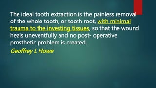
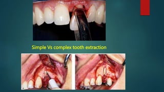
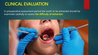
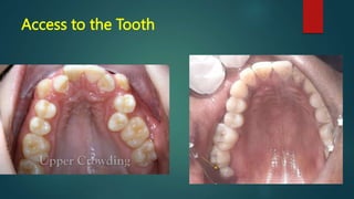
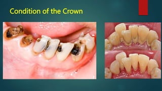
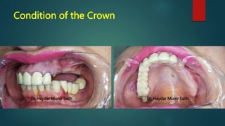
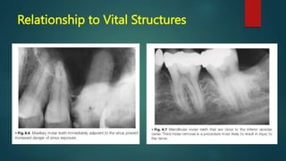
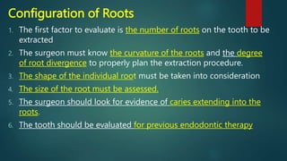
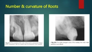
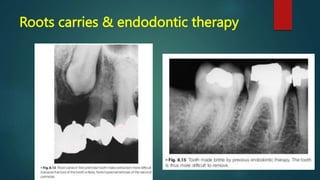
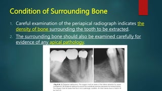
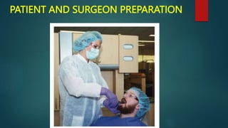
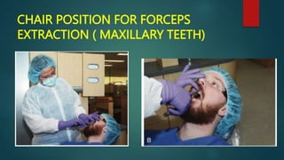
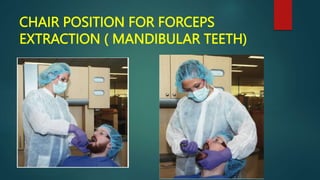
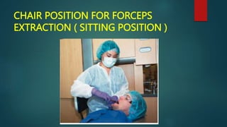
EXODONTIA
( Chapter One )
Principles of exodontia
DR. HAYDAR MUNIR SALIH ALNAMER
BDS. PHD (BOARD CERTIFIED)
Dental extraction has
always been
considered to be an
unpleasant procedure
for the patients due to
pain phobia.
The ideal tooth extraction is the painless removal
of the whole tooth, or tooth root, with minimal
trauma to the investing tissues, so that the wound
heals uneventfully and no post- operative
prosthetic problem is created.
Geoffrey L Howe
Extraction procedure can be:
1. Simple exodontia or closed method of
extraction or intraalveolar extraction
2. Complicated exodontia or open method of
extraction or transalveolar extraction
Simple Vs complex tooth extraction
In general, according to difficulty faced the
cases are classified into four types:
Type 1—easy patient easy case
Type 2—easy patient difficult case
Type 3—difficult patient easy case
Type 4—difficult patient difficult case
GENERAL PRINCIPLES INVOLVED IN
EXODONTIA
• Clinical evaluation
• Radiographic evaluation
• Patient and surgeon preparation
• Patient position
• Operator position
• Principles of extraction
• Principles of elevators
• Postoperative instructions
BASIC REQUIREMENTS
1. A good radiograph
2. Adequate anesthesia
3. Instruments
4. Adequate illumination
5. Efficient assistance
6. Suction apparatus
CLINICAL EVALUATION
In preoperative assessment period the tooth to be extracted should be
examined carefully to assess the difficulty of extraction
Access to the Tooth
 The first factor to be examined in preoperative assessment is
the extent to which the patient can open the mouth. Any
limitation of opening may compromise the ability of the
surgeon to give local anesthesia or perform a routine
extraction
 The location and position of the tooth to be extracted within a
dental arch should be examined.
 A properly aligned tooth has a normal access for placement of
elevators and forceps. However, crowded or otherwise
malposed teeth may present difficulty in positioning the
usually used forceps
Access to the Tooth
Mobility of the Tooth
 If the teeth are excessively mobile, uncomplicated tooth
removal should be expected, but soft tissue management
after the extraction may be more involved
 Teeth that have less-than-normal mobility should be
carefully assessed for the presence of hypercementosis or
ankylosis of the roots
Mobility of the Tooth
Condition of the Crown
 the presence of large caries or restorations in the crown.
If large portions of the crown have been destroyed by caries, the
likelihood of crushing the crown during the extraction is increased
 In addition, an endodontically treated tooth becomes desiccated and
typically becomes brittle and crumbles easily when force is applied.
 If the tooth to be extracted has a large accumulation of calculus, the
gross accumulation should be removed with a scaler or ultrasonic
cleaner before extraction
 If adjacent teeth have large restorations, the surgeon should use
elevators with extreme caution because fracture or displacement of
the restorations may occur
Condition of the Crown
Dr. Haydar Munir Salih Dr. Haydar Munir Salih
RADIOGRAPHIC EXAMINATION
 It is essential that proper radiographs be taken of
any tooth to be removed.
 In general, periapical radiographs provide the
most accurate and detailed information
concerning the tooth, its roots, and the
surrounding tissue.
 Panoramic radiographs are used frequently, but
their greatest usefulness is for impacted teeth as
opposed to erupted teeth.
Relationship to Vital Structures
Configuration of Roots
1. The first factor to evaluate is the number of roots on the tooth to be
extracted
2. The surgeon must know the curvature of the roots and the degree
of root divergence to properly plan the extraction procedure.
3. The shape of the individual root must be taken into consideration
4. The size of the root must be assessed.
5. The surgeon should look for evidence of caries extending into the
roots.
6. The tooth should be evaluated for previous endodontic therapy
Number & curvature of Roots
Roots carries & endodontic therapy
Condition of Surrounding Bone
1. Careful examination of the periapical radiograph indicates the
density of bone surrounding the tooth to be extracted.
2. The surrounding bone should also be examined carefully for
evidence of any apical pathology.
PATIENT AND SURGEON PREPARATION
 The concept of universal precautions states that all patients
must be viewed as having blood borne diseases that can be
transmitted to the surgical team.
 Before extraction: Patients should vigorously rinse their
mouths with an antiseptic mouth rinse such as chlorhexidine.
 To prevent teeth or fragments of teeth from falling into the
mouth and potentially being swallowed or aspirated into the
lungs, it is preferable to place a 4 x 4 inch gauze loosely into
the back of the mouth. However, it should not make the
patient gag.
PATIENT AND SURGEON PREPARATION
CHAIR POSITION FOR FORCEPS
EXTRACTION ( MAXILLARY TEETH)
CHAIR POSITION FOR FORCEPS
EXTRACTION ( MANDIBULAR TEETH)
CHAIR POSITION FOR FORCEPS
EXTRACTION ( SITTING POSITION )
exodontia

exodontia

  • 1.
    EXODONTIA ( Chapter One) Principles of exodontia DR. HAYDAR MUNIR SALIH ALNAMER BDS. PHD (BOARD CERTIFIED)
  • 2.
    Dental extraction has alwaysbeen considered to be an unpleasant procedure for the patients due to pain phobia.
  • 3.
    The ideal toothextraction is the painless removal of the whole tooth, or tooth root, with minimal trauma to the investing tissues, so that the wound heals uneventfully and no post- operative prosthetic problem is created. Geoffrey L Howe
  • 4.
    Extraction procedure canbe: 1. Simple exodontia or closed method of extraction or intraalveolar extraction 2. Complicated exodontia or open method of extraction or transalveolar extraction
  • 5.
    Simple Vs complextooth extraction
  • 6.
    In general, accordingto difficulty faced the cases are classified into four types: Type 1—easy patient easy case Type 2—easy patient difficult case Type 3—difficult patient easy case Type 4—difficult patient difficult case
  • 8.
    GENERAL PRINCIPLES INVOLVEDIN EXODONTIA • Clinical evaluation • Radiographic evaluation • Patient and surgeon preparation • Patient position • Operator position • Principles of extraction • Principles of elevators • Postoperative instructions
  • 9.
    BASIC REQUIREMENTS 1. Agood radiograph 2. Adequate anesthesia 3. Instruments 4. Adequate illumination 5. Efficient assistance 6. Suction apparatus
  • 10.
    CLINICAL EVALUATION In preoperativeassessment period the tooth to be extracted should be examined carefully to assess the difficulty of extraction
  • 11.
    Access to theTooth  The first factor to be examined in preoperative assessment is the extent to which the patient can open the mouth. Any limitation of opening may compromise the ability of the surgeon to give local anesthesia or perform a routine extraction  The location and position of the tooth to be extracted within a dental arch should be examined.  A properly aligned tooth has a normal access for placement of elevators and forceps. However, crowded or otherwise malposed teeth may present difficulty in positioning the usually used forceps
  • 12.
  • 13.
    Mobility of theTooth  If the teeth are excessively mobile, uncomplicated tooth removal should be expected, but soft tissue management after the extraction may be more involved  Teeth that have less-than-normal mobility should be carefully assessed for the presence of hypercementosis or ankylosis of the roots
  • 14.
  • 15.
    Condition of theCrown  the presence of large caries or restorations in the crown. If large portions of the crown have been destroyed by caries, the likelihood of crushing the crown during the extraction is increased  In addition, an endodontically treated tooth becomes desiccated and typically becomes brittle and crumbles easily when force is applied.  If the tooth to be extracted has a large accumulation of calculus, the gross accumulation should be removed with a scaler or ultrasonic cleaner before extraction  If adjacent teeth have large restorations, the surgeon should use elevators with extreme caution because fracture or displacement of the restorations may occur
  • 16.
  • 17.
    Condition of theCrown Dr. Haydar Munir Salih Dr. Haydar Munir Salih
  • 18.
    RADIOGRAPHIC EXAMINATION  Itis essential that proper radiographs be taken of any tooth to be removed.  In general, periapical radiographs provide the most accurate and detailed information concerning the tooth, its roots, and the surrounding tissue.  Panoramic radiographs are used frequently, but their greatest usefulness is for impacted teeth as opposed to erupted teeth.
  • 19.
  • 20.
    Configuration of Roots 1.The first factor to evaluate is the number of roots on the tooth to be extracted 2. The surgeon must know the curvature of the roots and the degree of root divergence to properly plan the extraction procedure. 3. The shape of the individual root must be taken into consideration 4. The size of the root must be assessed. 5. The surgeon should look for evidence of caries extending into the roots. 6. The tooth should be evaluated for previous endodontic therapy
  • 21.
  • 22.
    Roots carries &endodontic therapy
  • 23.
    Condition of SurroundingBone 1. Careful examination of the periapical radiograph indicates the density of bone surrounding the tooth to be extracted. 2. The surrounding bone should also be examined carefully for evidence of any apical pathology.
  • 25.
    PATIENT AND SURGEONPREPARATION  The concept of universal precautions states that all patients must be viewed as having blood borne diseases that can be transmitted to the surgical team.  Before extraction: Patients should vigorously rinse their mouths with an antiseptic mouth rinse such as chlorhexidine.  To prevent teeth or fragments of teeth from falling into the mouth and potentially being swallowed or aspirated into the lungs, it is preferable to place a 4 x 4 inch gauze loosely into the back of the mouth. However, it should not make the patient gag.
  • 26.
  • 27.
    CHAIR POSITION FORFORCEPS EXTRACTION ( MAXILLARY TEETH)
  • 28.
    CHAIR POSITION FORFORCEPS EXTRACTION ( MANDIBULAR TEETH)
  • 29.
    CHAIR POSITION FORFORCEPS EXTRACTION ( SITTING POSITION )