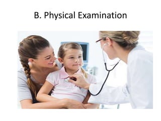
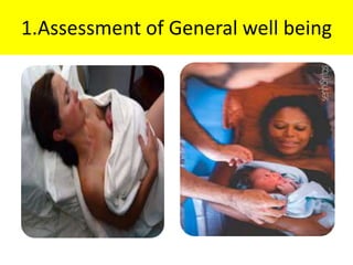
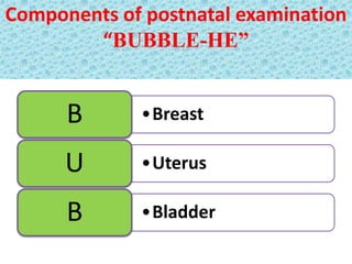
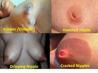
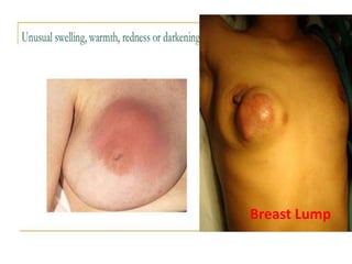
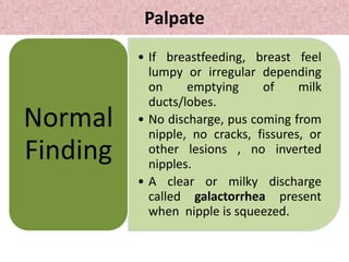
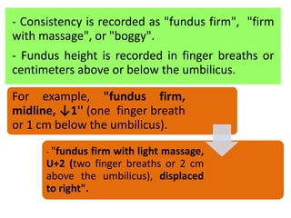
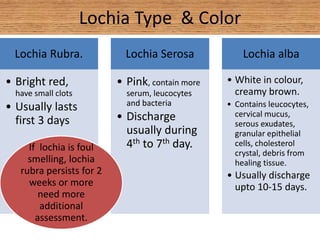
Postnatal Assessment
By-Isha Thapa Magar
Nursing Instructor
B.Sc 3th Year
History Taking
Postnatal Mother
Examination
Postnatal Mother Examination
Objective
• To observe the general condition of the
mother.
• To find out postnatal problem and manage.
• To provide necessary health teaching to
mother and family.
• To improve mental and physical health of
mother.
Equipments
• Thermometer tray
• BP instrument
• Measuring tape
• Clean glove
• Kidney tray
• Clean swabs and gauze piece
• Weighing scale
• Bed pan for mother unable to move
• Screen
• Torch
Procedure
Procedure
A. Getting Ready
• Prepare the necessary equipment.
• Greet the women respectfully and with
kindness.
• Explain mother about the procedure
• Ask the mother to empty her bladder.
• Maintain Privacy.
• Wash hands.
B. Physical Examination
1.Assessment of General well being
Gait and Movement
- Normal walk without a limp
- Gait and movements are steady
and moderately paced.
Behavior &Facial expression
- Alert, responsive, cooperative,
calm
General cleanliness, noting visible dirt
and odor.
Check skin noting lesions and bruises
- Normally , skin free from lesions and
bruises.
Check conjunctiva
– If conjunctiva appears white or very
pale see anemia for additional
information
2. Vital sign Measurement
2. Vital Signs measurement:
Temperature
- Temperature during the first 24
hours postpartum is within the
normal range.
- If fever, up to 100.4◦F (38◦C)→
indicate dehydration
- Temperature should be normal
after 24 hours with replacement of
fluids
- A temperature above100.4◦F
(38◦C) at any time or an abnormal
temperature after first 24 hours →
indicate infection
Pulse
- Puerperal bradycardia (40 to 80
beats per minute) → normal during
the first week after birth
- Orthostatic hypotension
- Tachycardia → indicate anxiety,
excitement, fatigue, pain, excessive
blood loss, infection, cardiac
problems.
Blood pressure
• Immediately after childbirth, the
blood pressure should remain
the same as during labor.
• An increase in blood pressure
→indicate gestational
hypertension
A decrease in blood pressure →
indicate shock, orthostatic
hypotension, dehydration, a side
effect of epidural anesthesia.
Blood pressure vary based
on the woman's position,
so assess blood pressure in
the same position every
time.
Respirations
- Normal respiratory rate of 12 to
20 breaths per minute should be
maintained.
- No important to assess breath
sounds → if the mother has had a
normal vaginal delivery, is
ambulatory, and is without signs
of respiratory distress.
• Breath sound always should be auscultated if
the birth was cesarean or the mother is
receiving magnesium sulfate, is a smoker, or
has a history of frequent or recent upper
respiratory tract infections, or asthma.
-Important for auscultation of breath
sound →if birth was cesarean,
mother is receiving magnesium
sulfate, is a smoker, or has a history
of frequent or recent upper
respiratory tract infections, or
asthma.
Pain
• Pain is fifth vital sign
• Ask the woman about the type
of pain and its location and
severity using a numeric scale
from 0 to 10 points
Components of postnatal examination
“BUBBLE-HE”
•BreastB
•UterusU
•BladderB
•Homan’s signH
•Emotional StatusE
•BowelB
•LochiaL
•EpisiotomyE
3. Breast examination
Inspection
Inspect breasts for;
- Size, asymmetry
- Contour
- Erythema
- Engorgement
- Note any
abnormalities.
Inspect nipples for;
- Cracks
- Redness
- Lesions
- Sores, rashes
- Fissures or bleeding
- Erect, flat or
inverted
• One breast is slightly larger than
other.
• If breastfeeding, breasts look
lumpy or irregular than usual.
• Veins larger and darker, more
visible beneath the skin.
• Regular with no dimpling, no
visible lumps, skin is smooth with
no puckering, no redness, no
lesion sores or rashes.
• Tenderness and lumpiness in both
breasts during the menstrual cycle.
• Areolas larger and darker.
Normal
Breast
• Changes in colour of breast
or nipple, wrinkling,
dimpling, thickening,
puckering.
• A nipple sink into breast.
• A red, scaly rash or sore
on breast &nipple.
Abnormal
findings
Punker /Dimpling Inverted nipple
Dripping Nipple Cracked Nipples
Breast Lump
Contd………
Palpate
• If breastfeeding, breast feel
lumpy or irregular depending
on emptying of milk
ducts/lobes.
• No discharge, pus coming from
nipple, no cracks, fissures, or
other lesions , no inverted
nipples.
• A clear or milky discharge
called galactorrhea present
when nipple is squeezed.
Normal
Finding
Galactorrhea
• Redness, warmth, painful
lump or on entire breast
→indicate abscess or
mastitis.
• A bloody discharge or milk
discharge occur without
stimulation
Abnormal
Finding
4. Abdominal Examination
Inspection
Inspect the shape, size, movement of abdomen
with respiration, scarred gravid, linea nigra,
caesarean section, old and new incision on the
abdomen.
Caesarean section incision sites →healing
process, discharge, redness and signs of
infections.
Linea nigra
Scarred gravida
Palpate Fundus
2.Place mother in a supine position with her
knees slightly flexed.
1. Palpate fundus for consistency and location.
It should be firmly contracted and at or near the
level of the umbilicus.
4. Place non-dominant hand above
mother’s symphysis pubis. This supports
and anchors the lower uterine segment
during palpation or massage of the fundus.
3. Put on clean gloves and lower the
perineal pads to observe lochia as the
fundus is palpated.
6. Palpate gently at umbilicus until the fundus
is located.
Determine the firmness and location of the
fundus.
This should be firmly contracted, in the midline
and approximately at the level of the
umbilicus.
5.Use flat part of fingers (not the finger tips)
for palpation. Palpation may be painful, for
the mother who had a cesarean birth.
8.The location of fundus should be rechecked
after emptying bladder.
If fundus is difficult to locate or is soft or boggy,
keep non dominant hand above symphysis pubis
and massage fundus with dominant hand until
fundus is firm.
7.If uterus is above the expected level or shifted
from the middle of the abdomen (usually to the
right), the bladder may be distended.
- Removing clots allows the uterus to contract properly. A firm fundus and pressure over the lower uterine segment help prevent uterine inversion.
9.After boggy fundus is massaged until it is firm, press
firmly to expel clots.
Do not attempt to expel clots before the fundus is firm.
Keep one hand pressed just above the symphysis (over
the lower uterine segment) throughout.
- Removing clots allows the uterus to
contract properly.
- A firm fundus and pressure over
the lower uterine segment help
prevent uterine inversion.
11. Document the consistency and location of
the fundus.
10.Measure fundus height in centimeters or
use fingers breaths.
Generally fundal height decreases about 1cm
per day for first 9-10 days post-partum
- Consistency is recorded as "fundus firm", "firm
with massage", or "boggy".
- Fundus height is recorded in finger breaths or
centimeters above or below the umbilicus.
For example, "fundus firm,
midline, ↓1'' (one finger breath
or 1 cm below the umbilicus).
- "fundus firm with light massage,
U+2 (two finger breaths or 2 cm
above the umbilicus), displaced
to right".
5. Bladder examination
- Ask to pass urine frequently the first few
days.
- Normal if bladder is not palpable.
- Women is able to urinate when the urge is
felt.
- Monitor clients for signs of UTI, including
fever, urinary frequency and/ or urgency,
difficult or painful urination.
- Infrequent or insufficient voiding (less than
200 ml) discomfort, burning urgency, or foul
smelling urine suggest infection
6. Bowel examination
Inspect the woman's abdomen for distention,
auscultation for bowel sounds in all four
quadrants prior to palpating the uterine
fundus, and palpate for tenderness.
Ask the patient about daily bowel movement
or has passed gas since giving birth.
She must no become constipated.
Explain that she should wipe from front to back
after voiding or defecating.
Normal assessment findings are active bowel
sounds, passing gas, and a non-distended
abdomen.
7. Lochia Examination
Check and
note
colour,
order and
amount of
lochia.
To assess amount
- ask her how many perineal
pads she has used in the
past 1 to 2 hours and
- how much drainage was on
each pad. (pad completely,
or was only half of pad
covered with drainage)
- Ask about
color of
drainage,
odor, and
presence of
any clots.
Lochia increases with maternal activity
and breastfeeding which is normal.
Lochia Type & Color
Lochia Rubra.
• Bright red,
have small clots
• Usually lasts
first 3 days
Lochia Serosa
• Pink, contain more
serum, leucocytes
and bacteria
• Discharge
usually during
4th to 7th day.
Lochia alba
• White in colour,
creamy brown.
• Contains leucocytes,
cervical mucus,
serous exudates,
granular epithelial
cells, cholesterol
crystal, debris from
healing tissue.
• Usually discharge
upto 10-15 days.
If lochia is foul
smelling, lochia
rubra persists for 2
weeks or more
need more
additional
assessment.
Lochia Odor
Lochia should have" no foul odor".
A truly foul odor may be a sign of
infection.
Lochia Amount
• 5cm saturation of pad in
one hour =10 ml.Scant
• 10 cm saturation of pad
within 1 hour =10 to 25mlLight
The average amount of discharge for the
first 5-6 days is estimated to be 250 ml.
• Moderate; 15cm
saturation with in 1 hour
=25 to 50 ml.
Moderate
• Heavy; pad is completely
saturated within 1 hour =
50 to 80 ml.
Heavy
• Postpartum hemorrhage is
clinically defined as a pad
saturated within 15-30
minutes.
PPH
Amount of lochia
During examination, the quantity, colour,
odor and consistency of lochia are
significant.
a. Persistence of red lochia → indicates
secondary postpartum hemorrhage.
b. Brown profuse lochia with bulky uterus →
sub-involution of the uterus
c. Excessive lochia → retained product of
conception.
d. Scanty lochia → indicate poor drainage.
e. When associated with pyrexia they are due
to localized uterine infection.
8. Episiotomy and perineum examination
Examine episiotomy and perineum
area through REEDA Assessment
R-Redness E-Edema E-Ecchymosis
D-Discharge
A-
Approximation
• Redness → infection or hematoma.
• Ecchymosis (excessive bruising) → vaginal
trauma and requires additional evaluation.
• Discharge→ should follow the expected lochia
pattern.
• Approximation→ episiotomy lines should be
well approximated.
Episiotomy line
Perineum
• Pull the labia from front to back.
• Check the episiotomy or areas of vaginal tearing.
• Look for hematoma formation, hemorrhoids,
vaginitis, perineal tearing.
Vulvar hematoma
9. Homan’s sign
Complain of pain in calf of the leg upon dorsi-
flexion of foot with leg extended is diagnostic of
Deep Vein Thrombosis (DVT) of the area.
A positive Homan's sign is indicative of DVT.
Homan’s sign
10. Emotional status
After delivery the woman may progress
through Rubin’s stages of taking in,
taking hold & letting go phases.
• May Begin with a refreshing sleep after
delivery.
• During first 24 to 48 hours after giving
birth, mother exhibits passive, dependent
behavior.
• New mothers spend time touching baby
commonly identifying specific features in
newborn such as " he has my nose" or his
fingers are long like his father's.
1.
Taking
In
Phase
• Starts on 2nd to 3rd day postpartum
and may last several weeks.
• Woman begins to initiate action and
to function more independently but
still show dependent behaviors.
• Woman may require more explanation
and reassurance that she is functioning
well, especially in caring for her infant.
• As the woman meets success in caring
for the newborn, her concern extends
to other family members and their
activities.
2.Taking
hold
phase
• It begins near end of 1st
weeks.
• Mother reestablishes
relationships with couple and
other people.
• She assumes responsibility and
care for newborn
independently.
3.
Letting
go
phase
11. Health Teaching
Health teaching should be given as per need
identification of mother.
  

Postnatal Mother Examination - BUBBLE-HE

  • 1.
    Postnatal Assessment By-Isha ThapaMagar Nursing Instructor B.Sc 3th Year
  • 2.
  • 3.
  • 4.
    Objective • To observethe general condition of the mother. • To find out postnatal problem and manage. • To provide necessary health teaching to mother and family. • To improve mental and physical health of mother.
  • 5.
    Equipments • Thermometer tray •BP instrument • Measuring tape • Clean glove • Kidney tray
  • 6.
    • Clean swabsand gauze piece • Weighing scale • Bed pan for mother unable to move • Screen • Torch
  • 7.
  • 8.
    A. Getting Ready •Prepare the necessary equipment. • Greet the women respectfully and with kindness. • Explain mother about the procedure
  • 9.
    • Ask themother to empty her bladder. • Maintain Privacy. • Wash hands.
  • 10.
  • 11.
  • 12.
    Gait and Movement -Normal walk without a limp - Gait and movements are steady and moderately paced. Behavior &Facial expression - Alert, responsive, cooperative, calm
  • 13.
    General cleanliness, notingvisible dirt and odor. Check skin noting lesions and bruises - Normally , skin free from lesions and bruises. Check conjunctiva – If conjunctiva appears white or very pale see anemia for additional information
  • 14.
    2. Vital signMeasurement
  • 15.
    2. Vital Signsmeasurement: Temperature - Temperature during the first 24 hours postpartum is within the normal range. - If fever, up to 100.4◦F (38◦C)→ indicate dehydration
  • 16.
    - Temperature shouldbe normal after 24 hours with replacement of fluids - A temperature above100.4◦F (38◦C) at any time or an abnormal temperature after first 24 hours → indicate infection
  • 17.
    Pulse - Puerperal bradycardia(40 to 80 beats per minute) → normal during the first week after birth - Orthostatic hypotension - Tachycardia → indicate anxiety, excitement, fatigue, pain, excessive blood loss, infection, cardiac problems.
  • 18.
    Blood pressure • Immediatelyafter childbirth, the blood pressure should remain the same as during labor. • An increase in blood pressure →indicate gestational hypertension
  • 19.
    A decrease inblood pressure → indicate shock, orthostatic hypotension, dehydration, a side effect of epidural anesthesia. Blood pressure vary based on the woman's position, so assess blood pressure in the same position every time.
  • 20.
    Respirations - Normal respiratoryrate of 12 to 20 breaths per minute should be maintained. - No important to assess breath sounds → if the mother has had a normal vaginal delivery, is ambulatory, and is without signs of respiratory distress.
  • 21.
    • Breath soundalways should be auscultated if the birth was cesarean or the mother is receiving magnesium sulfate, is a smoker, or has a history of frequent or recent upper respiratory tract infections, or asthma. -Important for auscultation of breath sound →if birth was cesarean, mother is receiving magnesium sulfate, is a smoker, or has a history of frequent or recent upper respiratory tract infections, or asthma.
  • 22.
    Pain • Pain isfifth vital sign • Ask the woman about the type of pain and its location and severity using a numeric scale from 0 to 10 points
  • 23.
    Components of postnatalexamination “BUBBLE-HE” •BreastB •UterusU •BladderB
  • 24.
  • 25.
  • 26.
    Inspection Inspect breasts for; -Size, asymmetry - Contour - Erythema - Engorgement - Note any abnormalities. Inspect nipples for; - Cracks - Redness - Lesions - Sores, rashes - Fissures or bleeding - Erect, flat or inverted
  • 27.
    • One breastis slightly larger than other. • If breastfeeding, breasts look lumpy or irregular than usual. • Veins larger and darker, more visible beneath the skin. • Regular with no dimpling, no visible lumps, skin is smooth with no puckering, no redness, no lesion sores or rashes. • Tenderness and lumpiness in both breasts during the menstrual cycle. • Areolas larger and darker. Normal Breast
  • 28.
    • Changes incolour of breast or nipple, wrinkling, dimpling, thickening, puckering. • A nipple sink into breast. • A red, scaly rash or sore on breast &nipple. Abnormal findings
  • 29.
    Punker /Dimpling Invertednipple Dripping Nipple Cracked Nipples
  • 30.
  • 31.
  • 32.
    Palpate • If breastfeeding,breast feel lumpy or irregular depending on emptying of milk ducts/lobes. • No discharge, pus coming from nipple, no cracks, fissures, or other lesions , no inverted nipples. • A clear or milky discharge called galactorrhea present when nipple is squeezed. Normal Finding
  • 33.
  • 34.
    • Redness, warmth,painful lump or on entire breast →indicate abscess or mastitis. • A bloody discharge or milk discharge occur without stimulation Abnormal Finding
  • 35.
  • 36.
    Inspection Inspect the shape,size, movement of abdomen with respiration, scarred gravid, linea nigra, caesarean section, old and new incision on the abdomen. Caesarean section incision sites →healing process, discharge, redness and signs of infections.
  • 37.
  • 38.
  • 39.
    Palpate Fundus 2.Place motherin a supine position with her knees slightly flexed. 1. Palpate fundus for consistency and location. It should be firmly contracted and at or near the level of the umbilicus.
  • 40.
    4. Place non-dominanthand above mother’s symphysis pubis. This supports and anchors the lower uterine segment during palpation or massage of the fundus. 3. Put on clean gloves and lower the perineal pads to observe lochia as the fundus is palpated.
  • 41.
    6. Palpate gentlyat umbilicus until the fundus is located. Determine the firmness and location of the fundus. This should be firmly contracted, in the midline and approximately at the level of the umbilicus. 5.Use flat part of fingers (not the finger tips) for palpation. Palpation may be painful, for the mother who had a cesarean birth.
  • 42.
    8.The location offundus should be rechecked after emptying bladder. If fundus is difficult to locate or is soft or boggy, keep non dominant hand above symphysis pubis and massage fundus with dominant hand until fundus is firm. 7.If uterus is above the expected level or shifted from the middle of the abdomen (usually to the right), the bladder may be distended.
  • 44.
    - Removing clotsallows the uterus to contract properly. A firm fundus and pressure over the lower uterine segment help prevent uterine inversion. 9.After boggy fundus is massaged until it is firm, press firmly to expel clots. Do not attempt to expel clots before the fundus is firm. Keep one hand pressed just above the symphysis (over the lower uterine segment) throughout. - Removing clots allows the uterus to contract properly. - A firm fundus and pressure over the lower uterine segment help prevent uterine inversion.
  • 45.
    11. Document theconsistency and location of the fundus. 10.Measure fundus height in centimeters or use fingers breaths. Generally fundal height decreases about 1cm per day for first 9-10 days post-partum
  • 46.
    - Consistency isrecorded as "fundus firm", "firm with massage", or "boggy". - Fundus height is recorded in finger breaths or centimeters above or below the umbilicus. For example, "fundus firm, midline, ↓1'' (one finger breath or 1 cm below the umbilicus). - "fundus firm with light massage, U+2 (two finger breaths or 2 cm above the umbilicus), displaced to right".
  • 47.
    5. Bladder examination -Ask to pass urine frequently the first few days. - Normal if bladder is not palpable. - Women is able to urinate when the urge is felt.
  • 48.
    - Monitor clientsfor signs of UTI, including fever, urinary frequency and/ or urgency, difficult or painful urination. - Infrequent or insufficient voiding (less than 200 ml) discomfort, burning urgency, or foul smelling urine suggest infection
  • 49.
    6. Bowel examination Inspectthe woman's abdomen for distention, auscultation for bowel sounds in all four quadrants prior to palpating the uterine fundus, and palpate for tenderness. Ask the patient about daily bowel movement or has passed gas since giving birth.
  • 50.
    She must nobecome constipated. Explain that she should wipe from front to back after voiding or defecating. Normal assessment findings are active bowel sounds, passing gas, and a non-distended abdomen.
  • 51.
    7. Lochia Examination Checkand note colour, order and amount of lochia. To assess amount - ask her how many perineal pads she has used in the past 1 to 2 hours and - how much drainage was on each pad. (pad completely, or was only half of pad covered with drainage) - Ask about color of drainage, odor, and presence of any clots. Lochia increases with maternal activity and breastfeeding which is normal.
  • 52.
    Lochia Type &Color Lochia Rubra. • Bright red, have small clots • Usually lasts first 3 days Lochia Serosa • Pink, contain more serum, leucocytes and bacteria • Discharge usually during 4th to 7th day. Lochia alba • White in colour, creamy brown. • Contains leucocytes, cervical mucus, serous exudates, granular epithelial cells, cholesterol crystal, debris from healing tissue. • Usually discharge upto 10-15 days. If lochia is foul smelling, lochia rubra persists for 2 weeks or more need more additional assessment.
  • 53.
    Lochia Odor Lochia shouldhave" no foul odor". A truly foul odor may be a sign of infection.
  • 54.
    Lochia Amount • 5cmsaturation of pad in one hour =10 ml.Scant • 10 cm saturation of pad within 1 hour =10 to 25mlLight The average amount of discharge for the first 5-6 days is estimated to be 250 ml.
  • 55.
    • Moderate; 15cm saturationwith in 1 hour =25 to 50 ml. Moderate • Heavy; pad is completely saturated within 1 hour = 50 to 80 ml. Heavy • Postpartum hemorrhage is clinically defined as a pad saturated within 15-30 minutes. PPH
  • 56.
  • 57.
    During examination, thequantity, colour, odor and consistency of lochia are significant. a. Persistence of red lochia → indicates secondary postpartum hemorrhage. b. Brown profuse lochia with bulky uterus → sub-involution of the uterus c. Excessive lochia → retained product of conception.
  • 58.
    d. Scanty lochia→ indicate poor drainage. e. When associated with pyrexia they are due to localized uterine infection.
  • 59.
    8. Episiotomy andperineum examination Examine episiotomy and perineum area through REEDA Assessment R-Redness E-Edema E-Ecchymosis D-Discharge A- Approximation
  • 60.
    • Redness →infection or hematoma. • Ecchymosis (excessive bruising) → vaginal trauma and requires additional evaluation. • Discharge→ should follow the expected lochia pattern. • Approximation→ episiotomy lines should be well approximated.
  • 61.
  • 62.
    Perineum • Pull thelabia from front to back. • Check the episiotomy or areas of vaginal tearing. • Look for hematoma formation, hemorrhoids, vaginitis, perineal tearing.
  • 63.
  • 64.
    9. Homan’s sign Complainof pain in calf of the leg upon dorsi- flexion of foot with leg extended is diagnostic of Deep Vein Thrombosis (DVT) of the area. A positive Homan's sign is indicative of DVT.
  • 65.
  • 66.
    10. Emotional status Afterdelivery the woman may progress through Rubin’s stages of taking in, taking hold & letting go phases. • May Begin with a refreshing sleep after delivery. • During first 24 to 48 hours after giving birth, mother exhibits passive, dependent behavior. • New mothers spend time touching baby commonly identifying specific features in newborn such as " he has my nose" or his fingers are long like his father's. 1. Taking In Phase
  • 67.
    • Starts on2nd to 3rd day postpartum and may last several weeks. • Woman begins to initiate action and to function more independently but still show dependent behaviors. • Woman may require more explanation and reassurance that she is functioning well, especially in caring for her infant. • As the woman meets success in caring for the newborn, her concern extends to other family members and their activities. 2.Taking hold phase
  • 68.
    • It beginsnear end of 1st weeks. • Mother reestablishes relationships with couple and other people. • She assumes responsibility and care for newborn independently. 3. Letting go phase
  • 69.
    11. Health Teaching Healthteaching should be given as per need identification of mother.
  • 70.