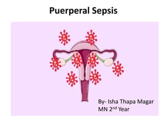
3. Introduction
⢠Puerperal sepsis is one of the five leading causes of
maternal mortality worldwide, and accounts for 15%
of all maternal deaths.
⢠In 1992, WHO defined puerperal sepsis as an
infection of the genital tract occurring at any time
between the rupture of membranes or labour and
the 42nd day post partum; in which, two or more of
the following are present:
â pelvic pain
â fever
4. Contd..
â abnormal vaginal discharge and
â delay in the reduction of the size of the uterus.
⢠At the same time, the WHO introduced the term
puerperal infections, which also include non-genital
infections in the obstetric population.
⢠Recent epidemiological data shows that puerperal
sepsis and non-genital tract infections are a major
area of concern.
⢠In puerperal sepsis, group A streptococcus (GAS) is
the most feared pathogen.
5. ⢠In non-genital tract infections, influenza viruses and
the HIV pandemic in the developing part of the world
are responsible for many maternal deaths, and
demand our attention.
6. ⢠It is the infection of the genital tract resulting from
the bacterial invasion into the uterus or surrounding
tissues during or after the labor within first 6 weeks
of delivery or abortion. It normally does not occur
within first 24 hours.
7. ⢠If it does not only cause death, puerperal sepsis
can cause long-term health problems such as
chronic pelvic inflammatory disease (PID) and
infertility.
⢠It is preventable with provision of adequate
antenatal care, referral and timely treatment of
complications of pregnancy, promoting
institutional delivery and postnatal care.
8. Incidence
⢠A study conducted in London in 2015 reported
that the prevalence and associated risk
factors of puerperal sepsis is high in South
Asia i.e. 3-4% of total deliveries. It is an
leading cause of preventable maternal deaths,
accounting for up to 11% of maternal deaths
worldwide.
9. ⢠Puerperal sepsis affects 2-10% of the patient
â 1 to 3 % in normal vaginal deliveries
â 5 to 15 % in scheduled cesarean deliveries
performed before labor begins
â 15 to 20 % in non-scheduled cesarean deliveries
performed after labor begins
10. ⢠A retrospective study on Puerperal Sepsis in Patan
Hospital in 2015 showed the causes of puerperal
pyrexia were
â urinary tract infection (47.5%),
â wound infection (20.5%),
â endometritis (19.7%)
â retained product of conception (8.2%),
â pyoperitoneum (2.5%) and
â septicemia (1.6%).
12. ⢠More than one type of bacteria may be involved
when a woman develops puerperal sepsis.
⢠Bacteria may be either endogenous or exogenous.
13. ďąEndogenous bacteria
ď§ These are bacteria which normally live in the vagina
and rectum without causing harm (e.g. some types
of streptococci and staphylococci, E.coli, clostridium
welchii).
ď§ Even when a clean technique is used for delivery,
infection can still occur from endogenous bacteria.
14. ďąEndogenous bacteria can become harmful and
cause infection if:
ď§ they are carried into the uterus, usually from the
vagina, by the examining finger or by instruments
during pelvic examinations
ď§ there is tissue damage, i.e. bruised, lacerated or
dead tissue (e.g. after a traumatic delivery or
following obstructed labour)
ď§ there is prolonged rupture of membranes because
microorganisms can then enter the uterus.
15. ďąExogenous bacteria
⢠These are bacteria which are introduced into the
vagina from the outside (streptococci, staphylococci,
clostridium tetani, etc.).
⢠Exogenous bacteria can be introduced into the vagina:
â by unclean hands and unsterile instruments
â by droplet infection (e.g. a health provider sneezing,
coughing onto own hands immediately prior to
examination)
â by foreign substances that are inserted into the vagina
(e.g. herbs, oil, cloth) by sexual activity.
16. Predisposing Factor
⢠Ante-partum factors:
ď Malnutrition
ď Anemia
ď diabetes
ď Pre-eclampsia
ď Premature rupture of membrane
ď Sexual intercourse during late pregnancy.
17. Intra partum factors
ďRepeated vaginal examination
ďTraumatic operative delivery
ďRetained bits of placental tissues or membranes
ďPlacenta previa
ďHemorrhage
ďCaesarean delivery
ďProlonged labor
ďLacerations
ďPremature rupture of the membrane.
18. ⢠In 2015, the systematic review conducted in south Asian
countries, identified associated risk factors with
puerperal sepsis were:
â maternal factors such as pre-existing conditions,
labor factors including peripartum bleeding, stillbirth,
perineal tears, prolonged delivery and unhygienic
practices.
â health service factors such as access to antenatal and
obstetric care, and
â community factors such as lower socioeconomic
status and unhygienic postpartum traditions
19. HOW PUERPERAL SEPSIS OCCURS
⢠The uterine infection may start before the onset of
labour i.e. in cases of pre-labour rupture of the
membranes, during labour, or in the early postnatal
period before healing of lacerations in the genital
tract and the placental site have taken place.
20. ⢠In cases of pre-labour rupture of membranes,
antibiotics should be given either to treat amnionitis,
if the woman has fever and foul-smelling vaginal
discharge, or as a prophylactic measure to reduce
the risk of infection.
21. ⢠Following delivery, puerperal sepsis may be localized
in the perineum, vagina, cervix or uterus.
⢠Infection of the uterus can spread rapidly if due to
virulent organisms, or if the motherâs resistance is
impaired.
⢠It can extend beyond the uterus to involve the
fallopian tubes and ovaries, to the pelvic cellular
tissue causing parametritis, to the pelvic
peritoneum, causing peritonitis, and into the blood
stream causing septicaemia.
22.
23.
24.
25. ⢠Thrombophlebitis of the uterine veins can transport
infected clots to other organs.
⢠Severe infection can be further complicated by
septic shock and coagulation failure which gives rise
to bleeding problems.
⢠Puerperal sepsis can be rapidly fatal.
26.
27. Clinical Features
ďąLocal Infection
ď§ Slight rise in temperature, generalized malaise and
headache
ď§ Redness and the swelling of the local wound
ď§ Pus formation and disruption of wound.
ďąUterine infection:
ď§ Pyrexia of variable degree and tachycardia.
ď§ Red, copious and offensive lochia.
ď§ Sub involution ,tender and soft uterus.
28. ďą Sever Infection
ď§ Fever with chills and rigor
ď§ Rapid pulse
ď§ Scanty, odorless lochia
ď§ Sub-Involution of uterus
ďąParametritis
ď§ Sustained rise in temperature(7th to 10th day)
ď§ Constant pelvic pain
ď§ Tenderness on either side of the hypo-gastrium
ď§ Unilateral, tender mass felt on vaginal examination
ď§ Leukocytosis.
29. ďą Pelvic Peritonitis
ďPyrexia with increased pulse rate
ďLower abdominal pain and tenderness
ďCollection of the pus in pouch of douglas.
ďą Generalized Peritonitis
ďHigh fever with rapid pulse
ďVomiting
ďAbdominal pain
ďTender and distended abdomen
30. ďą Thrombophlebitis
ďSwinging fever with chills and rigor
ďFeatures of pyemia.
ďąSepticemia
ďHigh temperature with rigor
ďRapid pulse
ďHeadache, insomnia or mental confusion
ďPositive blood culture
ďSign and symptoms of infection of lungs ,
meninges or joint .
31. Diagnosis
⢠History taking ( preexisting infection before
labor, symptoms of infection ,complicated labor
as PROM , instrument, prolonged)
⢠General examination
Temperature, pulse, blood pressure, tonsils,
lower limbs for signs of thrombophlebitis.
⢠Abdominal examination (loin tenderness,
abdominal rigidity and tenderness, uterine size)
to note involution of uterus whether uterus is
tender or not, presence of any features of
peritonitis or pelvic abcess.
32. ⢠Internal examination
to note character of lochia, condition of the perineal
wound, repaired or pelvic abscess, infected episiotomy.
⢠Bi-manual examination
to find out ay pelvic abscess such as Uterine size,
consistency, position , tenderness,
Cervix: closed ,open, contents felts through it or
laceration.
⢠Douglas pouch: bogginess
⢠Limbs: legs are examined to detect thrombophlebitis
or thrombosis.
33. Investigations
ďźHigh vaginal and endo-cervical Swab for
Culture in aerobic and anaerobic media and
sensitivity test to antibiotics.
ďźClean catch mid stream specimen of Urine for
urinalysis and culture.
ďźBlood test for total and differential white cell
count, haemoglobin estimation.
ďźBlood culture, if fever is associated with chills
and rigor.
34. ďźOther specific investigations
⢠Pelvic ultrasound
â to detect any retained bits of conception within uterus
â To locate any abscess with the pelvis
â To collect samples i.e. pus or fluid from the pelvis for
culture and sensitivity.
⢠Color flow doppler study to detect venous
thrombosis.
⢠CT ad MRI to detect pelvic vein thrombosis
⢠Chest X-ray to detect any lung pathology.
35. Treatment
ďąGeneral treatment
⢠Assess out for signs of shock, septicaemia, pallor,
anaemia, and treat accordingly.
⢠Perform an abdominal examination and assess
uterine size.
⢠Assess uterine haemorrhage and attempt to
control it.
⢠Failure to control uterine bleeding managed with
special treatment.
36. ⢠Start antibiotics after taking samples for
microbiology.
â Amoxycillin- clavulinic acid â 1.2 gms. intravenous 8
hourly or 625mg. oral 8 hourly/twice a day,
Or
â Ampicillin 500mg. intravenous 6 hourly,
Or
â Amoxycillin 500mg. intravenous 8 hourly,
Or
â Gentamicin 5mg./kg body weight/day in a single dose
or in two divided doses.
37. ⢠Giving penicillin with gentamicin and metronidazole
provides the broadest coverage.
⢠Give IV fluids: 1 litre of 5% dextrose in saline or
normal saline rapidly, every 24 hours.
⢠Check vital signs and urinary output every 6 hours.
⢠Reassess every 24 hours: if there is no improvement
start special treatment.
38. ⢠If there is improvement, continue intravenous
(IV) antibiotics for 3 days and then follow-up
with oral antibiotics.
⢠At this point, if the woman is much better,
send her home on oral antibiotics for 4-7 days,
after having checked her haemoglobin level
and given her treatment for anaemia if found.
39. ⢠Inform her to return if she develops fever,
vaginal bleeding or abdominal pain.
⢠If the woman is not better after three days on
intravenous antibiotics, treat her with special
treatment.
40. ďąTreat with special treatment if Condition of
the patient is
⢠If initial assessment indicate septicaemia,
repeated body temperatures above 101 F,
hypotension,
⢠Tender abdomen/abdominal mass, purulent
vaginal discharge,
⢠Patient not responding to initial treatment
and condition getting worse.
41. ⢠Perform a physical examination to rule out
pelvic abscesses, pelvic thrombophlebitis,
anaemia etc.,
⢠Take vaginal swabs for gram stain and culture
and antibiotic sensitivity test and blood for
culture and antibiotic sensitivity test.
42. ⢠Broad-spectrum antibiotic coverage must be
initiated immediately after collection of cultures.
â Amoxycillin- clavulinic acid (Amoxyclav) â 1.2 gms.
intravenous 8 hourly with or without gentamycin.
â If response is poor, Imipenem 500 mg. intravenous 8
hourly
Or
Ticarcillin-clavulinic acid 3.2 gms. intravenous 8
hourly may be used in place of amoxycillin-clavulinic
acid (Amoxyclav).
43. ⢠Manage complications appropriately.
â 1. Retained placental fragments,
â 2. Presence of blood clots,
â 3. Pelvic abscess;
⢠If the infection is complicated by the presence
of an infected focus, it may be necessary to
surgically drain the infected site/abscess.
⢠Continuing adequate and effective antibiotic
therapy in this situation is mandatory.
44. ďąThrombophlebitis;
⢠If thrombophlebitis occurs in a superficial
vein, self-care steps that include applying heat
to the painful area, elevating the affected leg
and using a non-steroidal anti-inflammatory
drug may be recommended. The condition
usually subsides within a week or two.
45. ⢠In the presence of deep vein thrombosis use of
anticoagulants, such as heparin, will prevent clots
from growing. After the heparin therapy, use of
warfarin (Coumarin) for several months continues
to prevent clots from growing.
⢠Support stockings- these help prevent recurrent
swelling and reduces the chances of
complications of deep vein thrombosis.
46. ďąAnaemia;
⢠- If haemoglobin level is <8mg./dl. â blood
transfusion is recommended.
⢠- If haemoglobin level is 8-10 mg./dl. â oral or
parenteral haematinics are recommended.
47. Surgical Management
ďąPerineal wound
ď§ Stitches are removed to facilitate drainage of
pus & relieve pain
ď§ Cleaned with sitz bath
ď§ The wound is to be dressed with hot
compress with mild dressed with mild
antiseptic solution. After infection is
controlled secondary suture after control of
infection
48.
49. ďąRetained uterine products
⢠Surgical evacuation if diameter more than 3
cm
⢠Antibiotic coverage for 24 hrs to avoid
septicemia
ďąSeptic pelvic thrombophlebitis then treat
with IV heparin for 7-10days
52. ďąWound dehiscence
⢠Dehiscence of episiotomy or abdominal
wound following cesarean section
⢠Scrubbing the wound twice daily
⢠Debridement of all necrotic tissue
⢠Closing wound with secondary suture
⢠Appropriate antibiotic after culture and
sensitivity
53. ďąPeritonitis > maintenance of electrolyte balance
by IV fluids with appropriate antibiotic therapy
ďąUnresponsive peritonitis
ď§ Pus drainage may be effective
ď§ Ruptured tubo-ovarian abscess should be
removed
ď§ Laparotomy is indicated for Unresponsive
peritonitis.
ď§ Hysterectomy indicated if rupture or perforation,
presence of multiple abscess, gangrenous uterus
or gas gangrene infection
54. Nursing Management
ďąNursing Diagnosis
â Potential risk for infection related to repeated PV
examination, prolong labour pain, prolonged
rupture of membrane, traumatic delivery,
obstructed labour, use of non-aseptic technique
during CS, episiotomy.
â Acute pain related to infections of wound.
â Risk for altered parent-infant attachment
â Imbalanced nutrition: less than body
requirements
55. Nursing Interventions
ďąGeneral Nursing interventions
⢠Keep in separate room. The room must have
adequate light and ventilation.
⢠Articles used to are the mother should be kept
separately.
⢠Midwives must wear gown, mask while caring the
mother.
⢠The patientâs food or diet must be nutritious and
digestively plenty of oral fluid and electrolyte should
be given.
56. ⢠if there is fluid loss and patient cannot take
adequate fluid orally ,give IV infusion of 5%
dextrose ,R/L ,Normal saline etc.
⢠Maintain the personal hygiene of patient by
providing oral care, perineal care, hair comb,
changing clean clothes etc.
57. ⢠Patient should be provided adequate rest and
sleep so she should provided clean comfortable
bed with sound environment.
⢠Advice mother to empty the bladder frequently
(2-4hourly)
⢠To relieve from constipation, mother should be
provide adequate liquid diet and plenty of fluid,
roughage food etc.
58. ⢠Intake output chart should be maintained.
⢠Check vital sign every 4-6 hrly.
⢠Fundal height and lochia should be checked daily to
rule out retained placental fragments.
⢠Appropriate treatment should be started to prevent
spread of infection outside the pelvis.
⢠Pain is relieved by adequate analgesic.
⢠Anemia is corrected by oral iron and if needed blood
transfusion.
⢠Provide newborn care.
59. ďąSpecific nursing Interventions
⢠Send blood and vaginal swabs for culture and
sensitivity test then give antibiotic according to the
result .
⢠Administration of antibiotics as prescribed.
⢠The most commonly used antibiotic are Inj. Ampicillin,
Inj. Gentamycin, Cefotaxime ,Metronidazole .
⢠Note - The treatment should be continued until the
infection is controlled for at least 10days.
60. Prevention
⢠Antenatal
ďImprovement of the general condition
ďTreatment of streptococci.
ďAbstinence from sexual intercourse in last 2
months
ďCare about personal hygiene
ďAvoiding contact with people having skin
infection.
ďAvoid unnecessary vaginal examination in later
months.
61. Intra partum
ďStaff attending on labor client should be free of
infection
ďFull surgical asepsis to be taken while conducting
delivery
ďWomen having respiratory tract infection or skin
infection should be admitted in single room or
separate ward.
ďMembranes should be kept intact as long as
possible and vaginal examination should be
restricted to minimum.
62. ďTraumatic vaginal delivery and intra uterine
manipulation should be preferably avoided .If
required, should be done using fresh
(sterile)gloves with liberal use of strong antiseptic
solution.
ďLaceration of the genital should be repaired
promptly
ďExcessive blood loss during delivery should be
replaced by transfusion to improve resistance.
ďProphylactic antibiotic must be administered in
cases of premature rupture of membranes.
63. Postpartum
ďTake aseptic precaution while dressing the
perineal wound.
ďRestriction of the visitor in the postpartum
room.
ďMothers to be instructed to use clean sterile
pads and change frequently.
ďVulva and perineum should be cleaned .
ďFloor should be kept clean.
64. References
⢠Arulkumaran, N., & Singer, M. (2013). Puerperal sepsis.
Best Practice and Research: Clinical Obstetrics and
Gynaecology, 27(6), 893â902.
https://doi.org/10.1016/j.bpobgyn.2013.07.004
⢠Buddeberg, B. S., & Aveling, W. (2015). Puerperal sepsis
in the 21st century: Progress, new challenges and
the situation worldwide. Postgraduate Medical
Journal, 91(1080), 572â578.
https://doi.org/10.1136/postgradmedj-2015-133475
⢠World Health Organization [WHO]. (2008). Education
material for teachers of midwifery Midwifery education
modules-second edition.
65. ⢠S.Durga & G .Saraswati (2011).Midwifery
Nursing part (3rd edition).Baneshwor.
Medhavi Publication
⢠Dutta,D.C.(2001).Text book of Obstetrics(fifth
edition.).Calcutta. New Central Book
Agency.LTP