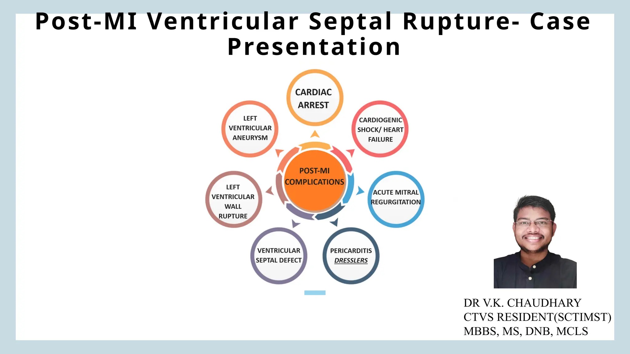
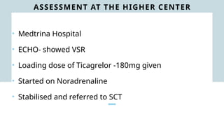
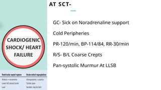
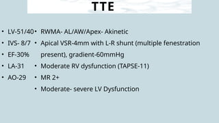
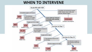
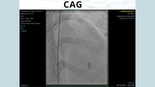
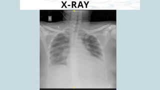
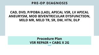
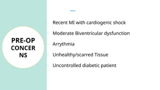
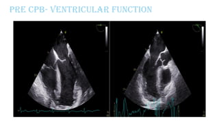
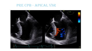
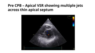
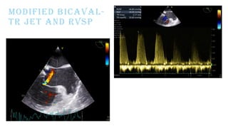
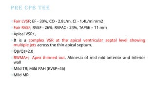
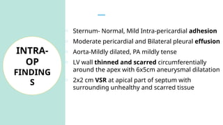
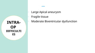
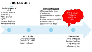
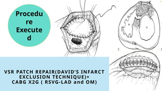
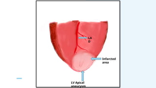
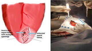
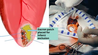
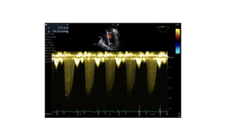
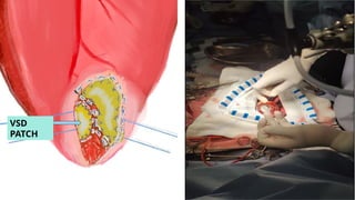
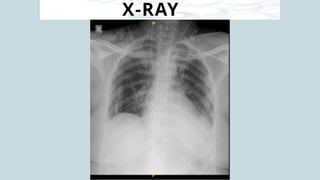
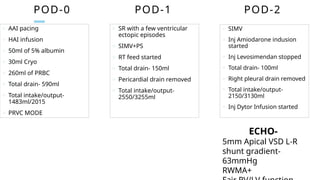
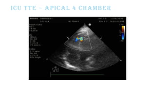
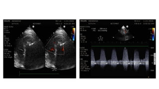
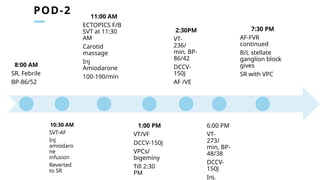
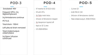
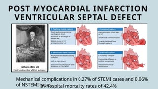
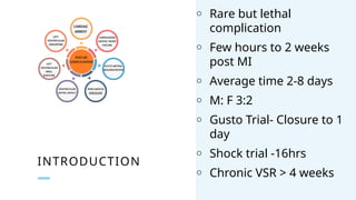
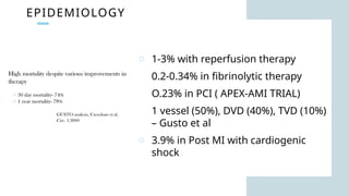
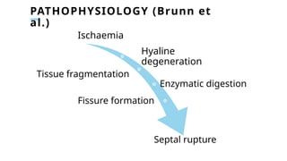
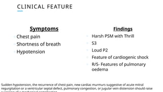
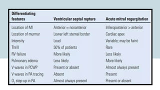
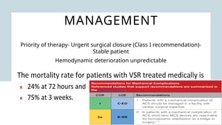
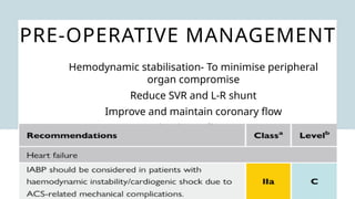
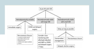
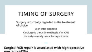
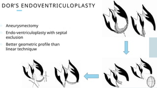
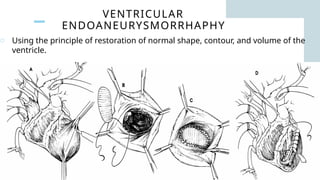
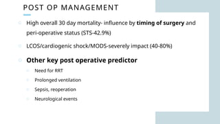
Post-MI Ventricular Septal Rupture- Case
Presentation
DR V.K. CHAUDHARY
CTVS RESIDENT(SCTIMST)
MBBS, MS, DNB, MCLS
DEMOGRAPHY
o 42 Y/F
o DM X 6months
o DLP X 6months
o No Previous History
Of ACS
o No Other
Comorbidities
HOPI
• Chest pain since 2:00 PM 3/1/2026
• Nearby hospital- ECG done- symptomatic management
• Returned to home
• Persistent chest pain with extreme fatiguability and restlessness- Royal
Health Centre
• ECG- STE in V1-V5, Trop I-18.32
• Loading dose of Aspirin/Clopilet/Atorvas given and referred
Asymptoma
tic
Typical/atypical chest
pain
Pulmonary
oedema/hypotension/shock
Sudden cardiac
death
ASSESSMENT AT THE HIGHER CENTER
• Medtrina Hospital
• ECHO- showed VSR
• Loading dose of Ticagrelor -180mg given
• Started on Noradrenaline
• Stabilised and referred to SCT
AT SCT-
o GC- Sick on Noradrenaline support
o Cold Peripheries
o PR-120/min, BP-114/84, RR-30/min
o R/S- B/L Coarse Crepts
o Pan-systolic Murmur At LLSB
ECG
120/Min, STE- V2-V5 And AvL,
X-RAY
TTE
TTE
• LV-51/40
• IVS- 8/7
• EF-30%
• LA-31
• AO-29
• RWMA- AL/AW/Apex- Akinetic
• Apical VSR-4mm with L-R shunt (multiple fenestration
present), gradient-60mmHg
• Moderate RV dysfunction (TAPSE-11)
• MR 2+
• Moderate- severe LV Dysfunction
WHEN TO INTERVENE
CAG
X-RAY
PRE-OP DIAGNOSIS
CAD, DVD, P/POBA (LAD), APICAL VSR, LV APICAL
ANEURYSM, MOD BIVENTRICULAR DYSFUNCTION,
MILD MR, MILD TR, SR, DM, HTN, DLP
Procedure Plan
VSR REPAIR + CABG X 2G
o Recent MI with cardiogenic shock
o Moderate Biventricular dysfunction
o Arrythmia
o Unhealthy/scarred Tissue
o Uncontrolled diabetic patient
PRE-OP
CONCER
NS
Pre CPB- Ventricular function
Pre CPB - Apical VSR
Pre CPB – Apical VSR showing multiple jets
across thin apical septum
MODIFIED BICAVAL-
TR JET AND RVSP
PRE CPB TEE
oFair LVSF; EF - 30%, CO - 2.8L/m, CI - 1.4L/min/m2
oFair RVSF; RVEF - 26%, RVFAC - 24%, TAPSE – 11 mm
oApical VSR+,
oIt is a complex VSR at the apical ventricular septal level showing
multiple jets across the thin apical septum.
oQp/Qs=2.0
oRWMA+; Apex thinned out. Akinesia of mid mid-anterior and inferior
wall
oMild TR; Mild PAH (RVSP=46)
oMild MR
CPB DATA AND MYOCARDIAL PROTECTION
o Core cooling-28 degree Celsius
o 5- Blood cardioplegia
o Aortic root and RUPV vent
o CPB Time-193mins
o ACC time-127mins
o CUFF- 1100ml
o Net fluid balance- +265ml
o Core cooling- 30 degrees
celsius
o 2-blood cardioplegia
o Aortic root vent
o CPB time-99mins
o ACC time-74mins
o Net fluid balance- +265ml
CPB-I CPB-II
Roller pump, Membrane oxygenator, Crystalloid prime,
Protamine-heparin chemistry
Aortic
cannula
24Fr
Edwards
SVC 20Fr DLP
IVC 32Fr RMI
Aortic
cannula
24Fr
Edwards
RA 32/40Fr
o Sternum- Normal, Mild Intra-pericardial adhesion
o Moderate pericardial and Bilateral pleural effusion
o Aorta-Mildly dilated, PA mildly tense
o LV wall thinned and scarred circumferentially
around the apex with 6x5cm aneurysmal dilatation
o 2x2 cm VSR at apical part of septum with
surrounding unhealthy and scarred tissue
INTRA-
OP
FINDING
S
o Large Apical aneurysm
o Fragile tissue
o Moderate Biventricular dysfunction
INTRA-
OP
DIFFICULTI
ES
PROCEDURE
Establishment of
CPB
Standard midline
sternotomy
Aorto-Bicaval
bypass
Root cardioplegia
Arrest in diastole
1st Procedure
Distal anastomosis
of RSVG-LAD & OM
Infarct exclusion
Coming off bypass
ACC removed after deair
VOO@84/min
Inj Livosidemendan, Inj vaso,
Inj Adre
Proximal anastomosis of RSVG
Protamine reversal
Decannulation in stages
2nd
Procedure
Aorto-RA bypass
Root cardioplegia
VSR patch-pasty
Infarct exclusion
TEE- Residual
VSD with
significant
shunt
VSR PATCH REPAIR(DAVID’S INFARCT
EXCLUSION TECHNIQUE)+
CABG X2G ( RSVG-LAD and OM)
Procedu
re
Execute
d
LA
D
LV Apical
aneurysm
Infarcted
area
Longitudinal
ventriculoto
my
Infarcted
septal
tissue
Ventricular
septal defect
with multiple
openings
Dacron patch
placed for
infarct
exclusion
POST CPB 1
Post CPB 1
Post CPB 1
RA AND PA SPO2
o Going on CPB - U/E,
o CPB 1- CABG * 2G, VSR patch
o Coming off CPB1 - NSR with supports Levosimendan@0.075, Adr@0.05 & Vaso @0.0003
o Post CPB 1 TEE- initially showed a color jet within the RV in the transgastric images which were
not traced to LV in the midesophageal images. Continued imaging revealed this colour jet to be
getting prominent.
o PA sO2 - 71%, RA sO2 - 67%
o Went on CPB-2 @ protamine 75%
o For CPB 2- Inj.Heparin 5mg/kg repeated.
CPB DATA AND MYOCARDIAL PROTECTION
o Core cooling-28 degree Celsius
o 5- Blood cardioplegia
o Aortic root and RUPV vent
o CPB Time-193mins
o ACC time-127mins
o CUFF- 1100ml
o Net fluid balance- +265ml
o Core cooling- 30 degrees
celsius
o 2-blood cardioplegia
o Aortic root vent
o CPB time-99mins
o ACC time-74mins
o Net fluid balance- +265ml
CPB-I CPB-II
Roller pump, Membrane oxygenator, Crystalloid prime,
Protamine-heparin chemistry
Aortic
cannula
24Fr
Edwards
SVC 20Fr DLP
IVC 32Fr RMI
Aortic
cannula
24Fr
Edwards
RA 32/40Fr
VSD
PATCH
POST CPB 2
Post CPB 2
Post CPB 2
POST CPB TEE
oP/VSR+LV apical exclusion+CABG
oFair LVSF, EF=40%, CO=4.7 L/min, CI-2.4 L/min/m2
oContractility improved with inotropes
oMild MR/TR
POSTop in icu
X-RAY
POD-0
o AAI pacing
o HAI infusion
o 50ml of 5% albumin
o 30ml Cryo
o 260ml of PRBC
o Total drain- 590ml
o Total intake/output-
1483ml/2015
o PRVC MODE
o SR with a few ventricular
ectopic episodes
o SIMV+PS
o RT feed started
o Total drain- 150ml
o Pericardial drain removed
o Total intake/output-
2550/3255ml
POD-1
o SIMV
o Inj Amiodarone indusion
started
o Inj Levosimendan stopped
o Total drain- 100ml
o Right pleural drain removed
o Total intake/output-
2150/3130ml
o Inj Dytor Infusion started
POD-2
ECHO-
5mm Apical VSD L-R
shunt gradient-
63mmHg
RWMA+
ICU TTE – APICAL 4 CHAMBER
POD-2
8:00 AM
SR, Febrile
BP-86/52
10:30 AM
SVT-AF
Inj
amiodaro
ne
infusion
Reverted
to SR
11:00 AM
ECTOPICS F/B
SVT at 11:30
AM
Carotid
massage
Inj
Amiodarone
100-190/min
1:00 PM
VT/VF
DCCV-150J
VPCs/
bigeminy
Till 2:30
PM
2:30PM
VT-
236/
min, BP-
86/42
DCCV-
150J
AF /VE
6:00 PM
VT-
273/
min, BP-
48/38
DCCV-
150J
Inj.
7:30 PM
AF-FVR
continued
B/L stellate
ganglion block
gives
SR with VPC
POD-3
o Extubated- NIV
o Frequent VPCs- Inj
MgSo4/Xylocard
o Inj Amiodarone continue
o PCT-6.6
o Total drain- 100ml
o Left pleural drain removed
o Total intake/output-
2350/5350ml
o Inj Dytor Infusion
continued
POST MYOCARDIAL INFARCTION
VENTRICULAR SEPTAL DEFECT
Mechanical complications in 0.27% of STEMI cases and 0.06%
of NSTEMI cases
In-hospital mortality rates of 42.4%
INTRODUCTION
o Rare but lethal
complication
o Few hours to 2 weeks
post MI
o Average time 2-8 days
o M: F 3:2
o Gusto Trial- Closure to 1
day
o Shock trial -16hrs
o Chronic VSR > 4 weeks
EPIDEMIOLOGY
o 1-3% with reperfusion therapy
o 0.2-0.34% in fibrinolytic therapy
o O.23% in PCI ( APEX-AMI TRIAL)
o 1 vessel (50%), DVD (40%), TVD (10%)
– Gusto et al
o 3.9% in Post MI with cardiogenic
shock
PATHOPHYSIOLOGY (Brunn et
al.)
Ischaemia
Hyaline
degeneration
Tissue fragmentation
Enzymatic digestion
Fissure formation
Septal rupture
CLINICAL FEATURE
Symptoms
o Chest pain
o Shortness of breath
o Hypotension
Findings
o Harsh PSM with Thrill
o S3
o Loud P2
o Feature of cardiogenic shock
o R/S- Features of pulmonary
oedema
Sudden hypotension, the recurrence of chest pain, new cardiac murmurs suggestive of acute mitral
regurgitation or a ventricular septal defect, pulmonary congestion, or jugular vein distension should raise
ECG
No specific ECG features are diagnostic of post-MI VSR
May reveal AV block in 1/3rd
of patients.
Persistent ST elevation – Associated with
ventricular aneurysm
ECHO
• RWMA
• VSR- Location, Size,
• Basal- PLAX
• Apical- A4CV
• L-R shunt magnitude
• Ventricular function
• Doppler- 100% Sensitive
and specific in
differentiating VSR from
CAG
• Class I Recom.- if patient is stable before definitive
treatment of mechanical complication
• LOE- B
• Coronary intervention
LOCATION
o AWMI (LAD)- Apical VSR
o IWNI (RCA/LCx)- Basal
septum
o Basal septum VSR is worse
than that of AWMI apical VSR
o Simple- Through and through
defect, usually located anteriorly
o Complex- Serpiginous
dissection, remote form primary
septal defect, most commonly in
inferior VSR
ANATOMY
SIMPLE VS COMPLEX VSR
BECKERS AND MONTEGEM CLASSIFICATION
1975
Type-I
o Abrupt, slit-like
tear
o Associated with an
acute infarct <24h
Type-II
o Erosion of the
infarcted
myocardium
o Clinically correlated
with Subacute
presentation
Type-III
o Concomitant aneurysm
formation
o Significant thinning of
the septum
o Clinically correlated
with chronic
presentation
IMPORTANT TRIAL
o Median time from symptoms to
VSD diagnosis- 1 day
o High Risk factor- Age, Female sex
o 30-day Mortality- 74%
o 1 year mortality- 78%
CONCLUSION-
o Thrombolysis with 6 hrs reduce
risk of VSR
o VSR occur soon in thrombolytic era
IMPORTANT TRIAL
CONCLUSION-
o Earlier presentation of Post-MI VSR when thrombolytic therapy is
used
o VSRs occur soon in the thrombolytic era
IMPORTANT TRIAL
o Frequency of VSR 0.17%
o 90-day survival rate- 20% in VSR
o Factor associated with
Mechanical complication- Old
age, female, Q wave, X-ray-
pulmonary oedema,
CONCLUSION-
Rates of mechanical complication
are lower in P-PCI than those after
fibrinolytic therapy
MANAGEMENT
Priority of therapy- Urgent surgical closure (Class I recommendation)-
Stable patient
Hemodynamic deterioration unpredictable
PRE-OPERATIVE MANAGEMENT
Hemodynamic stabilisation- To minimise peripheral
organ compromise
Reduce SVR and L-R shunt
Improve and maintain coronary flow
Maintain cardiac out
TIMING OF SURGERY
Soon after diagnosis
Cardiogenic shock- Immediately after CAG
Hemodynamically unstable- Urgent basis
Surgery is currently regarded as the treatment
of choice
Surgical VSR repair is associated with high operative
DAGGET’S PATCH REPAIR
o Closes VSR by placing a patch over
the defect and suturing it to RV/LV
DAVID’S TECHNIQUE
o Infarct exclusion technique
o All sutures placed in the left
ventricle.
SURGICAL MANAGEMENT OF LV ANEURYSM
1944 Beck Fascia lata reinforcement
1955 Likoff-
Bailey
1st dosed resection
1958 Cooley 1st open resection
1973 Stoney In coat" plicature
1977 Dagget Posterior patch
1979 Levitsky Antterior patch
1980 Hutchkins Influence of cardiac geometry
1984 Jatene Circular reduction
Trend towards LV Reconstruction
DOR’S ENDOVENTRICULOPLASTY
o Aneurysmectomy
o Endo-ventriculoplasty with septal
exclusion
o Better geometric profile than
linear techniquw
VENTRICULAR
ENDOANEURYSMORRHAPHY
o Using the principle of restoration of normal shape, contour, and volume of the
ventricle.
POST OP MANAGEMENT
Early
complicat
ion
Bleeding
7-11%
Close monitoring
Blood Transfusion
Re-exploration (7-
11%)
Residual
VSR/shunt
23-43%
Can worsen HF
Persistent shock
Difficulty to wean
from
CPB/Ventilator
Low cardiac
output
syndrome
Due Extensive
myocardial
necrosis
Poor ventricular
function
May require IABP/
ECMO
Arrythmias
20-30% ( First 48
hrs)
VT/VF- Most
common 15-20%
Originating form
incision site
Due to reperfusion
injury and scar
tissue
AF-20%- Due to
right heart strain
POST OP MANAGEMENT
complicati
on
AKI
Upto-16%
Requiring dialysis
More common in
CKD
SEPSIS
Upto- 18%
Due to prolonged
ventilation/mecha
nican support
Stroke
Stroke or
neurological
events- 16 %
Respiratory
failure
Due to prolonged
ventilation
POST OP MANAGEMENT
o High overall 30 day mortality- influence by timing of surgery and
peri-operative status (STS-42.9%)
o LCOS/cardiogenic shock/MODS-severely impact (40-80%)
o Other key post operative predictor
o Need for RRT
o Prolonged ventilation
o Sepsis, reoperation
o Neurological events
THANK YOU

Post-MI Ventricular Septal Rupture- Case Presentation.pptx

  • 1.
    Post-MI Ventricular SeptalRupture- Case Presentation DR V.K. CHAUDHARY CTVS RESIDENT(SCTIMST) MBBS, MS, DNB, MCLS
  • 2.
    DEMOGRAPHY o 42 Y/F oDM X 6months o DLP X 6months o No Previous History Of ACS o No Other Comorbidities
  • 3.
    HOPI • Chest painsince 2:00 PM 3/1/2026 • Nearby hospital- ECG done- symptomatic management • Returned to home • Persistent chest pain with extreme fatiguability and restlessness- Royal Health Centre • ECG- STE in V1-V5, Trop I-18.32 • Loading dose of Aspirin/Clopilet/Atorvas given and referred Asymptoma tic Typical/atypical chest pain Pulmonary oedema/hypotension/shock Sudden cardiac death
  • 5.
    ASSESSMENT AT THEHIGHER CENTER • Medtrina Hospital • ECHO- showed VSR • Loading dose of Ticagrelor -180mg given • Started on Noradrenaline • Stabilised and referred to SCT
  • 7.
    AT SCT- o GC-Sick on Noradrenaline support o Cold Peripheries o PR-120/min, BP-114/84, RR-30/min o R/S- B/L Coarse Crepts o Pan-systolic Murmur At LLSB
  • 8.
  • 9.
  • 10.
  • 11.
    TTE • LV-51/40 • IVS-8/7 • EF-30% • LA-31 • AO-29 • RWMA- AL/AW/Apex- Akinetic • Apical VSR-4mm with L-R shunt (multiple fenestration present), gradient-60mmHg • Moderate RV dysfunction (TAPSE-11) • MR 2+ • Moderate- severe LV Dysfunction
  • 12.
  • 13.
  • 14.
  • 15.
    PRE-OP DIAGNOSIS CAD, DVD,P/POBA (LAD), APICAL VSR, LV APICAL ANEURYSM, MOD BIVENTRICULAR DYSFUNCTION, MILD MR, MILD TR, SR, DM, HTN, DLP Procedure Plan VSR REPAIR + CABG X 2G
  • 16.
    o Recent MIwith cardiogenic shock o Moderate Biventricular dysfunction o Arrythmia o Unhealthy/scarred Tissue o Uncontrolled diabetic patient PRE-OP CONCER NS
  • 17.
  • 18.
    Pre CPB -Apical VSR
  • 19.
    Pre CPB –Apical VSR showing multiple jets across thin apical septum
  • 20.
  • 21.
    PRE CPB TEE oFairLVSF; EF - 30%, CO - 2.8L/m, CI - 1.4L/min/m2 oFair RVSF; RVEF - 26%, RVFAC - 24%, TAPSE – 11 mm oApical VSR+, oIt is a complex VSR at the apical ventricular septal level showing multiple jets across the thin apical septum. oQp/Qs=2.0 oRWMA+; Apex thinned out. Akinesia of mid mid-anterior and inferior wall oMild TR; Mild PAH (RVSP=46) oMild MR
  • 22.
    CPB DATA ANDMYOCARDIAL PROTECTION o Core cooling-28 degree Celsius o 5- Blood cardioplegia o Aortic root and RUPV vent o CPB Time-193mins o ACC time-127mins o CUFF- 1100ml o Net fluid balance- +265ml o Core cooling- 30 degrees celsius o 2-blood cardioplegia o Aortic root vent o CPB time-99mins o ACC time-74mins o Net fluid balance- +265ml CPB-I CPB-II Roller pump, Membrane oxygenator, Crystalloid prime, Protamine-heparin chemistry Aortic cannula 24Fr Edwards SVC 20Fr DLP IVC 32Fr RMI Aortic cannula 24Fr Edwards RA 32/40Fr
  • 23.
    o Sternum- Normal,Mild Intra-pericardial adhesion o Moderate pericardial and Bilateral pleural effusion o Aorta-Mildly dilated, PA mildly tense o LV wall thinned and scarred circumferentially around the apex with 6x5cm aneurysmal dilatation o 2x2 cm VSR at apical part of septum with surrounding unhealthy and scarred tissue INTRA- OP FINDING S
  • 24.
    o Large Apicalaneurysm o Fragile tissue o Moderate Biventricular dysfunction INTRA- OP DIFFICULTI ES
  • 25.
    PROCEDURE Establishment of CPB Standard midline sternotomy Aorto-Bicaval bypass Rootcardioplegia Arrest in diastole 1st Procedure Distal anastomosis of RSVG-LAD & OM Infarct exclusion Coming off bypass ACC removed after deair VOO@84/min Inj Livosidemendan, Inj vaso, Inj Adre Proximal anastomosis of RSVG Protamine reversal Decannulation in stages 2nd Procedure Aorto-RA bypass Root cardioplegia VSR patch-pasty Infarct exclusion TEE- Residual VSD with significant shunt
  • 26.
    VSR PATCH REPAIR(DAVID’SINFARCT EXCLUSION TECHNIQUE)+ CABG X2G ( RSVG-LAD and OM) Procedu re Execute d
  • 27.
  • 28.
  • 29.
  • 30.
  • 31.
  • 32.
  • 33.
  • 36.
  • 37.
    o Going onCPB - U/E, o CPB 1- CABG * 2G, VSR patch o Coming off CPB1 - NSR with supports Levosimendan@0.075, Adr@0.05 & Vaso @0.0003 o Post CPB 1 TEE- initially showed a color jet within the RV in the transgastric images which were not traced to LV in the midesophageal images. Continued imaging revealed this colour jet to be getting prominent. o PA sO2 - 71%, RA sO2 - 67% o Went on CPB-2 @ protamine 75% o For CPB 2- Inj.Heparin 5mg/kg repeated.
  • 38.
    CPB DATA ANDMYOCARDIAL PROTECTION o Core cooling-28 degree Celsius o 5- Blood cardioplegia o Aortic root and RUPV vent o CPB Time-193mins o ACC time-127mins o CUFF- 1100ml o Net fluid balance- +265ml o Core cooling- 30 degrees celsius o 2-blood cardioplegia o Aortic root vent o CPB time-99mins o ACC time-74mins o Net fluid balance- +265ml CPB-I CPB-II Roller pump, Membrane oxygenator, Crystalloid prime, Protamine-heparin chemistry Aortic cannula 24Fr Edwards SVC 20Fr DLP IVC 32Fr RMI Aortic cannula 24Fr Edwards RA 32/40Fr
  • 39.
  • 40.
  • 41.
  • 42.
  • 43.
    POST CPB TEE oP/VSR+LVapical exclusion+CABG oFair LVSF, EF=40%, CO=4.7 L/min, CI-2.4 L/min/m2 oContractility improved with inotropes oMild MR/TR
  • 44.
  • 45.
  • 46.
    POD-0 o AAI pacing oHAI infusion o 50ml of 5% albumin o 30ml Cryo o 260ml of PRBC o Total drain- 590ml o Total intake/output- 1483ml/2015 o PRVC MODE o SR with a few ventricular ectopic episodes o SIMV+PS o RT feed started o Total drain- 150ml o Pericardial drain removed o Total intake/output- 2550/3255ml POD-1 o SIMV o Inj Amiodarone indusion started o Inj Levosimendan stopped o Total drain- 100ml o Right pleural drain removed o Total intake/output- 2150/3130ml o Inj Dytor Infusion started POD-2 ECHO- 5mm Apical VSD L-R shunt gradient- 63mmHg RWMA+
  • 47.
    ICU TTE –APICAL 4 CHAMBER
  • 49.
    POD-2 8:00 AM SR, Febrile BP-86/52 10:30AM SVT-AF Inj amiodaro ne infusion Reverted to SR 11:00 AM ECTOPICS F/B SVT at 11:30 AM Carotid massage Inj Amiodarone 100-190/min 1:00 PM VT/VF DCCV-150J VPCs/ bigeminy Till 2:30 PM 2:30PM VT- 236/ min, BP- 86/42 DCCV- 150J AF /VE 6:00 PM VT- 273/ min, BP- 48/38 DCCV- 150J Inj. 7:30 PM AF-FVR continued B/L stellate ganglion block gives SR with VPC
  • 50.
    POD-3 o Extubated- NIV oFrequent VPCs- Inj MgSo4/Xylocard o Inj Amiodarone continue o PCT-6.6 o Total drain- 100ml o Left pleural drain removed o Total intake/output- 2350/5350ml o Inj Dytor Infusion continued
  • 51.
    POST MYOCARDIAL INFARCTION VENTRICULARSEPTAL DEFECT Mechanical complications in 0.27% of STEMI cases and 0.06% of NSTEMI cases In-hospital mortality rates of 42.4%
  • 52.
    INTRODUCTION o Rare butlethal complication o Few hours to 2 weeks post MI o Average time 2-8 days o M: F 3:2 o Gusto Trial- Closure to 1 day o Shock trial -16hrs o Chronic VSR > 4 weeks
  • 53.
    EPIDEMIOLOGY o 1-3% withreperfusion therapy o 0.2-0.34% in fibrinolytic therapy o O.23% in PCI ( APEX-AMI TRIAL) o 1 vessel (50%), DVD (40%), TVD (10%) – Gusto et al o 3.9% in Post MI with cardiogenic shock
  • 54.
    PATHOPHYSIOLOGY (Brunn et al.) Ischaemia Hyaline degeneration Tissuefragmentation Enzymatic digestion Fissure formation Septal rupture
  • 55.
    CLINICAL FEATURE Symptoms o Chestpain o Shortness of breath o Hypotension Findings o Harsh PSM with Thrill o S3 o Loud P2 o Feature of cardiogenic shock o R/S- Features of pulmonary oedema Sudden hypotension, the recurrence of chest pain, new cardiac murmurs suggestive of acute mitral regurgitation or a ventricular septal defect, pulmonary congestion, or jugular vein distension should raise
  • 57.
    ECG No specific ECGfeatures are diagnostic of post-MI VSR May reveal AV block in 1/3rd of patients. Persistent ST elevation – Associated with ventricular aneurysm
  • 58.
    ECHO • RWMA • VSR-Location, Size, • Basal- PLAX • Apical- A4CV • L-R shunt magnitude • Ventricular function • Doppler- 100% Sensitive and specific in differentiating VSR from
  • 59.
    CAG • Class IRecom.- if patient is stable before definitive treatment of mechanical complication • LOE- B • Coronary intervention
  • 60.
    LOCATION o AWMI (LAD)-Apical VSR o IWNI (RCA/LCx)- Basal septum o Basal septum VSR is worse than that of AWMI apical VSR o Simple- Through and through defect, usually located anteriorly o Complex- Serpiginous dissection, remote form primary septal defect, most commonly in inferior VSR ANATOMY
  • 61.
  • 62.
    BECKERS AND MONTEGEMCLASSIFICATION 1975 Type-I o Abrupt, slit-like tear o Associated with an acute infarct <24h Type-II o Erosion of the infarcted myocardium o Clinically correlated with Subacute presentation Type-III o Concomitant aneurysm formation o Significant thinning of the septum o Clinically correlated with chronic presentation
  • 64.
    IMPORTANT TRIAL o Mediantime from symptoms to VSD diagnosis- 1 day o High Risk factor- Age, Female sex o 30-day Mortality- 74% o 1 year mortality- 78% CONCLUSION- o Thrombolysis with 6 hrs reduce risk of VSR o VSR occur soon in thrombolytic era
  • 65.
    IMPORTANT TRIAL CONCLUSION- o Earlierpresentation of Post-MI VSR when thrombolytic therapy is used o VSRs occur soon in the thrombolytic era
  • 66.
    IMPORTANT TRIAL o Frequencyof VSR 0.17% o 90-day survival rate- 20% in VSR o Factor associated with Mechanical complication- Old age, female, Q wave, X-ray- pulmonary oedema, CONCLUSION- Rates of mechanical complication are lower in P-PCI than those after fibrinolytic therapy
  • 67.
    MANAGEMENT Priority of therapy-Urgent surgical closure (Class I recommendation)- Stable patient Hemodynamic deterioration unpredictable
  • 68.
    PRE-OPERATIVE MANAGEMENT Hemodynamic stabilisation-To minimise peripheral organ compromise Reduce SVR and L-R shunt Improve and maintain coronary flow Maintain cardiac out
  • 70.
    TIMING OF SURGERY Soonafter diagnosis Cardiogenic shock- Immediately after CAG Hemodynamically unstable- Urgent basis Surgery is currently regarded as the treatment of choice Surgical VSR repair is associated with high operative
  • 71.
    DAGGET’S PATCH REPAIR oCloses VSR by placing a patch over the defect and suturing it to RV/LV
  • 72.
    DAVID’S TECHNIQUE o Infarctexclusion technique o All sutures placed in the left ventricle.
  • 73.
    SURGICAL MANAGEMENT OFLV ANEURYSM 1944 Beck Fascia lata reinforcement 1955 Likoff- Bailey 1st dosed resection 1958 Cooley 1st open resection 1973 Stoney In coat" plicature 1977 Dagget Posterior patch 1979 Levitsky Antterior patch 1980 Hutchkins Influence of cardiac geometry 1984 Jatene Circular reduction Trend towards LV Reconstruction
  • 74.
    DOR’S ENDOVENTRICULOPLASTY o Aneurysmectomy oEndo-ventriculoplasty with septal exclusion o Better geometric profile than linear techniquw
  • 75.
    VENTRICULAR ENDOANEURYSMORRHAPHY o Using theprinciple of restoration of normal shape, contour, and volume of the ventricle.
  • 76.
    POST OP MANAGEMENT Early complicat ion Bleeding 7-11% Closemonitoring Blood Transfusion Re-exploration (7- 11%) Residual VSR/shunt 23-43% Can worsen HF Persistent shock Difficulty to wean from CPB/Ventilator Low cardiac output syndrome Due Extensive myocardial necrosis Poor ventricular function May require IABP/ ECMO Arrythmias 20-30% ( First 48 hrs) VT/VF- Most common 15-20% Originating form incision site Due to reperfusion injury and scar tissue AF-20%- Due to right heart strain
  • 77.
    POST OP MANAGEMENT complicati on AKI Upto-16% Requiringdialysis More common in CKD SEPSIS Upto- 18% Due to prolonged ventilation/mecha nican support Stroke Stroke or neurological events- 16 % Respiratory failure Due to prolonged ventilation
  • 78.
    POST OP MANAGEMENT oHigh overall 30 day mortality- influence by timing of surgery and peri-operative status (STS-42.9%) o LCOS/cardiogenic shock/MODS-severely impact (40-80%) o Other key post operative predictor o Need for RRT o Prolonged ventilation o Sepsis, reoperation o Neurological events
  • 80.

Editor's Notes

  • #22 It is a complex VSR at the apcial ventricular level showing multiple jets across the thin apical septum
  • #23 VSR at the apicial ventricular septal level showing multiple jets across the thin apical septum
  • #36 The jet was soon seen in the midesophageal views as well
  • #37 Confirmed in a biplane view
  • #38 The VSD was confirmed to arise from the septum towards the upper part of the patch