Post MI Ventricular Septal
Defects
Dr. Asma Iqbal
PGR
Services
Hospital,Lahore
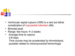
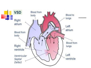
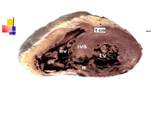
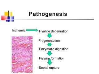
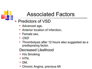
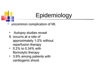
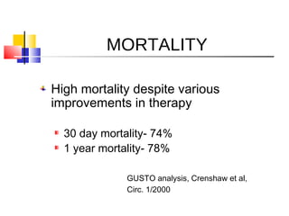
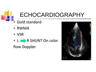
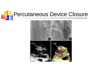
• Ventricular septal rupture (VSR) is a rare but lethal
complication of myocardial infarction (MI).
• Bimodal peak
• Range: few hours  2 weeks
• Average time to rupture
2-8 days
Time course may be accelerated by thrombolysis,
possible related to intramyocardial hemorrhage
PATHOPHYSIOLOGY
Ischemia Hyaline degenration
Fragmentation
Enzymatic digestion
Fissure formation
Septal rupture
Pathogenesis
Associated Factors
Predictors of VSD
Advanced age,
Anterior location of infarction,
Female sex,
CKD
Thrombolysis after 12 hours also suggested as a
predisposing factor.
Decreased Likelihood
 H/o Smoking
 HTN,
 DM,
 Chronic Angina, previous MI
EPIDEMIOLOGY
Epidemiology
• uncommon complication of MI. 
•  Autopsy studies reveal
It  occurrs at a rate of
approximately 1-2% without
reperfusion therapy
• 0.2% to 0.34% with
fibrinolytic therapy
• 3.9% among patients with
cardiogenic shock
MORTALITY
High mortality despite various
improvements in therapy
30 day mortality- 74%
1 year mortality- 78%
GUSTO analysis, Crenshaw et al,
Circ. 1/2000
Relative Improvement in survival due to
Earlier diagnosis
Earlier flow restoration
More aggressive surgical intervention
More aggressive BP control post MI
ANATOMICAL SITES AND
CLASSIFICATION
Septal blood Supply
The septal blood supply comes from branches of
•the left anterior descending coronary artery,
•the posterior descending branch of the right
coronary artery, or the circumflex artery when it is
dominant
There are three types of VSR (the original
classification made by Becker and van Mantgem
was for free-wall rupture):
Type I: there is an abrupt tear in the wall without
thinning,Associated with Acute infarcts
Type II, the infarcted myocardium erodes before
rupture and is covered by a thrombus;
Type III represents the perforation of a previously
formed aneurysm,process associated with older
infarcts
BECKER AND MANTGEM
CLASSIFICATION
Anatomical Types of VSR
Two types of VSD (According to site )
Simple: through and through defect usually
located anteriorly, About 60% of VSRs occur
with infarction of the anterior wall,LAD is
Culprit
Complex: serpentiginous dissection tract
remote from the primary septal defect-
most commonly an inferior VSD, 40% with
infarction of the posterior or inferior wall
DIAGNOSIS
History
Chest Pain
SOB
Hypotension
Clinical Findings
Loud S2
S3
Pulmonary edema
RV/LV failure
Loud/harsh pansystolic murmur
Within the first week post AMI
Best heard at Lt. Lower sternal border
Less loud at the apex
Associated with a palpable thrill
Depending on the location, may radiate to the axilla
mimicking MR
Diagnosis
Up to 50% of patients experience chest
pain associated with the development
of murmur
CHF and shock often associated with
the development of murmur
INVESTIGATIONS
 IMAGING MODALITIES
 ECG
 Catheterization and Pressure Measurement
ECG
No electrocardiographic (ECG) features are
diagnostic of postinfarction VSR, though ECG
indeed provides some useful information.
Persistent ST-segment elevation associated with
ventricular aneurysm
may reveal atrioventricular block in one third of
patients.
anatomic location of the septal rupture.
RADIOGRAPHS
On plain chest radiography, 82% of patients
with postinfarction ventricular septal
rupture (VSR) demonstrate left ventricular
enlargement,
78% have pulmonary edema,
and 64% have a pleural effusion
ECHOCARDIOGRAPHY
Gold standard
RWMA
VSR
L R SHUNT On color
flow Doppler
Color Flow
Doppler
100% sensitive and
specific in
differentiating VSR
from acute MR
CATHETERIZATION AND PRESSURE
MEASUREMENT
Left-heart catheterization with coronary
angiography is recommended in all stable
patients
An important diagnostic test for differentiating
VSR from mitral valve insufficiency is
catheterization of the right heart with a Swan-
Ganz catheter.
Left- and right-side pressure measurements help
estimate the degree of biventricular failure
Need for cardiac catheterization
2/3 of the patients have multivessel
coronary artery disease
Decreased operative mortality and
improved late survival has been shown in
patients with multivessel disease
Cardiogenic shock not a deterrent to Cath
=> Coronary angiography
should be performed
TREATMENT MODALITIES
Medical Therapy
Percutaneous Device closure
Surgical Repair
Pre-Operative Management
Hemodynamic stabilization so as to
minimize peripheral organ compromise
Reduce Systemic vascular resistance, and
thus, the left-to-right shunt
Maintain or improve coronary artery blood
flow
Maintain cardiac output and arterial
pressure to ensure peripheral organ
perfusion
Supportive medical
management
Vasodilators
Vasopressors
The profound level of cardiogenic shock in
some patients precludes vasodilator
treatment, often necessitating vasopressor
support.
Intra-aortic balloon counterpulsation
(IABCP)
WHAT IS IABP
COP
L R SHUNT
Perfusion
Percutaneous Device Closure
Timing of Surgery
Controversial (in the past)
Non-randomized studies showing:
Early repair, 40% - 50% mortality
Late repair (past 3 weeks), 10% mortality
=>
Aggressive Medical management aimed
at delaying surgical intervention
Timing of Surgery
Surgery should be performed soon after
diagnosis in most patients
Patients is cardiogenic shock should be
operated on immediately after
anigography
Hemodynamically stable patients
should have surgery on an urgent basis
The relative safety of repair 2-3 weeks or
more after perforation has been
established. Because the edges of the
defect have become firmer and fibrotic,
repair is more secure and is easily
accomplished. A successful clinical outcome
is related to the adequacy of the closure of
the VSR;
GOALs OF SURGERY
Exclusion or Removal of Infarcted
Myocardium
Elimination of L to R shunt
Operative techniques
Resection of Infarcted Myocardium
Double Patch Repair of VSR
3-D Patch Technique
Operative Technique
Classical approach to
antero-septal rupture
Infarctectomy, and
Reconstruction of the
ventricular septum with
Dacron patches
Operative Technique
Classical approach
to infro-posterior
rupture
Infarctectomy, and
Reconstruction of
infroposterior VSD,
ReconstructionReconstruction free wall
with Dacron patches.
Outcome
Six month survival without
surgical intervention stated to be
less than 10%.
Kirklin, Churchill Livingston 1993
Outcome
In patients with cardiogenic shock mortality
reported to be the highest
Posterior VSD (IMI) is another factor strongly
associated with poor surgical outcome due to
Difficulty of exposure, and
Frequent concomitant infarction of the postero-
medial papillary muscle
THANK YOU

Ischemic ventricular septal_defects_dr.asma

  • 1.
    Post MI VentricularSeptal Defects Dr. Asma Iqbal PGR Services Hospital,Lahore
  • 2.
    • Ventricular septalrupture (VSR) is a rare but lethal complication of myocardial infarction (MI). • Bimodal peak • Range: few hours  2 weeks • Average time to rupture 2-8 days Time course may be accelerated by thrombolysis, possible related to intramyocardial hemorrhage
  • 5.
  • 6.
    Ischemia Hyaline degenration Fragmentation Enzymaticdigestion Fissure formation Septal rupture Pathogenesis
  • 7.
    Associated Factors Predictors ofVSD Advanced age, Anterior location of infarction, Female sex, CKD Thrombolysis after 12 hours also suggested as a predisposing factor. Decreased Likelihood  H/o Smoking  HTN,  DM,  Chronic Angina, previous MI
  • 8.
  • 9.
    Epidemiology • uncommon complicationof MI.  •  Autopsy studies reveal It  occurrs at a rate of approximately 1-2% without reperfusion therapy • 0.2% to 0.34% with fibrinolytic therapy • 3.9% among patients with cardiogenic shock
  • 10.
    MORTALITY High mortality despitevarious improvements in therapy 30 day mortality- 74% 1 year mortality- 78% GUSTO analysis, Crenshaw et al, Circ. 1/2000
  • 11.
    Relative Improvement insurvival due to Earlier diagnosis Earlier flow restoration More aggressive surgical intervention More aggressive BP control post MI
  • 12.
  • 13.
    Septal blood Supply Theseptal blood supply comes from branches of •the left anterior descending coronary artery, •the posterior descending branch of the right coronary artery, or the circumflex artery when it is dominant
  • 14.
    There are threetypes of VSR (the original classification made by Becker and van Mantgem was for free-wall rupture): Type I: there is an abrupt tear in the wall without thinning,Associated with Acute infarcts Type II, the infarcted myocardium erodes before rupture and is covered by a thrombus; Type III represents the perforation of a previously formed aneurysm,process associated with older infarcts BECKER AND MANTGEM CLASSIFICATION
  • 15.
    Anatomical Types ofVSR Two types of VSD (According to site ) Simple: through and through defect usually located anteriorly, About 60% of VSRs occur with infarction of the anterior wall,LAD is Culprit Complex: serpentiginous dissection tract remote from the primary septal defect- most commonly an inferior VSD, 40% with infarction of the posterior or inferior wall
  • 16.
  • 17.
  • 18.
    Clinical Findings Loud S2 S3 Pulmonaryedema RV/LV failure Loud/harsh pansystolic murmur Within the first week post AMI Best heard at Lt. Lower sternal border Less loud at the apex Associated with a palpable thrill Depending on the location, may radiate to the axilla mimicking MR
  • 19.
    Diagnosis Up to 50%of patients experience chest pain associated with the development of murmur CHF and shock often associated with the development of murmur
  • 20.
    INVESTIGATIONS  IMAGING MODALITIES ECG  Catheterization and Pressure Measurement
  • 21.
    ECG No electrocardiographic (ECG)features are diagnostic of postinfarction VSR, though ECG indeed provides some useful information. Persistent ST-segment elevation associated with ventricular aneurysm may reveal atrioventricular block in one third of patients. anatomic location of the septal rupture.
  • 22.
    RADIOGRAPHS On plain chestradiography, 82% of patients with postinfarction ventricular septal rupture (VSR) demonstrate left ventricular enlargement, 78% have pulmonary edema, and 64% have a pleural effusion
  • 23.
  • 24.
    Color Flow Doppler 100% sensitiveand specific in differentiating VSR from acute MR
  • 25.
    CATHETERIZATION AND PRESSURE MEASUREMENT Left-heartcatheterization with coronary angiography is recommended in all stable patients An important diagnostic test for differentiating VSR from mitral valve insufficiency is catheterization of the right heart with a Swan- Ganz catheter. Left- and right-side pressure measurements help estimate the degree of biventricular failure
  • 26.
    Need for cardiaccatheterization 2/3 of the patients have multivessel coronary artery disease Decreased operative mortality and improved late survival has been shown in patients with multivessel disease Cardiogenic shock not a deterrent to Cath => Coronary angiography should be performed
  • 27.
  • 28.
    Pre-Operative Management Hemodynamic stabilizationso as to minimize peripheral organ compromise Reduce Systemic vascular resistance, and thus, the left-to-right shunt Maintain or improve coronary artery blood flow Maintain cardiac output and arterial pressure to ensure peripheral organ perfusion
  • 29.
    Supportive medical management Vasodilators Vasopressors The profoundlevel of cardiogenic shock in some patients precludes vasodilator treatment, often necessitating vasopressor support. Intra-aortic balloon counterpulsation (IABCP)
  • 30.
    WHAT IS IABP COP LR SHUNT Perfusion
  • 32.
  • 33.
    Timing of Surgery Controversial(in the past) Non-randomized studies showing: Early repair, 40% - 50% mortality Late repair (past 3 weeks), 10% mortality => Aggressive Medical management aimed at delaying surgical intervention
  • 35.
    Timing of Surgery Surgeryshould be performed soon after diagnosis in most patients Patients is cardiogenic shock should be operated on immediately after anigography Hemodynamically stable patients should have surgery on an urgent basis
  • 36.
    The relative safetyof repair 2-3 weeks or more after perforation has been established. Because the edges of the defect have become firmer and fibrotic, repair is more secure and is easily accomplished. A successful clinical outcome is related to the adequacy of the closure of the VSR;
  • 37.
    GOALs OF SURGERY Exclusionor Removal of Infarcted Myocardium Elimination of L to R shunt
  • 38.
    Operative techniques Resection ofInfarcted Myocardium Double Patch Repair of VSR 3-D Patch Technique
  • 39.
    Operative Technique Classical approachto antero-septal rupture Infarctectomy, and Reconstruction of the ventricular septum with Dacron patches
  • 40.
    Operative Technique Classical approach toinfro-posterior rupture Infarctectomy, and Reconstruction of infroposterior VSD, ReconstructionReconstruction free wall with Dacron patches.
  • 41.
    Outcome Six month survivalwithout surgical intervention stated to be less than 10%. Kirklin, Churchill Livingston 1993
  • 42.
    Outcome In patients withcardiogenic shock mortality reported to be the highest Posterior VSD (IMI) is another factor strongly associated with poor surgical outcome due to Difficulty of exposure, and Frequent concomitant infarction of the postero- medial papillary muscle
  • 43.

Editor's Notes

  • #40 Post MI surgical pictures
  • #41 Scan in the pictures ant. MI