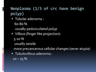
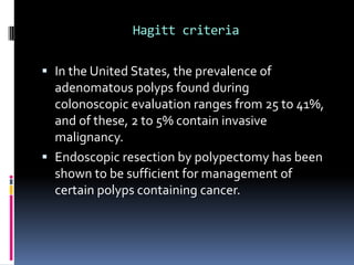
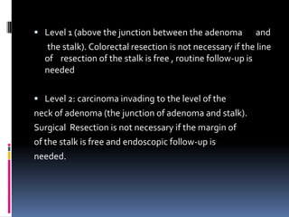
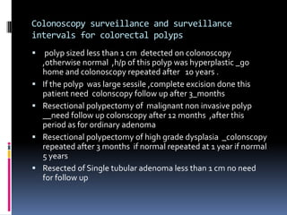
Colorectal                     polyp
definition,types,colonscopic survillence


        National cancer institute –misrata
        Surgery department
        Dr . Mohamed alhashmi sidoun
   Colorectal polyp is projecting mass into the
    lumen of bowel above the surface of epithelium.
   Polyp is word descriptive for the shape of lesion
    (not related to the pathological process)
   Polyposis :multiple polyps
   Polypoid
   Colorectal polyps are considerd slow growing
    tumour
   The term “malignant polyp” refers to a macroscopically
    benign appearing adenoma in which the invasive
    carcinoma is detected after histologic examination of the
    resected specimen.
Classification of polyps

 According to the shape
 According to the histology
According to the shape

1. Pedunculated ( with stalk)
2. Sessile (no stalk ,no neck )
According to the histopathology
 Polyps such as hyperplasic,juvenile and
inflammatory polyps have no malignantpotential,
although polyps do have malignant potentia

Type            Benign           Malignant

Epithelial      Neoplasma        Adenocarcinoma
                Inflammatory
                Hamartoma
                Metaplastic

mesenchymal     Lipoma           Sarcoma
                Fibroma          lymphoma
                Leiomyoma
                haemangioma
Neoplasma (1/3 of crc have benign
polyp)
 Tubular adenoma :
  60-80 %
  usually pedunculated polyp
 Villous (finger like projection):
 5-10 %
  usually sessile
 more precancerous cellular changes (sever atypia)
 Tubulovillous adenoma :
 10 – 25 %
The prognostic factors :
(polyps transformed to carcinoma)
The presence of poor prognostic features should
lead the physician to favor colectomy
1.   Polyp size more than 1 cm are found to harbor cancers more
     than the cells
     size less than 4 mm usually hyperplastic (no malignant
     changes but should be excised
     sessile polyp more than 2 cm usually villous with high
     malignant changes and high recurrence rate

2.Shape of the polyp : the invasion of adenocrcinoma in
    pedunculated polyp need travel during head,neck,stalk
    before reaching the submucosa (polypectomy not
    appropriate) contrast with sessile where just travel the
    basement membrane
3.Histopatholo of the polyp :
   the villous subdivision are associated with the highest
   malignant potential because they have the largest
   surface area ,no stalk and no neck.
  2 cm sized tubular adenoma _35%risk of malignancy
 2 cm sized villous adenoma _50 % risk
4.Multiple polyps more prognostic than single (familial
    adenomatous polyposis) 100% risky
5. Missed polypectomy or patient refuse even smaller
    one(polypectomy of small polyps decreases the cancer
    incidence up to 70%.
6.risk of age ,family history,smoking
    (the peak incidence of crc is 60 years of age ,the peak
    incidence of colorectal polyp discovering is 50 years of
    age ,this means 10 years time span for progression )
Hagitt criteria

 In the United States, the prevalence of
  adenomatous polyps found during
  colonoscopic evaluation ranges from 25 to
  41%, and of these, 2 to 5% contain invasive
  malignancy.
 Endoscopic resection by polypectomy has been
  shown to be sufficient for management of
  certain polyps containing cancer.
 Depth of invasion has been shown to correlate with the
  risk of lymph node metastasis. Other unfavorable
  histologic features include lymphovascular invasion, poor
  differentiation, inability to assess margin (piecemeal
  resection), and positive resection margin (<2 mm); these
  are important factors to consider in management. For
  these patients formal oncologic surgical resection is
  indicated.
 Polypectomy is usually performed during colonoscopy
  using snare polypectomy techniques. High-quality
  polypectomy is the complete excision of the polyp, which
  should include the entire stalk to its base. The
  submucosa of the bowel wall should be included to allow
  optimal histologic evaluation of the margins.
 Level 0 indicates carcinoma in situ or intramucosal
  carcinoma. These lesions are not invasive, and therefore
  behave as benign adenomas due to an absence of
  lymphatics in the mucosal laye
 Level 0 Carcinoma in situ or intramucosal carcinoma. Not
  invasive.
  Level 1 Carcinoma invading through muscularis mucosa
  into submucosa, but limited to the head of the polyp.
  Level 2 Carcinoma invading the level of the neck of the
  adenoma.
  Level 3 Carcinoma invading any part of the stalk .
  Level 4 Carcinoma invading into the submucosa of the
  bowel wall below the stalk of the polyp, but above the
  muscularis propria.
 Level 1 (above the junction between the adenoma          and
  the stalk). Colorectal resection is not necessary if the line
  of resection of the stalk is free , routine follow-up is
  needed

 Level 2: carcinoma invading to the level of the
neck of adenoma (the junction of adenoma and stalk).
Surgical Resection is not necessary if the margin of
of the stalk is free and endoscopic follow-up is
needed.
 Level 3: carcinoma invading any part of the stalk.
A free margin of resection precludes the necessarily
for any formal colorectal resection.
 Level 4: carcinoma invading the submucosa of
bowel wall below the stalk of the polyps but above the
 muscular propria. This is invasive cancer and a formal
bowel resection is necessary,

 By definition all sessile polyps with invasive carcinoma
  are level 4. Hence, resection of bowel wall is indicated.
Colonoscopy surveillance and surveillance
intervals for colorectal polyps
    polyp sized less than 1 cm detected on colonoscopy
    ,otherwise normal ,h/p of this polyp was hyperplastic _go
    home and colonoscopy repeated after 10 years .
   If the polyp was large sessile ,complete excision done this
    patient need colonscopy follow up after 3_months
   Resectional polypectomy of malignant non invasive polyp
    __need follow up colonscopy after 12 months ,after this
    period as for ordinary adenoma
   Resectional polypectomy of high grade dysplasia _colonscopy
    repeated after 3 months if normal repeated at 1 year if normal
    5 years
   Resected of Single tubular adenoma less than 1 cm no need
    for follow up
 Resection of adenoma +3 polyps +previous h/o
  other cancer __need colonscopy every 5 years
 For patient who have had surgical resection of
  colon cancer need follow up with colonscopy
  every 6 months for the initial 2 years
 Low risk (size <1cm,no.1-2 )repeated after 5
  years
 Intermediated risk (size >1cm ,no. 3-4 ) :after 3
  years
 High risk(>1cm ,no. more than 5 ) : after one year
 N.b the three risk degrees are exchangable .
References :
Current medical diagnosis and treatment 2008
Muir`s textbook of pathology
Nms surgery (fifth edition)
Website adress : clinics in colon and rectal
Surgery .

Colorectal polyp

  • 1.
    Colorectal polyp definition,types,colonscopic survillence National cancer institute –misrata Surgery department Dr . Mohamed alhashmi sidoun
  • 2.
    Colorectal polyp is projecting mass into the lumen of bowel above the surface of epithelium.  Polyp is word descriptive for the shape of lesion (not related to the pathological process)  Polyposis :multiple polyps  Polypoid  Colorectal polyps are considerd slow growing tumour  The term “malignant polyp” refers to a macroscopically benign appearing adenoma in which the invasive carcinoma is detected after histologic examination of the resected specimen.
  • 3.
    Classification of polyps According to the shape  According to the histology
  • 4.
    According to theshape 1. Pedunculated ( with stalk) 2. Sessile (no stalk ,no neck )
  • 5.
    According to thehistopathology Polyps such as hyperplasic,juvenile and inflammatory polyps have no malignantpotential, although polyps do have malignant potentia Type Benign Malignant Epithelial Neoplasma Adenocarcinoma Inflammatory Hamartoma Metaplastic mesenchymal Lipoma Sarcoma Fibroma lymphoma Leiomyoma haemangioma
  • 6.
    Neoplasma (1/3 ofcrc have benign polyp)  Tubular adenoma : 60-80 % usually pedunculated polyp  Villous (finger like projection): 5-10 % usually sessile more precancerous cellular changes (sever atypia)  Tubulovillous adenoma : 10 – 25 %
  • 7.
    The prognostic factors: (polyps transformed to carcinoma) The presence of poor prognostic features should lead the physician to favor colectomy 1. Polyp size more than 1 cm are found to harbor cancers more than the cells size less than 4 mm usually hyperplastic (no malignant changes but should be excised sessile polyp more than 2 cm usually villous with high malignant changes and high recurrence rate 2.Shape of the polyp : the invasion of adenocrcinoma in pedunculated polyp need travel during head,neck,stalk before reaching the submucosa (polypectomy not appropriate) contrast with sessile where just travel the basement membrane
  • 10.
    3.Histopatholo of thepolyp : the villous subdivision are associated with the highest malignant potential because they have the largest surface area ,no stalk and no neck. 2 cm sized tubular adenoma _35%risk of malignancy 2 cm sized villous adenoma _50 % risk
  • 11.
    4.Multiple polyps moreprognostic than single (familial adenomatous polyposis) 100% risky 5. Missed polypectomy or patient refuse even smaller one(polypectomy of small polyps decreases the cancer incidence up to 70%. 6.risk of age ,family history,smoking (the peak incidence of crc is 60 years of age ,the peak incidence of colorectal polyp discovering is 50 years of age ,this means 10 years time span for progression )
  • 12.
    Hagitt criteria  Inthe United States, the prevalence of adenomatous polyps found during colonoscopic evaluation ranges from 25 to 41%, and of these, 2 to 5% contain invasive malignancy.  Endoscopic resection by polypectomy has been shown to be sufficient for management of certain polyps containing cancer.
  • 13.
     Depth ofinvasion has been shown to correlate with the risk of lymph node metastasis. Other unfavorable histologic features include lymphovascular invasion, poor differentiation, inability to assess margin (piecemeal resection), and positive resection margin (<2 mm); these are important factors to consider in management. For these patients formal oncologic surgical resection is indicated.  Polypectomy is usually performed during colonoscopy using snare polypectomy techniques. High-quality polypectomy is the complete excision of the polyp, which should include the entire stalk to its base. The submucosa of the bowel wall should be included to allow optimal histologic evaluation of the margins.
  • 14.
     Level 0indicates carcinoma in situ or intramucosal carcinoma. These lesions are not invasive, and therefore behave as benign adenomas due to an absence of lymphatics in the mucosal laye  Level 0 Carcinoma in situ or intramucosal carcinoma. Not invasive. Level 1 Carcinoma invading through muscularis mucosa into submucosa, but limited to the head of the polyp. Level 2 Carcinoma invading the level of the neck of the adenoma. Level 3 Carcinoma invading any part of the stalk . Level 4 Carcinoma invading into the submucosa of the bowel wall below the stalk of the polyp, but above the muscularis propria.
  • 15.
     Level 1(above the junction between the adenoma and the stalk). Colorectal resection is not necessary if the line of resection of the stalk is free , routine follow-up is needed  Level 2: carcinoma invading to the level of the neck of adenoma (the junction of adenoma and stalk). Surgical Resection is not necessary if the margin of of the stalk is free and endoscopic follow-up is needed.
  • 16.
     Level 3:carcinoma invading any part of the stalk. A free margin of resection precludes the necessarily for any formal colorectal resection.  Level 4: carcinoma invading the submucosa of bowel wall below the stalk of the polyps but above the muscular propria. This is invasive cancer and a formal bowel resection is necessary,  By definition all sessile polyps with invasive carcinoma are level 4. Hence, resection of bowel wall is indicated.
  • 17.
    Colonoscopy surveillance andsurveillance intervals for colorectal polyps  polyp sized less than 1 cm detected on colonoscopy ,otherwise normal ,h/p of this polyp was hyperplastic _go home and colonoscopy repeated after 10 years .  If the polyp was large sessile ,complete excision done this patient need colonscopy follow up after 3_months  Resectional polypectomy of malignant non invasive polyp __need follow up colonscopy after 12 months ,after this period as for ordinary adenoma  Resectional polypectomy of high grade dysplasia _colonscopy repeated after 3 months if normal repeated at 1 year if normal 5 years  Resected of Single tubular adenoma less than 1 cm no need for follow up
  • 18.
     Resection ofadenoma +3 polyps +previous h/o other cancer __need colonscopy every 5 years  For patient who have had surgical resection of colon cancer need follow up with colonscopy every 6 months for the initial 2 years  Low risk (size <1cm,no.1-2 )repeated after 5 years  Intermediated risk (size >1cm ,no. 3-4 ) :after 3 years  High risk(>1cm ,no. more than 5 ) : after one year N.b the three risk degrees are exchangable .
  • 19.
    References : Current medicaldiagnosis and treatment 2008 Muir`s textbook of pathology Nms surgery (fifth edition) Website adress : clinics in colon and rectal Surgery .