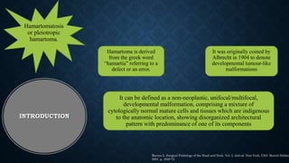
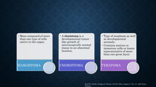
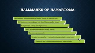
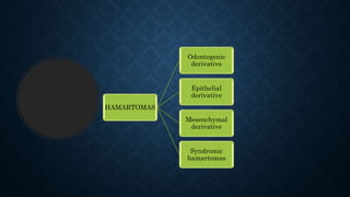
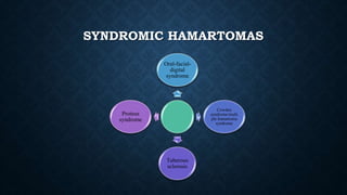
3. Hamartoma is derived
from the greek word
“hamartia” referring to a
defect or an error.
It was originally coined by
Albrecht in 1904 to denote
developmental tumour-like
malformations
Hamartomatosis
or pleiotropic
hamartoma.
It can be defined as a non-neoplastic, unifocal/multifocal,
developmental malformation, comprising a mixture of
cytologically normal mature cells and tissues which are indigenous
to the anatomic location, showing disorganized architectural
pattern with predominance of one of its components
INTRODUCTION
Barnes L. Surgical Pathology of the Head and Neck. Vol. 2. 2nd ed. New York, USA: Marcel Dekker
2001. p. 1649-72
4. • Mass composed of more
than one type of cells
native to the organ.
HAMARTOMA
• A choristoma is a
developmental tumor-
like growth of
microscopically normal
tissue in an abnormal
location.
CHORISTOMA
• Type of neoplasm as well
as developmental
anomaly.
• Contains mature or
immature cells or tissue
representative of more
than one germ layer.
TERATOMA
Brad W. Neville, Douglas D. Damm, Carl M. Allen, Angela C. Chi, 12 - Soft Tissue
Tumors,
5. HALLMARKS OF HAMARTOMA
•Developmental malformation may be present at birth, but manifests later
•Self-limited growth, coordinated with that of the surrounding tissues
•Can present as solitary or multiple masses
Usually not encapsulated with ill-defined margins
May regress spontaneously
Not a true neoplasm, but a true neoplasm may develop in a hamartoma
Association with chromosomal abnormalities and syndromes.
8. DENS INVAGINATUS/DENS IN
DENTE
• Dens invaginatus is a developmental anomaly resulting in
invagination of the enamel organ into the dental papilla
before the mineralization of the dental tissues begin.
Dens in dente, invaginated odontome,
dilated gestant odontoma, dilated
composite odontome,
William's syndrome,
Nance-Huran syndrome
9. PREVALENCE
• Reported prevalence of adult teeth affected with dens
invaginatus is between 0.3% and 10% with the problem
observed in 0.25 % to 26. 1% of individuals examined.
10. Kornfeld (1934) -
invagination results from a
focal failure of growth of
the internal enamel
epithelium while the
surrounding normal
epithelium continues to
proliferate and engulf the
static area
Rushton (1937) -
invagination is a result of
rapid and aggressive
proliferation of a part of
the internal enamel
epithelium invading the
dental papilla. He regarded
this as benign neoplasm of
limited growth
Oehlers (1957) considered
that distortion of the
enamel organ during tooth
development and
subsequent protrusion of a
part of the enamel organ
will lead to the formation of
an enamel-lined channel
ending at the cingulum or
occasionally at the incisal
tip.
Kinson (1943) suggested
that the problem was the
result of external forces
exerting an effect on the
tooth germ during
development . Such forces
could be from adjacent tooth
germs,
ETIOLOGY
11. Type I: An enamel-lined
minor form occurring
within the confines of the
crown not extending
beyond the cemento-
enamel junction.
Type II: An enamel-lined
form which invades the root
but remains confined as a
blind sac. It may or may
not communicate with the
dental pulp.
Type III A: A form which
penetrates through the root
and communicates laterally
with the periodontal ligament
space through a pseudo-
foramen. There is usually no
communication with the pulp,
which lies compressed within
the root.
TYPE III B: A form which
penetrates through the root and
perforating at the apical area
through a pseudoforamen. The
invagination may be completely
lined by enamel, but frequently
cementum will be found lining the
invagination
OHELER’
S
CLASSIFI
CATION
12. CLINICAL
FEATURES
• Due to the deep invagination, which allows entry of irritants into
an area which is separated from pulpal tissue by only a thin layer
of enamel and dentine,
• It is more susceptible for the development of dental caries.
13. RADIOGRAPHIC
FEATURES
• The nature of the peri-invagination is
wide and present in a blunder-buss
formation.
• The root canal almost definitely loses
its vitality, if infected, due to the
intricate communications between the
invagination and the root canal.
• As it invaginates the coronal and
radicular portion, the pulpal outline
may change, resulting in the blunting‟
of the pulp horns.
15. DENS EVAGINATUS
• Tuberculated cusp,
accessory tubercle,
occlusal tuberculated
premolar, Leong's
premolar, evaginatus
odontoma, occlusal
pearl
•Dens evaginatus (DE) or evaginated odontoma is a
developmental anomaly characterized by the presence of
an accessory cusp, abnormal tubercle, or elevation that
occurs in human dentition.
17. CLINICAL
CONSIDERATIONS
• Malocclusion with the
opposing tooth upon the cusp-
like elevation occurs as
involved teeth erupt into the
dental arches.
• The resultant occlusal
traumatic force causes
abnormal wear or fracture of
the tubercle, and is the usual
manner of pulp exposure for
this anomaly
20. ORIGIN
a developmental localized activity of hertwig
epithelial root sheath (HERS) remnants that are
adherent to the root surface during the root
development.
After root formation HERS eventually fragments
to the epithelial cell rests of malassez (ecrms) it
suggested that erms could differentiate into
ameloblast-like cells..
Producing enamel organic matrix deposits on the
root
22. CLINICAL FEATURES
The size and location of
enamel pearls are variable,
ranging in size from 0.3 mm to
4 mm in diameter or even
greater to measurements of
1.8 mm wide and 8 mm long.
Enamel pearls are small
rounded nodules that most
frequently develop in the
furcation/bifurcation or
trifurcation of teeth
The majority occur on the
roots near the cementoenamel
junction area of molars,
premolars and incisors rarely
are affected, the involvement
of deciduous molars has been
reported.
In most of the cases, one pearl
is found in the bifurcation but
it’s been documented four
pearls on a single tooth
23. TREATMENT
• Odontoplasty can be
performed if required to gain
adequate access to the defect
and to reduce or eliminate
cervical enamel projections or
enamel pearls
24. ODONTOMA
The term odontoma was coined by Paul Broca in
1867.
Broca defined the term as tumors formed by the
overgrowth or transitory of complete dental tissue.
In a broad sense, it means a growth with both the
epithelial and mesenchymal components
exhibiting complete differentiation resulting in
functional ameloblasts and odontoblasts.
These cells in turn form variable amounts of enamel
and dentin and pulpal tissue of the odontoma.
28. SIGNS AND
SYMPTOMS
Asymptomatic,
although
occasionally
signs and
symptoms
relating to their
presence do
occur.
Unerupted or impacted
teeth, retained deciduous
teeth, swelling, and evidence
of infection.
Compound
odontomas
seldom cause
bony expansion
but complex
odontomas often
cause slight or
even marked
bony expansion.
The presence of odontomas
may lead to mal-positioning
or displacement of adjacent
teeth, aplasia, malformation
and devitalization of
adjacent teeth.
29. TREATMENT
• As it is a capsulated tumor, its removal is a
simple surgical procedure but special care
should be taken to remove it totally in order to
avoid a relapse which is especially critical in
immature complex odontomas.
• Odontomas are easily enucleated and adjacent
teeth that may have been displaced are seldom
harmed by surgical excision because they are
usually separated by a septum of bone.
31. ORAL AND LABIAL
MELANOTIC MACULE
• They represent a well-circumscribed flat
area of brown to black mucosal
pigmentation. There is an increase in
melanin production by normal mature
melanocytes (without increase in their
number).
• It is associated with Peutz–Jeghers
syndrome and Addison's disease
32. ORAL MELANOCYTIC
NEVI
• Nevi or mole represents collection of
nevus cells which are derivatives of
melanocytes or their precursor neural
crest cells. Oral nevi are usually small
and show regular symmetrical outline
with no change in colour, shade or
texture over time. Static nevi do not
require excision and may be followed up
34. CONGENITAL AND INFANTILE
HAEMANGIOMA
Congenital haemangioma (CH) and infantile haemangioma (IH) are
present at birth or develop in the infancy period.
Majority of them involute spontaneously or gradually over the years. Microscopically,
the proliferative phase of IH and CH comprises complex cellular mixtures, chiefly the
endothelial cells.
Associated syndromes include PHACES and LUMBAR syndrome.
35. VASCULAR
MALFORMATIONS
• Vascular malformation (VM) refers
to congenital morphogenic
anomalies of the various vessels.
Histologically, they comprise of
normal vascular components.
• VM may occur as primary or in
association with regional or diffuse
syndromes such as Sturge–Weber,
Klippel–Trénaunay, proteus
syndrome, Bannayan–Riley–
Ruvalcaba syndrome, and Osler–
Weber–Rendu, to mention a few.
36. GLOMUVENOUS
MALFORMATIONS
• Glomuvenous malformation (GVM) occurs
more often in children. Clinically, it
appears as red-to-blue nodules or
multifocal plaque-like lesions.
• Microscopically, GVM is composed of
varying proportion of blood vessels and
glomus cells. The familial cases have been
linked to mutations in the glomulin gene
located in chromosome 1p21-22
37. EXOSTOSES
• They are described as peripheral
localized overgrowth of the bone.
• Based on the anatomic location in the
jaws, they are termed as buccal bone
exostoses, torus palatinus, and torus
mandibularis.
• Surgical intervention is required only
in case of tissue trauma, periodontal or
prosthodontic impediment.
38. RHABDOMYOMATOUS
MESENCHYMAL HAMARTOMA
• It is an exceptionally rare congenital lesion of the
oral cavity.
• It chiefly comprises of striated muscle tissue.
• It is associated with Delleman, amniotic band and
Goldenhar syndromes.
39. LEIOMYOMATOUS
HAMARTOMA
• Leiomyomatous hamartoma is another rare entity
which commonly involves the midline of palate
and tongue.
• Oral leiomyoma is more frequent in men than in
women, with a 1.43:1 ratio.
• Peak age incidence reported is 40-49 years old.
40. NEUROFIBROMA
• As the name indicates, it is an admixture of
perineural fibroblasts and Schwann cells.
• It is associated with von Recklinghausen's
neurofibromatosis syndrome.
• Approximately 12% of cases are associated
with the syndrome tend to develop malignancy.
41. FIBROLIPOMATOUS
HAMARTOMA OF
NERVE
• Neural fibrolipoma or fibrolipomatous
hamartoma of nerve (FLHN) is a
tumour-like lipomatous process.
• FLHN was reported in the pharyngeal
mucosa by Kumar et al.
• Some lesions may represent carpal
tunnel syndrome as a late complication
42. ORAL NEUROVASCULAR
HAMARTOMA
• The hamartomatous nature of oral neurovascular
hamartoma can be supported by the characteristics
such as limited growth potential, ill-defined borders
and histologically consisting of closely packed
groups of well-formed nerve bundles and vessels.
45. ADENOMATOID ODONTOGENIC TUMOUR
• The hamartomatous nature of adenomatoid
odontogenic tumor is supported by its limited
growth potential and lack of recurrence.
46. CONTROVERSIAL HAMARTOMAS
Adenomatoid
hyperplasia
of
minor
salivary
gland
It is a rare lesion of the
minor salivary glands.
Clinically it presents as
a solitary, painless mass
or nodule.
Microscopically, it
comprises of lobular
aggregates of normal
mucus acini. Recurrence
and malignancy are not
reported.
Hyperplastic
dental
follicle
It has been referred as an
odontogenic
hamartomatous lesion
associated with
single/multiple
unerupted teeth. It
commonly involves
permanent first and
second molars.
Microscopically, it
comprises of
odontogenic epithelium
and calcifications
Ameloblastic
fibroma,
ameloblastic
fibro-dentinoma
and
fibro-odontoma
Reichart and Philipsen
proposed a neoplastic
and hamartomatous line
of development for the
mixed odontogenic
tumours. But currently,
there is no substantial
evidence to prove either
of the above hypothesis.
47. CONTROVERSIAL
HAMARTOMA
• Mucosal neuroma
• It is commonly associated with multiple endocrine
neoplasia (MEN) IIB syndrome.
• Clinically, it occurs in multiple small masses.
• Microscopically, it is partially encapsulated and
contains aggregation or proliferation of histologically
normal nerves.
Congenital granular cell tumour
Congenital granular cell tumour is thought to be a variant of
granular cell tumour, but the exact nature of the lesion is
unclear.
It exclusively occurs in infants or immediately after birth.
Most of the lesions cease to grow or regress spontaneously
without intervention
48.
49. 1. Pilch BZ. Larynx and hypopharynx. In: Pilch BZ,
editor. Head and Neck Surgical Pathology.
Philadelphia, USA: Lippincott Williams and Wilkins;
2001. p. 230-83.
2. Craig O. Hamartoma. Postgrad Med J 1965;41:636-8.
3. Barnes L. Surgical Pathology of the Head and Neck.
Vol. 2. 2nd ed. New York, USA: Marcel Dekker; 2001. p.
1649-72.
4. Kumar V, Abbas AK, Aster JC, editors. Robbins Basic
Pathology. 9th ed. Philadelphia, USA: Saunders,
Elsevier; 2013. p. 257.
5. Allon I, Allon DM, Hirshberg A, Shlomi B, Lifschitz-
Mercer B, Kaplan I. Oral neurovascular hamartoma: A
lesion searching for a name. J Oral Pathol Med
2012;41:348-53.
The occurrence of multiple hamartomas in the same patient is often referred as
Osseous choristoma Lingua1 thyroid choristoma Lingual sebaceous choristoma Glial choristoma Gastric mucosal choristoma
A teratoma is a rare type of germ cell tumor that may contain immature or fully formed tissue, including teeth, hair, bone and muscle
Dens invagination in a human tooth was first described by a dentist named 'Socrates' in 1856.
The prevalence is 0.3–10%. tooth inclusion, dentoid in dente, gestant odontome, dents telescopes
The permanent maxillary lateral incisor appears to be the most frequently affected.
In some cases, the enamel-lining is deficient and channels may also exist between the invagination and the pulp.
Hence, pulp necrosis often occurs at early stages, within a few years of eruption, sometimes even before root end closure
The defect may differ in shape and size from a loop like, pear-shaped or slightly radiolucent structure to a severe type resembling tooth within a tooth.
The outline of the invagination is generally well defined with an opaque layer of enamel
It consists of enamel covering a dentinal core that usually contains pulp tissue.
The presence of pulp within the cusp-like tubercle has clinical significance and distinguishes it from supplemental cusps, such as cusp of carabelli .
Most commonly associated with premolars.
There is typically a bilateral, symmetric distribution, with a slight sexual predilection for female
Because the DE tubercle may extend above the occlusal surface up to 3.5 mm (6.0 mm for anterior teeth),
The prevalence of enamel pearls is variable in the literature it ranges from 1.1% to 9.7% The prevalence of enamel pearls varies according to the population studied and is highest in Asians
In the literature, Enamel pearls are described as three typesA], which are composed by enamel only
B], which contain a core of tubular dentine;
C], which contain a pulp horn that may be an extension from the pulp chamber or root canal
This enamel and dentin were usually laid down in an abnormal pattern because the organization of odontogenic cells failed to reach the normal state of morph differentiation. So they are considered as developmental anomalies rather than true neoplasm.
(P)osterior fossa and other structural brain malformations; large (H)emangiomas of the face, neck, and/or scalp; anatomical anomalies of the cerebral or cervical (A)rteries; (C)ardiac anomalies/(C)oarctation of the aorta; and (E)ye abnormalities with sternal anomalies.
Lower body hemangioma, Urogenital abnormalities / ulceration, Myelopathy, Bony deformities, Anorectal malformations / arterial anomalies, and Rectal anomalies.
Glomus cells are the cells located in carotid bodies and aortic bodies, they sense carbon dioxide, oxygen and Ph levels of blood.
Microtia (a partially formed or completely absent ear) and other ear abnormalities
Underdeveloped facial muscles which may be associated with weakness
Underdeveloped jaw, cheekbone and/or temple bone
Cleft lip and/or palate
Abnormalities of the eyes, such as anophthalmia/microphthalmia, epibulbar tumors (noncancerous growths in the eyes), retinal abnormalities, and vision loss
An unusually large or small mouth
Dental abnormalities
The majority of the cases reported indicated an asymptomatic slow-growing mass, firm to touch and the color of the lesions depended on their depth and on their vascularity. Lucas[9] published a case of a gingival leiomyoma, which appeared as purple swelling. Most of the lesions are asymptomatic masses but some authors have reported symptomatic lesions, most frequently having difficulty in chewing or swallowing, and loose teeth, induced due to local growth
Tuberous sclerosis (TS) is a rare syndrome characterized by the classic triad of seizures, mental deficiency and angiofibromas, affecting about 1 in 6000 people.[34] Oral hamartomas in TS were reported by Celenk et al. (2005) and Amin and O’Callaghan (2012).[34,35]
Cowden syndrome represents the principal PTEN (phosphatase and tensin homolog) gene-related disorder which occurs in 1 in 200,000 people.[34] The oral manifestations include multiple papules involving the gingivae, buccal mucosa and dorsum of tongue.
Proteus syndrome is a rare congenital hamartomatous condition with an incidence of less than 1 in a population of 1 million.[34] Reported associated oral hamartomas include exostoses of facial bones and lymphangiomas.[36]
Oral-facial-digital syndrome (OFDS) comprises a group of heterogeneous disorders with an incidence of 1 in 50,000–250,000 newborns.[34] The oral hamartomatous findings include lingual hamartomas (in 70% cases of OFDS I). Microscopically, they are composed of muscles, adipose tissues and salivary glands
It is also known as ‘two-thirds tumor,’ because 2/3rd of cases occur in the maxilla, 2/3rd occur in young females, 2/3rd of the cases are associated with un-erupted teeth, and 2/3rd of the affected teeth are canines.
Oral hamartomas are unique presentations of the head and neck region.
Hamartomas should promptly be included in the differential diagnosis of the tumours of oral cavity, essentially the paediatric tumours, to avoid aggressive treatment and morbidity