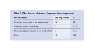
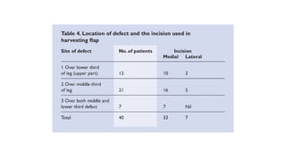
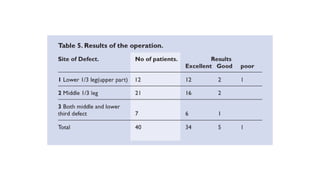
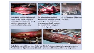
Hemisoleus muscle flap in the
reconstruction of exposed
bones in the lower limb
• Journal of Wound Care
• vol 22 , no 11 , NOVEMBER 2013
• I. Ahmad et al; JNMU, Aligarh
Introduction
• Severe lower limb traumas are common among victims of road
accidents.
• The relatively unprotected anteromedial portion of the tibia is at
risk of bone exposure following trauma
• The use of local skin flaps and fasciocutaneous flaps may become
limited especially if their pedicles come within the zone of injury.
• According to the authors, microvascular free-tissue transfer
increases the ability to close wounds, transfer vascularised bone,
and prevent amputation, yet remains a complex, invasive
procedure with significant complication rates, donor site
morbidity, and failure rates.
• In these cases, muscle flaps are a good option to cover
these defects in order to combat infection and to provide
good vascularity to the exposed area of bone for better
healing.
• In addition, the muscle flap provides bulk to the bony defect
• Mathes et al. described the proximally-based soleus muscle
flap,6 which has been used for the soft tissue coverage of
the middle third of the leg.
• It has also been widely employed for the treatment of
chronic osteomyelitis of the leg bones
• To further investigate the use of the proximallybased soleus muscle
flap for the treatment of complex wounds of the lower legs, this study
aimed to treat specifically patients with defects in the middle and
lower third of the leg using the hemisoleus muscle flap.
• In cases involving large soft defects, both hemisoleus and hemi-
gastrocnemius flaps were used
Methodolgy
• This prospective study conducted between October 2010 and January
2013 involved patients between the ages of 15–65 with defects
present in the middle and upper part of the lower third of the leg.
• For division of the leg into upper middle and lower leg, reference
points were taken as the femorotibial joint above and the lateral
malleolus below.
• Preoperative Doppler ultrasound was used to locate the perforator.
• Prior to the reconstructive procedure, the wound was thoroughly
debrided aseptically, with dressing applied on a daily basis.
• An external fixator was applied to stabilise the fractured segment
• Depending upon the position of the defect and ease of rotation, either
the medial or lateral soleus was used to cover the defects
Surgical anatomy
• The fibrous interosseous membrane connecting the tibia to the
fibula divides the leg into anterior and posterior compartments.
• The anterior compartment is further divided into anterior and lateral
compartments by an intermuscular fascia.
• The posterior compartment is divided into superficial and deep
compartments by a thin intermuscular fascia.
• The superficial compartment contains the gastrocnemius, soleus,
and plantaris muscles.
• The gastrocnemius and soleus join together to become the Achilles
tendon that inserts into the calcaneal bone.
• The soleus muscle is vascularised by the posterior tibial and
peroneal arteries, which delivers blood supply through several
perforators along its course
• The soleus muscle is a large, broad, bipennate muscle consisting of a
lateral and a medial head with independent neurovascular supply.
• It is categorised as a type II muscle flap. The muscle is supplied by large
and small vascular pedicles.
• The lateral and medial hemisoleus muscles are separated in the midline
by a septum, which is present in the distal part of the muscle.
• Intramuscular septum has a distinct watershed in the blood supply of
the proximal half of the muscle and many fine vascular communications
between intramuscular vascular territories.
• The two muscle heads join together to form the dorsolateral and
dorsomedial component of the Achilles tendon.
• This tendon, which is formed by the soleus and gastrocnemius muscles,
inserts on the calcaneum tuberosity.
Surgical technique
• The soleus muscle was harvested through a medial and lateral approach. The most
reliable perforator consistently found during the study using colour Doppler imaging
was located around 8–9cm below the joint line. This was taken as the upper limit of
dissection.
• Medial approach: First, the incision was drawn from the medial malleolus up to the
proximal part of the leg located about 1.5cm posterior to the medial aspect of the
tibia. If the skin defect was in the leg, the line was usually continuous with the defect.
• The saphenous nerve and the great saphenous vein were identified, and a
longitudinal incision was made posterior to these two structures. Dissection between
the gastrocnemius and soleus was performed by bluntly separating the two muscles.
• By sharp dissection, the medial head of the soleus (medial hemisoleus) was
separated from its tibial attachments. Care was taken not to damage the deep fascia
covering the posterior tibial neurovascular bundle.
• During this dissection, the posterior tibial neurovascular
pedicle became visible and was preserved
• For harvesting of a proximally-based soleus flap, the
dissection was continued by releasing the aponeurosis of
the soleus muscle from the aponeurosis of the
gastrocnemius (separate the component of each muscle
within the Achilles tendon), and at last the distal part of the
soleus with its aponeurosis component was cut
• Lateral approach:
• The incision was made longitudinally along the fibula bone from the
fibula neck down to the lateral malleolus. The lateral incision was
outlined on the donor leg along the fibula for the lower part and
reaching the popliteal area by an oblique extension.
• After incising the skin, subcutaneous tissue and fascia, in order to
achieve better exposure of the proximal part of the fibula, lateral
soleus and surrounding vessels and nerves, the superior attachment
of the lateral gastrocnemius was severed from the femur and the
muscle belly was split longitudinally
• Once the flap was inset, it was secured through absorbable suture.
Raw muscle belly was covered through split thickness skin grafts
preferably taken from non-traumatised limb.
• A suction drain was placed underneath the flap to drain out
collection. The flap was monitored at regular intervals.
• In the case of a distal lower leg soft tissue defect, the hemisoleus
required a few relaxing incisions, allowing it to advance more
distally and cover the defect easily.
• Postoperative care:
• Postoperatively, the limb was immobilised with a slab. Flap
immobilisation was maintained for 7 days. During the course of
treatment, the limb was kept in an elevated position.
• The flap was regularly evaluated through a small rent over the skin
graft. The appropriate uptake of the graft is an indication of
survival of the flap. Once bone was found to be united after
radiological evaluation, partial weight bearing commenced.
• The outcome and success of the flap surgery was evaluated on the basis of
flap survival, graft intake, functional gain and donor site morbidity.
• Flap survival was graded as excellent, good or poor.
• A grading of excellent depicts no flap lost and survival of the skin grafted
over the flap surface, good depicts where there is some loss of skin
grafted over the flap or marginal loss of the flap, and poor depicts the
complete loss of the flap or if there is necrosis of more than 50% of the
flap.
• The functional outcomes were evaluated on the patient’s capacity to gain
full flexion and extension, to bear weight, return to work and the
requirement for assisting devices.
RESULTS
• Post-operative functional gain was also analysed.
• Full flexion and extension were reported at the ankle joint in
30 patients.
• A further 9 patients had restricted movement at ankle
joints. The average time for partial weight gain was 3
months for a patient having a non-fractured tibia
Discussion
• In this study, we have successfully used the proximally-based hemisoleus flap
for the coverage of soft tissue defects over the middle and distal lower leg.
• Along with the hemisoleus muscle, we have used gastrocnemius in some
cases with large soft tissue defects.
• In the present study, we used both medial and lateral approaches for
hemisoleus flap harvesting.
• The lateral approach required dissection of an additional peroneal
compartment; the risk of damaging the common peroneal nerve is always
present.
• The most difficult dissection involved cases of large wound size, long
standing and severe cases of trauma. Additionally, chronic inflammation
resulted in dense fibrosis.
• The bulk available for the covering flap was insufficient in some cases; atrophy
of the muscle was significant in these type of defects.
• Pu et al in his series of four patients with an extensive mid-tibial wound of
the leg, successfully used the combined medial gastrocnemius and medial
hemisoleus muscle flaps for soft-tissue reconstruction.
• Limb salvage was achieved in all four patients during follow-up.
• In a subsequent study, patients with an open tibial wound in the junction
of the middle and distal thirds of the leg underwent a soft tissue coverage
using the proximally-based medial hemisoleus muscle flap, demonstrating
positive results.
• Kauffman et al21 reviewed the outcome of 12 patients who underwent
soleus flap reconstruction of distal third lower extremity defects. Nine
of the 12 patients achieved a healed, stable wound; however, several
flaps and multiple additional procedures were often required.
• One of the 12 patients experienced soleus flap loss and two of the
patients required below-knee amputations.
• Failure of limb salvage was related to traumatic injuries or comorbid
conditions such as peripheral vascular disease, smoking, and planned
radiation
Conclusion
• The soleus muscle flap is an excellent flap for the
reconstruction of the middle and lower third leg defects. It is
ideal for covering exposed, osteomyelitic and dead bones.
THANK YOU

Hemisoleus flap FOR LOWER LIMB DEFECTA .pptx

  • 1.
    Hemisoleus muscle flapin the reconstruction of exposed bones in the lower limb
  • 2.
    • Journal ofWound Care • vol 22 , no 11 , NOVEMBER 2013 • I. Ahmad et al; JNMU, Aligarh
  • 3.
    Introduction • Severe lowerlimb traumas are common among victims of road accidents. • The relatively unprotected anteromedial portion of the tibia is at risk of bone exposure following trauma • The use of local skin flaps and fasciocutaneous flaps may become limited especially if their pedicles come within the zone of injury. • According to the authors, microvascular free-tissue transfer increases the ability to close wounds, transfer vascularised bone, and prevent amputation, yet remains a complex, invasive procedure with significant complication rates, donor site morbidity, and failure rates.
  • 4.
    • In thesecases, muscle flaps are a good option to cover these defects in order to combat infection and to provide good vascularity to the exposed area of bone for better healing. • In addition, the muscle flap provides bulk to the bony defect • Mathes et al. described the proximally-based soleus muscle flap,6 which has been used for the soft tissue coverage of the middle third of the leg. • It has also been widely employed for the treatment of chronic osteomyelitis of the leg bones
  • 5.
    • To furtherinvestigate the use of the proximallybased soleus muscle flap for the treatment of complex wounds of the lower legs, this study aimed to treat specifically patients with defects in the middle and lower third of the leg using the hemisoleus muscle flap. • In cases involving large soft defects, both hemisoleus and hemi- gastrocnemius flaps were used
  • 6.
    Methodolgy • This prospectivestudy conducted between October 2010 and January 2013 involved patients between the ages of 15–65 with defects present in the middle and upper part of the lower third of the leg. • For division of the leg into upper middle and lower leg, reference points were taken as the femorotibial joint above and the lateral malleolus below. • Preoperative Doppler ultrasound was used to locate the perforator. • Prior to the reconstructive procedure, the wound was thoroughly debrided aseptically, with dressing applied on a daily basis. • An external fixator was applied to stabilise the fractured segment
  • 7.
    • Depending uponthe position of the defect and ease of rotation, either the medial or lateral soleus was used to cover the defects
  • 8.
    Surgical anatomy • Thefibrous interosseous membrane connecting the tibia to the fibula divides the leg into anterior and posterior compartments. • The anterior compartment is further divided into anterior and lateral compartments by an intermuscular fascia. • The posterior compartment is divided into superficial and deep compartments by a thin intermuscular fascia. • The superficial compartment contains the gastrocnemius, soleus, and plantaris muscles. • The gastrocnemius and soleus join together to become the Achilles tendon that inserts into the calcaneal bone. • The soleus muscle is vascularised by the posterior tibial and peroneal arteries, which delivers blood supply through several perforators along its course
  • 9.
    • The soleusmuscle is a large, broad, bipennate muscle consisting of a lateral and a medial head with independent neurovascular supply. • It is categorised as a type II muscle flap. The muscle is supplied by large and small vascular pedicles. • The lateral and medial hemisoleus muscles are separated in the midline by a septum, which is present in the distal part of the muscle. • Intramuscular septum has a distinct watershed in the blood supply of the proximal half of the muscle and many fine vascular communications between intramuscular vascular territories. • The two muscle heads join together to form the dorsolateral and dorsomedial component of the Achilles tendon. • This tendon, which is formed by the soleus and gastrocnemius muscles, inserts on the calcaneum tuberosity.
  • 10.
    Surgical technique • Thesoleus muscle was harvested through a medial and lateral approach. The most reliable perforator consistently found during the study using colour Doppler imaging was located around 8–9cm below the joint line. This was taken as the upper limit of dissection. • Medial approach: First, the incision was drawn from the medial malleolus up to the proximal part of the leg located about 1.5cm posterior to the medial aspect of the tibia. If the skin defect was in the leg, the line was usually continuous with the defect. • The saphenous nerve and the great saphenous vein were identified, and a longitudinal incision was made posterior to these two structures. Dissection between the gastrocnemius and soleus was performed by bluntly separating the two muscles. • By sharp dissection, the medial head of the soleus (medial hemisoleus) was separated from its tibial attachments. Care was taken not to damage the deep fascia covering the posterior tibial neurovascular bundle.
  • 11.
    • During thisdissection, the posterior tibial neurovascular pedicle became visible and was preserved • For harvesting of a proximally-based soleus flap, the dissection was continued by releasing the aponeurosis of the soleus muscle from the aponeurosis of the gastrocnemius (separate the component of each muscle within the Achilles tendon), and at last the distal part of the soleus with its aponeurosis component was cut
  • 12.
    • Lateral approach: •The incision was made longitudinally along the fibula bone from the fibula neck down to the lateral malleolus. The lateral incision was outlined on the donor leg along the fibula for the lower part and reaching the popliteal area by an oblique extension. • After incising the skin, subcutaneous tissue and fascia, in order to achieve better exposure of the proximal part of the fibula, lateral soleus and surrounding vessels and nerves, the superior attachment of the lateral gastrocnemius was severed from the femur and the muscle belly was split longitudinally • Once the flap was inset, it was secured through absorbable suture. Raw muscle belly was covered through split thickness skin grafts preferably taken from non-traumatised limb.
  • 13.
    • A suctiondrain was placed underneath the flap to drain out collection. The flap was monitored at regular intervals. • In the case of a distal lower leg soft tissue defect, the hemisoleus required a few relaxing incisions, allowing it to advance more distally and cover the defect easily. • Postoperative care: • Postoperatively, the limb was immobilised with a slab. Flap immobilisation was maintained for 7 days. During the course of treatment, the limb was kept in an elevated position. • The flap was regularly evaluated through a small rent over the skin graft. The appropriate uptake of the graft is an indication of survival of the flap. Once bone was found to be united after radiological evaluation, partial weight bearing commenced.
  • 14.
    • The outcomeand success of the flap surgery was evaluated on the basis of flap survival, graft intake, functional gain and donor site morbidity. • Flap survival was graded as excellent, good or poor. • A grading of excellent depicts no flap lost and survival of the skin grafted over the flap surface, good depicts where there is some loss of skin grafted over the flap or marginal loss of the flap, and poor depicts the complete loss of the flap or if there is necrosis of more than 50% of the flap. • The functional outcomes were evaluated on the patient’s capacity to gain full flexion and extension, to bear weight, return to work and the requirement for assisting devices.
  • 15.
  • 21.
    • Post-operative functionalgain was also analysed. • Full flexion and extension were reported at the ankle joint in 30 patients. • A further 9 patients had restricted movement at ankle joints. The average time for partial weight gain was 3 months for a patient having a non-fractured tibia
  • 22.
    Discussion • In thisstudy, we have successfully used the proximally-based hemisoleus flap for the coverage of soft tissue defects over the middle and distal lower leg. • Along with the hemisoleus muscle, we have used gastrocnemius in some cases with large soft tissue defects. • In the present study, we used both medial and lateral approaches for hemisoleus flap harvesting. • The lateral approach required dissection of an additional peroneal compartment; the risk of damaging the common peroneal nerve is always present. • The most difficult dissection involved cases of large wound size, long standing and severe cases of trauma. Additionally, chronic inflammation resulted in dense fibrosis. • The bulk available for the covering flap was insufficient in some cases; atrophy of the muscle was significant in these type of defects.
  • 23.
    • Pu etal in his series of four patients with an extensive mid-tibial wound of the leg, successfully used the combined medial gastrocnemius and medial hemisoleus muscle flaps for soft-tissue reconstruction. • Limb salvage was achieved in all four patients during follow-up. • In a subsequent study, patients with an open tibial wound in the junction of the middle and distal thirds of the leg underwent a soft tissue coverage using the proximally-based medial hemisoleus muscle flap, demonstrating positive results.
  • 24.
    • Kauffman etal21 reviewed the outcome of 12 patients who underwent soleus flap reconstruction of distal third lower extremity defects. Nine of the 12 patients achieved a healed, stable wound; however, several flaps and multiple additional procedures were often required. • One of the 12 patients experienced soleus flap loss and two of the patients required below-knee amputations. • Failure of limb salvage was related to traumatic injuries or comorbid conditions such as peripheral vascular disease, smoking, and planned radiation
  • 25.
    Conclusion • The soleusmuscle flap is an excellent flap for the reconstruction of the middle and lower third leg defects. It is ideal for covering exposed, osteomyelitic and dead bones.
  • 26.